Introduction:
Polymethyl-methacrylate (PMMA) constitutes usage in the widest spectrum of prosthodontic treatment options ranging from removable complete and partial dentures to implant retained prostheses. A prosthesis is subjected to a plethora of organisms in the oral cavity. The removal of biofilm deposited on a prosthesis can be accomplished with denture cleansers[1]. A wide variety of denture cleansing agents are used routinely and regularly by denture wearers. Hence, it becomes highly important to determine the effect of denture cleansers on the properties of acrylic resins. This study aimed to evaluate two important properties, colour stability and hardness of three different commercially available heat cure PMMA denture base resins caused by a denture cleansing agent containing sodium perborate.
Materials And Method:
This study was carried out to compare the colour stability and hardness of commonly used, three different commercially available polymethylmethacrylate heat cure denture base resins ( groups A,T and D).
Fabrication Of Specimens
For the fabrication of specimens, dimensions of the customized metallic mold were selected in accordance with the size of diffuse reflectance spectra (DRS) assembly in the spectrophotometer (illustration 1). Thus, total 45 disc shaped wax-patterns of 8mm diameter and 3mm thickness were prepared using metallic mold. 15 wax-patterns for each of the three groups of specimens were invested in a mixture of dental stone and dental plaster. Each of the three flasks contained 15 disc shaped wax patterns (illustration 2). After wax elimination, resin was packed using compression molding technique in the molds created by these wax patterns following manufacturer’s instructions. The resin was bench cured for 30 minutes, followed by heat polymerization cycle in water bath at 730C for 90 minutes, which was followed by final curing 940C for 30 minutes. The flasks were allowed to bench cool for 24 hours before opening. The specimens were removed from the molds and immersed in distilled water at 37 ± 10 C for 48 hours for residual monomer elimination. The excess resin was trimmed with a tungsten carbide bur using a handpiece at low speed (10,000 rpm), taking utmost care not to alter the dimensions. The specimens were checked for the dimensions using a digital verniercaliper (Insize, Amazon, India).
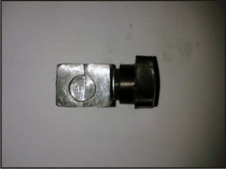 | Illustration 1 : Customized Metallic Mold
 |
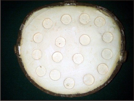 | Illustration 2 : Molds Created By Wax-patterns
|
Colour Analysis
The specimens of each of the three groups were numbered and analysis for colour and hardness was carried out to determine the baseline values. The spectrophotometer (Carrywin UV-Vis-NIR, Agilent Technologies,India) evaluated the amount of light transmitted through the specimens (optical density)[2]. For obtaining the CIE L*a*b* values of the specimens, a digital technique was followed[3]. All the specimens were placed on a black cardboard surface (illustration 3) in order to acquire digital images. A white standard photograph paper was also used with the specimens as a calibration material to eliminate the environmental factors. A digital camera (DSLR- Nikon D 90) was fixed on a tripod, with a 40 cm object–camera distance (without flash). It was oriented perpendicular to the test samples to acquire the digital images. The images were taken at 11:00 AM, on a clear day, were saved in the tagged image file format (TIFF), and were later resolved on a 24 bit resolution screen for further analysis using a commercial graphic software (Adobe Photoshop 6.0)[3],[4]. During the analysis, fixed circular areas with 74 pixels in diameter were selected from the middle third portion of each sample. The L*, a*, b* values of these areas were measured three times by application of the histogram function of the software, and the mean values were recorded. These values were divided by the L*, a*, b* values of the adjacent white photograph paper in order to eliminate the potential effects of the environmental factors, and the corrected L*, a*, b* values of each specimen were recorded as the baseline colour measurement.
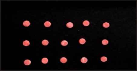 | Illustration 3 : Specimens Arranged On Black Cardboard
|
Hardness Testing
Hardness testing was done using Durometer hardness tester (Shore D) according to ASTM-D2240 (American Standards of Testing and Materials) (illustration 4). The hardness value is determined by the Durometer indenter foot into the sample. If the indenter completely penetrates the sample, a reading zero is obtained, and if no penetration occurs, a reading of 100 results. The reading is dimensionless. Three measurements for indentation hardness were done on each specimen and their average values were recorded. This served as the hardness value before immersion in the denture cleansing solution.
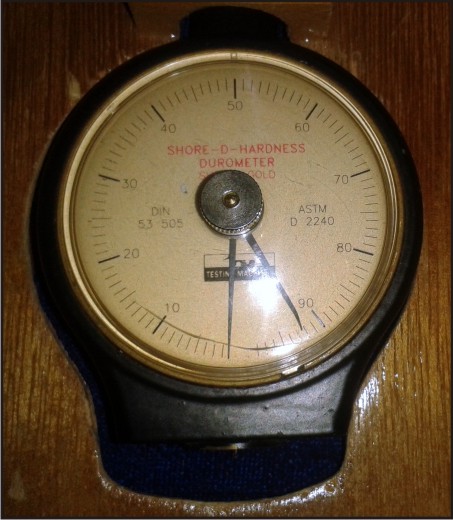 | Illustration 4 : Durometer Hardness Tester (Shore D)
|
Immersion Procedure
Immersion of specimens from the three groups (T, A and D) were carried out at the same time in three different containers containg the cleansing solution (illustration 5). The effervescent cleanser was prepared according to the manufacturer’s instructions, by adding one tablet to 200 ml of warm tap water and the immersion time being 30 minutes. After immersion for 30 minutes, the resin specimens were removed from the cleansing solutions, thoroughly washed in running water, dried with absorbent paper, and then the procedure of immersion was repeated. 180 immersions were performed over a period of 15 days, simulating 180 days of immersion, i.e.; 6 months of denture cleansing by the patient.
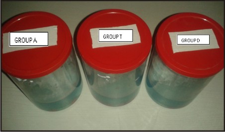 | Illustration 5 : Immersion Of The Specimens In Denture Cleanser
|
Following immersion process, colour and hardness analysis was carried out as mentioned previously. Colour differences (ΔE) between the measurements (before and after immersion in cleanser solution), in terms of L*, a* and b*, were calculated using following equation:[5]
in which ΔL, Δa and Δb are the differences of L, a and b values before and after immersion.
The levels of color change (ΔE) have been quantified by the National Bureau of Standards (NBS) with the NBS units of color difference. NBS units are expressed by the following formula: NBS unit = ΔE × 0.92
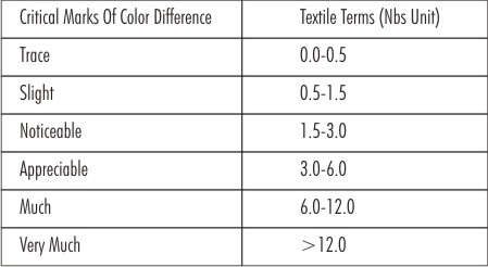 | Critical Marks Of Color Difference According To National Bureau Of Standard (1955)
|
Statistical Analysis
Data was summarized as Mean ± SD. Paired sample t-test was applied to groups [T, A and D] and comparison was done using One way Analysis of Variance (ANOVA), following which post hoc Tukey’s test was used. Paired sample t-test reveal significant change in %T before and after immersion in each group(P<0.001), which showed a reduction in optical density of the specimens after immersion in the denture cleanser (Table 1). Multiple comparisons, Tukey HSD test revealed that amongst the three groups, the color difference was not significant (P>0.3) (Table 2). Paired sample T-test in each group for hardness showed that there was no significant difference before and after immersion (P≥0.2) (Table 3).
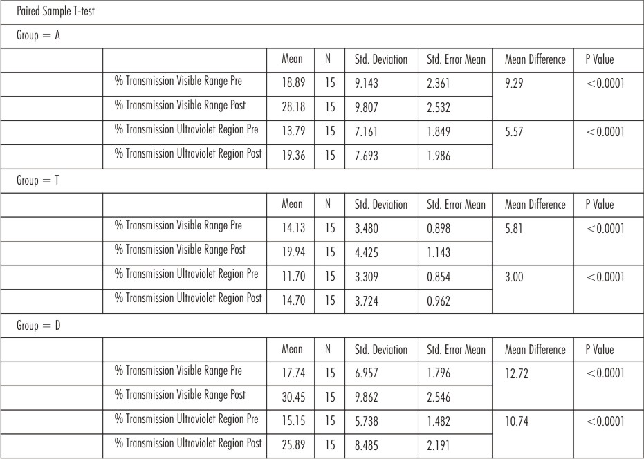 | Table 1 : Paired Sample T-test Reveal Significant Change In %t Before And After Immersion
|
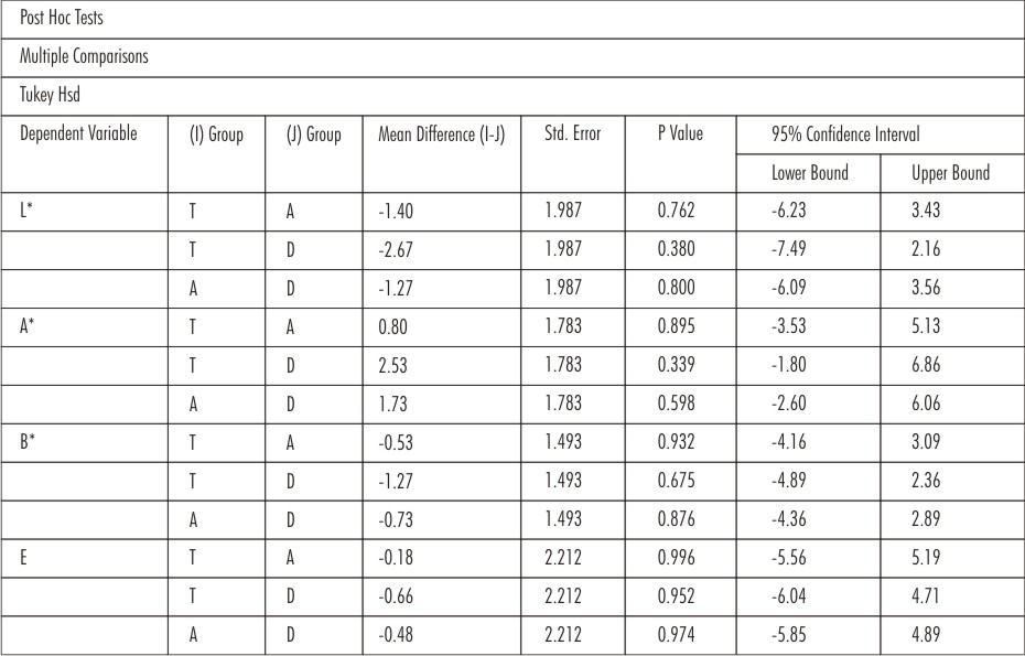 | Table 2 : Tukey Hsd Test Reveals That Amongst The Three Groups, The Colour Difference Is Not Significant (P>0.3)
|
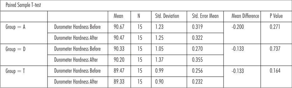 | Table 3 : Paired Sample T-test In Each Group For Hardness Shows That There Is No Significant Difference Before And After Immersion (P>0.2)
|
Results:
The results depicted that the change in optical density was least in group T, followed by group A and D respectively Moreover, the CIE L*a*b* values of the specimens that were obtained with the help of digital camera using a commercial graphic software also revealed the same color change. The change in hardness was not significant.
Discussion:
Mechanical denture cleansing methods are commonly aided by chemical denture cleansing agents which contain sodium perborate and alkaline peroxide in tablet form. These effervescent tablets are classified as chemical soak type products. When dissolved in water, sodium perborate decomposes to form an alkaline peroxide solution. This peroxide solution subsequently releases oxygen, enabling a mechanical cleaning by the oxygen bubbles in addition to the chemical cleaning. Efficacy of these agents in removing stains and reducing formation of biofilm on the irregularities of denture surfaces have been reported,[6]. Evaluating the effect of denture cleansers on the properties of the resin is highly important. Denture base polymers are susceptible to color shifting if the cleansing solutions are not used correctly. The whitening effect relates to the high temperature of the water used in the solution[7]. The effervescent tablets are efficient in removing biofilm and stains, but the alkaline peroxide solution can alter the resin properties if used inappropriately[7] .
Colour changes were evaluated using the CIE L*a*b* colorimetric system,[8],[9] a uniform 3-dimensional system that has been widely used for the determination of chromatic differences by translating combinations of differences into mathematical data. NBS units were also used in this study, because the NBS parameter is important for color comparison and quality control functions. For the acrylic resins evaluated in this study, L* values decreased for all the groups. The same change was noted with the help of spectrophotometer, which showed increase in light transmission (%T) through the specimens after immersion in the denture cleanser, due to the reduced optical density of the specimens. The spectrophotometer is an instrument that separates electromagnetic radiation into its component wavelengths and selectively measures the intensity of radiation after passing through a sample. The absorption or transmission of specific wavelengths is characteristic for a substance,[10], and a spectral analysis serves as a “fingerprint” of the compound.
Colour change is an indicator of aging or damage to dental materials,[11],[12], it increases as immersion time increases. Saraç et al and Purnaveja et al reported that denture cleanser can cause whitening or bleaching, loss of soluble components, or increase in water absorption of PMMA resin materials,[13],[14]. The PMMA resins evaluated in this study tend to change colour during long periods of immersion in distilled water. This could be a result of the leaching out of coloring material. Previous reports have shown that color change in denture base materials is caused by changes in the matrix of the material and the staining effect of external colorants. Solubility, water sorption, leakage, surface roughness, and chemical degradation of denture base material may also cause change in colour. This could explain why L*, a*, and b* values increased or decreased.
Hardness is a term used to describe the resistance of a material to plastic deformation typically measured under indentation load[15]. Concerning the results of the present study, a statistical analysis showed a non-significant difference before and after immersion in the denture cleanser (for each group). The absence of any effect of immersion solutions on the surface hardness of acrylic resins could be due to the presence of cross-linking material which reduces the denture base solubility to organic solvents,[16],[17].
Additional investigations regarding the influence of denture cleanser on the other properties like surface roughness, flexural strength of these materials are indicated. Furthermore, additional studies on the relationship between composition of denture base acrylic resins and their colour stability and hardness are necessary to understand the effects of aging of denture cleansers on the mechanism of colour change and change in hardness. Studies using different concentration of cleansing agents also need to be done.
Conclusion:
Within the limitations of the present study, it can be concluded that sodium perborate based denture cleansing agent causes a change in the color of the PMMA denture base material, when immersion is done over a long period of time, but does not cause a significant change in hardness. So this kind of denture cleansing agents should be used less frequently, and according to manufacturer’s instructions.
References:
1. Joseph R. Comparison of efficacy of sodium hypochlorite withsodiumperborate in the removal of stains from heat cured clear acrylic resin. Journal of Indian Prosthodontic Society 2009; 9:6-12
2. Komiyama O. Kawara M. Stress relaxation of heat-activated acrylic denture base resin in the mold after Processing. J Prosthet Dent 1998;79: 175-81.
3. Cal E., Sonugelen M., Guneri P., Kesercioglu A. , Kose T. Application of a digital technique in evaluating the reliability of shade guides. J Oral Rehabil 2004; 31: 483–491
4. Patel A., Sethuraman R., Prajapati P., Patel J. A comparative analysis of staining characteristics of mouthrinses on provisional resin: An in vitro study. Journal of Interdisciplinary Dentistry 2013 ;3 : 3
5. Hong G , Mundra H , Hamada T. Influence of denture cleansers on the color stability of three types of denture base acrylic resin J Prosthet Dent 2009 ; 101 : 205 – 213
6. Verran J., Maryan C. Retention of Candida albicans on acrylic resin and silicone of different surface topography. J Prosthet Dent 1997; 77: 535
7. Peracini A., Davi L., Ribeiro N., Paranhos H. Effect of denture cleansers on physical properties of heat-polymerized acrylic resin. Journal of Prosthodontic Research 2010; 54: 78-83.
8. CIE (CommisionInternationale de I’Eclaairge). Colorimetery- technical report. CIE Pub. No. 15, 3rd ed. Vienna: Bureau Central de la CIE; 2004. p. 16-20.
9. Wyszecki G, Stiles WS. Color science: concepts and methods, quantitative dataand formulae. 2nd ed. New York: Wiley-Interscience; 1982. P.13-116, 168, 223.
10. Hunter R S., Harold R. W. “ The measurement of appearance”, New York: Wiley; 1987
11. Polyiois C L., Yannikakis S A., Ziisis A J., Donietriou P P. Color changes of denture base materials after disinfection and sterilization immersion. Int J Prosthodont 1997; 10: 83-89.
12. Anil N, Hekimoglu C, Sahin S. Color stability of heat-polymerized and autopolymerized soft denture liners. J Prosthet Dent1999;81:481-4.
13. Saraç D, Saraç YS, Kurt M, Yüzbasioglu E.The effectiveness of denture cleansers on soft denture liners colored by food colorant solutions. J Prosthodont 2007;16: 185-91.
14. Purnaveja S, Fletcher AM, Ritchie GM,Amin WM, Moradians S, Dodd AW. Color stability of two self-curing denture base materials. Biomaterials 1982;3: 249-50.
15. Anusavice. Phillips’ Science of Dental Materials. Eleventh Edition. Elsevier P-73
16. Ogle RE, Sorensen SE, Lewis EA. A new visible light cure system applied to removable prosthodontics. J Prosthet Dent 1986; 56: 497-507.
17. Musuhara E, Hirasawa R. “Studies on reforming dental acrylic resin of the cross-linking PMMA”. Tokyo Medicine and Dental University 1957; 7: 77-81.
|