Introduction
One of the most unusual anomalies of shape of the tooth is fusion. It is union of two separated tooth germs into one. Clinically it appears as two separate crowns joined together or a crown of double the size of a normal tooth. It is usually confused with gemination, which is an attempt of division of a single tooth germ.
The presence of deep fissure in fused teeth predisposes them to dental caries and makes them unaesthetic. Radiographically, there are two separate pulp chambers and root canals to a common pulp chamber and root canal system.[1]
Here we report an unusual case of fused right primary mandibular lateral incisor & canine.
Case Report
A seven & half year old male patient reported to the Department of Pedodontics & Preventive Dentistry with a chief complaint of large mobile tooth. On intraoral examination it was found that 82 & 83 were fused together & was grade II mobile (Figure 1). His medical history was uneventful and the family history for any dental anomaly was non contributory. Since the tooth in question was mobile and the patient had pain during mastication so the choice of treatment was extraction. The fused tooth was extracted (Figure 2) & stainless steel k-files were inserted into the teeth & an IOPAR was taken which confirmed incomplete fusion between 82 & 83 (Figure 3) showing two different pulp chambers & root canals. The parents were informed about the expected delay in eruption of permanent successor & therefore periodic review was advised.
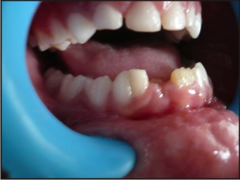 | Fig 1 : Fused Primary 82 & 83
 |
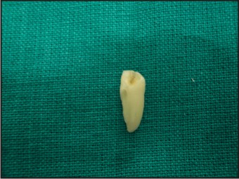 | Fig 2 : Extracted Fused Teeth
|
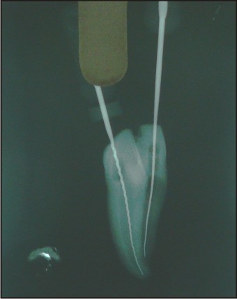 | Fig 3 : Radiograph With K-files
|
Discussion
To differentiate between fusion and gemination,‘Two tooth’ rule was introduced in 1979. According to this rule when fused tooth is counted as one and the number of teeth in the dental arch is reduced, then the term fusion is considered, and if not reduced then the term gemination is considered.[2] In our case the number of teeth in the dental arch is less[1], thus the anomaly was diagnosed as fusion.
Current data in the literature show that double teeth in primary dentition are observed in 0.1-1.6% with no sex predilection.[3]
Depending on the stage of development of teeth, fusion can be classified into two types: complete and partial. The complete fusion begins before calcification and the crown incorporates features of both participating teeth with regard to enamel, dentin, cementum and pulp, whereas the partial fusion occurs at a later stage and the tooth might exhibit separate crowns and fusion may be limited to the roots alone with pulp canals fused or separate. The crowns of fused teeth either appears large and single, or an inciso-cervical groove of varying depth or a bifid crown.[4]
Primary teeth anomalies can affect the permanent successors significantly which has been clearly demonstrated in this particular case. The presence of primary double tooth could also cause delayed resorption of root due to great root mass and increase area of root surface relative to the size of the permanent successor crown. This may lead to delayed or ectopic eruption of the permanent successor.[5] Gellin’s in 1984 reported that the influence of permanent successors was up to 100% when double primary teeth involved the lateral incisors and cuspids. In cases of double teeth involving the mandibular lateral incisors and canines, hypodontia of permanent successors is most common. In contrast, no effect on the permanent successors was observed when the double teeth occurred between the primary mandibular central and lateral incisors, and this result was also in agreement with previous studies. In cases of double teeth involving maxillary central and lateral incisors, hypodontia of permanent successors was observed in only 38% of the cases, not 100% as reported in Nik-Hussein’s study.
Several reports suggested that in order to intercept future malocclusion, further treatment, including extraction, partial removal, or separation of double teeth, should be considered. When dividing double teeth, the complicated dental canal system should be evaluated carefully. Orthodontic and prosthodontic management should be considered to ensure functional occlusion and improve esthetics. In a preventive concern, the labial and lingual vertical grooves of the double primary teeth may be pronounced and difficult to clean, and are highly susceptible to caries. Sealing the grooves with sealant or resin may decrease the risk of caries.[6]
References
1. Rao PK, Mascarenhas R, Jodalli P, Kumar V & Devadig D. Fusion and germination in a primary mandibular anterior teeth. Global journal of medical research dentistry and otolaryngology.2013;13:2
2. Rao PK,Mascarenhas R, Anita A. Fusion in Deciduous Mandibular Anterior Teeth – A Rare Case. Dentistry 2014;S2:001.
3. Sekerci AE, Sisman Y, Ekizer A, Sahman H, Gumus H, Aydinbelge M. Prevalence of double (fused/geminated) primary teeth in turkey — a study. Pakistan oral and dental journal. 2011;31:1.
4. More CB & Tailor MN. Tooth Fusion, a Rare Dental Anomaly: Analysis of Six Cases. International journal of oral and maxillofacial pathology.2012;4(1):50-53.
5. R Veerakumar, M Arul Pari, MN Prabhu. Caution! We are erupting as twins.Journal of clinical and diagnostic research 2011;5:1123-1124.
6. Wu CW, Lin YT & LIN YT. Double primary teeth in children under 17 years old and their correlation with permanent successor. Chang gung medical journal. 2010;33:2.
|