Introduction
In our day to day practice it is not unusual to see several patients with a complaint of trismus. According to Dorland’s Illustrated Medical Dictionary[1] trismus (Greek Trimos: ‘grating’, ‘grinding’) is a motor disturbance of the trigeminal nerve, especially spasm of the masticatory muscles, with difficulty in opening the mouth, a characteristic early symptom of tetanus.
Trismus has a number of potential causes, which ranges from simple and non – progressive to those that are potentially life threatening (Table 1).[2], [3], [4], [5], [6] This condition may impair eating, impede oral hygiene, restrict access for dental procedures and adversely affect speech and facial appearance. A patient is diagnosed to have trismus if the maximal interincisal distance is less than 40-45mm caused by contracture not by obstructive joint impingement. It’s the distance between the incisal edge of the maxillary and mandibular incisors and in edentulous patients, it is measured between the maxillary and mandibular alveolar ridges. Treatment for trismus should be directed at eliminating its cause which includes noninvasive and invasive (surgical) procedures. Noninvasive procedures includes manual manipulation with help of wooden splints such as tongue depressor and dynamic splinting has also been tried in literature effectively for the management. Surgical procedures are considered as rescue procedure but noninvasive procedures are preferred over invasive one.
Here, we describe a unique case of trismus caused by the unusual entrapment of loose miniplate into the extraction socket of upper third molar. A comprehensive literature search on pubmed / medline was performed by using the various combinations of keywords and we found no results. So, we believe, it is the first reported case in the literature with such an unusual etiology.
Table 1: Etiology of Trismus
Trismus
1) Intra articular
A) Ankylosis
B) Arthritis synovitis
C) Meniscus pathology
2) Extra articular
A) Infection
i) Odontogenic
I. Pulpal
II. Periodontal
III. Pericoronal
ii) Nonodontogenic
I. Tonsillitis
II. Tetanus
III. Meningitis
IV. Brain abscess
V. Parotid abscess
B) Trauma
i. Fracture mandible
ii. Fracture zygomatic arch
iii. Incorporation of foreign bodies
C) Dental treatment related
i. Post extraction
ii. Post local anaesthetic injection
D) TMJ
i. Trauma to TMJ
ii. MPDS
iii. Internal disk derangement
E) Tumors and premalignant conditions
i. Primary and secondary tumors of oral cavity involving muscle of mastication, parotid area and jaw joint
ii. Oral sub mucous fibrosis
F) Drugs
I. Phenothiazine
ii. Succinyl choline
iii. Tricyclic antidepressant
iv. Metoclopramide
v. Halothane
G) Radiotherapy and chemotherapy
I. Osteoradionecrois
ii. Post radiation fibrosis
H) Congenital
i. Hypertrophy of coronoid
ii. Trismus pseudo camptodactyly syndrome
I) Miscellaneous
i. Myositis ossificans
ii. Hysteria
iii. Lupus erythematosus
Case Report
A 31 year old male presented with the difficulty in opening the mouth from past 4 months and there was gradual decrease in mouth opening to attain the present situation. Despite many consultations with doctors and dentists, no diagnosis was made till this time. His past medical & dental history revealed that the patient met with a road traffic accident 8 years back and diagnosed to have fracture of right side of maxilla and mandible, for which he was operated with open reduction & internal fixation under GA. Patient was also giving the history of extraction with respect to upper right third molar 4 months back, after which he encountered gradual decrease in mouth opening.
His personal & family histories were non – contributory. Clinical examination revealed almost nil mouth opening (<5mm) (Figure1). His OPG revealed that ORIF had been done i.r.t to both mandible and maxilla. It also revealed the rotation of mandibular angle plate into 18 (Figure 2). So a diagnosis of trismus secondary to rotation of loose plate of mandible was made.
Our treatment included the removal of rotated plate under LA (Figure 3). Post operatively the patient was prescribed muscle relaxant in BD dose for 3 days. After 5th post operative day, the patient was advised to initiate physiotherapy for opening and closing the jaws with the help of dynamic appliance (Figure 4) and to perform lateral excursions of mandible for 5 minutes every 3 – 4 hours along with heat therapy consists of placing moist hot towel on the affected area for 15 – 20 minutes every hour. Sugarless chewing gum was also prescribed twice daily minimum for 15 minutes for 15 days, patient mouth opening was 40mm one month post operatively and 6 months follow up showed promising results (Figure 5).
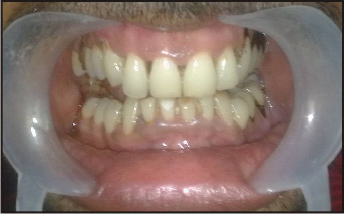 | Fig 1 : Extra Oral View Revealed Almost Nil Mouth Opening.
 |
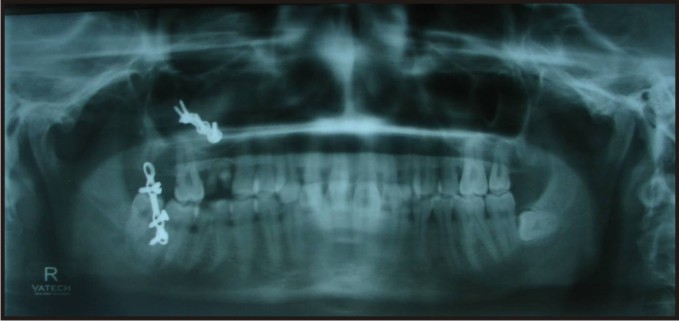 | Fig 2 : Opg Revealed Rotation Of Mandibular Angle Plate Into The Socket Of 18.
|
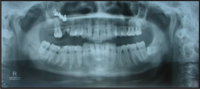 | Fig 3 : Post-operative Opg
|
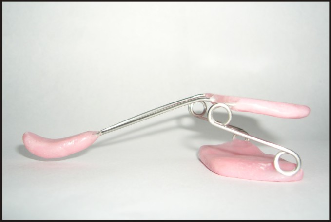 | Fig 4 : Showing The Picture Of Splint Used For This Patient Postoperatively
|
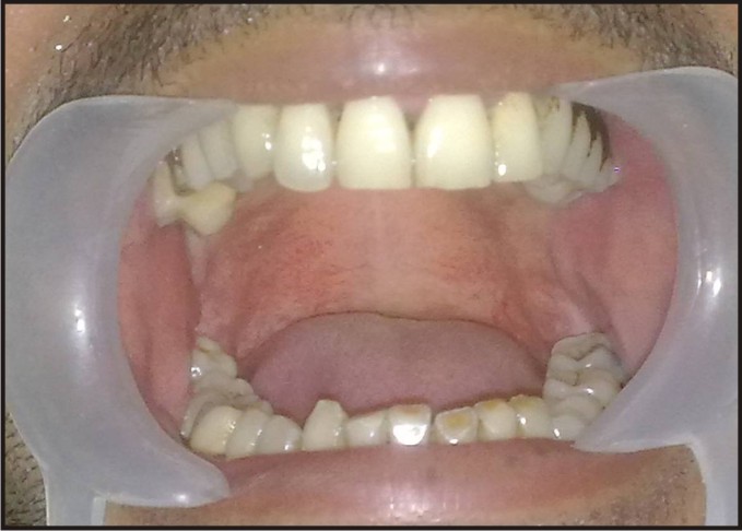 | Fig 5 : Mouth Opening After Six Month Post Op
|
Discussion
Trismus is a pathological condition of the muscles of mastication which commonly affects patients who have undergone dental procedures, oral surgical procedures and radiation therapy for head and neck cancer. Trismus is diagnosed from clinical examination of the maximal inter-incisal distance of less than 40-45mm [7].
For the clinician to diagnose trismus properly, he or she must be able to determine the cause from a variety of possibilities. It is important to obtain a complete history and to perform a thorough clinical examination. Radiographs should be done as deemed appropriate. After eliminating the cause, the practitioner should prescribe the heat therapy, analgesics,a soft diet; and &muscle relaxants to manage the initial phase of muscle spasm [5].
Heat therapy consists of placing moist towels on the affected area for 15-20 minutes per hour. NSAIDS are usually adequate in managing the pain associated with trismus; its anti-inflammatory properties are also beneficial. An anti-anxiety drugs may be required if the discomfort is more intense. If necessary, muscle relaxants can be prescribed with anti-anxiety drugs. When the acute phase is over, the patient should be advised to initiate physiotherapy for opening and closing the jaws and to perform lateral excursions of the mandible for 5 minutes every 3-4 hours. Sugarless chewing gum is another means of providing lateral excursion movement of the TMJ.
Other treatment methods for management includes ice-cream stick exercises, manual spring loaded devices and dynamic splinting. Dynamic splint is different from other two modalities as it uses a reproducible, calibrated and changeable tension. It also enhances the patient`s comfort and compliance [8].
In virtually all cases of trismus that are managed as outlined above, patients report improvement within 48 hours. Therapy should be continued until the patient is free of symptoms. If pain and dysfunction continue unabated beyond 48 hours, the possibility of infection should be considered. Antibiotics should be added to the treatment regimen and continued for 7 days.
Open reduction and internal fixation is the gold standard treatment for fractures. Loosening of screws and plates is one of common complication and that is why; many surgeons believes in removing the plate after six months. In the above mentioned case, patient was having history of open reduction and internal fixation eight years back and had not got his plates removed. This led to loosening of plate and unusual entrapment of plate into recent extraction socket. Thus, came into light an unexplained cause of trismus. This case report shows the improvement in reduced mouth opening on removing the cause and applying the treatment various modalities like prescribing muscle relaxant, heat therapy, physiotherapy in the form of chewing gums and dynamic splint.
In conclusion, the author reports a rare case of trismus. Based on the experience, previous literature and a thorough pubmed and google search, the author has concluded that still, it is not a reported cause of trismus & it should be added to the literature.
References:
1. Taylor EJ, ed. Dorland’s Illustrated Medical Dictionary, 27th ed. Philadelphia: W.B.Saunders, 1998; p.1759.
2. Malamed SF. Handbook of Local Anesthesia, 3rd ed. St. Louis: C.V. Mosby Co., 1990; p.248 – 249.
3. Marien M. Trismus: causes, differential diagnosis and treatment. Gen Dent 1997; 45(4): 350 – 355.
4. Peterson LJ, Ellis E III, Hupp JR, Tucker MR. Contemporary Oral and Maxillofacial Surgery. Toronto: C.V. Mosby Co., 1988; p.223, 257, 383 – 385.
5. PJ Dhanrajani , O Jonaidel. Trismus: Aetilogy, Differential Diagnosis & Treatment. Dent Update 2002; 29: 88-94.
6. Stone J, Kaban LB. Trismus after injection of local anesthetic. Oral Surg. Oral Med Oral Pathol 1979; 48: 29 – 32.
7. Leonard M. Trismus: What is it, what causes it and how to treat it? Dentistry Today 1999: June: 74 – 77.
8. David H Shulman, Barry Shipman, Frank B Willis. Treating trismus with dynamic splinting: a case report. Journal of oral science 2009; 51(1): 141-144.
|