Introduction
Aesthetics plays an important role in contemporary dentistry, especially because of increased demand of beautiful appearance of an individual. Smile considered beautiful if it imitates natural appearance with clear, well aligned teeth and defined anatomical shapes.[1]
Dental fluorosis is defined as hypo mineralization of enamel resulting from excessive ingestion of fluoride during tooth development. The degree to which enamel is affected is dependent upon duration, timing and intensity of the fluoride concentration. In its mild form small white enamel streaks and as severity of condition increase black to brown stains develop.[2] The discoloration of teeth caused by fluorosis can be of esthetic concern for many patients.
Presence of stains and superficial irregularities in dental enamel surface can be solved through use of an enamel micro abrasion technique; however these intrinsic alterations should have a hard texture and affect the superficial layer of enamel.[3]
Case History
Diagnosis And Treatment Planning: A 19 year old female reported to department of Prosthodontics, Mallareddy institute of dental sciences, Hyderabad, with slightly brownish intrinsic stains of a hard texture located to labial enamel surface of maxillary incisors, canines and premolars (Figure 1). History of the patient revealed the presence of yellowish brown and white patches on teeth since childhood. Patient wanted crowns for all six maxillary anterior teeth for better appearance.
Enamel micro abrasion technique with micro abrasion paste indigenously made in the clinic using a mixture of 10 percent hydrochloric acid and polishing pumice powder followed by in office bleaching was proposed to patient and for which she was convinced.
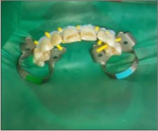 | Image 1: Pre Treatment View
 |
PROCEDURE: The patient was referred to department of Periodontia for oral prophylaxis and polishing of teeth. The enamel micro abrasion procedure was performed by polishing the enamel of each of the maxillary anterior tooth using a slurry made with pumice polishing powder (Neelkanth Minechem, India) and 10 percent hydrochloric acid (Nice Chemical Ltd., India), under rubber dam isolation and light activated gingival barrier (Figure 2).
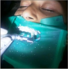 | Image 2: Enamel Micro Abrasion Procedure
|
The micro abrasion material was applied on each tooth by cotton pellets and micro abrasion was done using hand piece driven rubbing action.
The whole procedure was repeated three times for two minutes each, and washing of tooth surface is done using water after each application. After final application, teeth were cleaned with copious water and dried for in office bleaching procedure.
Bleaching procedure was performed using a commercially available (hydrogen peroxide based) in-office bleaching system, Polaoffice (SDI Limited, Australia). After drying the tooth surface, a thin layer of gel obtained from mixing the powder and gel component of the bleaching system was applied on the tooth surface and left for 8 minutes each. Light curing of the gel is done as per manufacturer’s instructions (Figure 3).
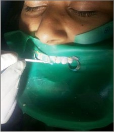 | Image 3: In Office Bleaching After Enamel Microabrasion
|
Bleaching material is cleaned by using suction and wet squeezed cotton. After the desired shade was obtained, the surfaces of teeth were polished by using fluoridated polishing paste and patient was instructed not to have any food substance that might cause sensitivity and discoloration of the teeth. The obtained esthetic outcome was maintained in the subsequent follow up visits (Figure 4).
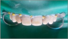 | Image 4: Post Treatment View
|
Discussion
Mottled enamel was first described by Eager in 1901, which was later designated as dental or enamel fluorosis, a condition in which permanent teeth are mainly affected, through it occasionally affects primary teeth.[4] Dental fluorosis is easily identifiable. It is a clinical manifestation of chronic exposure of high intakes of fluoride through drinking water. The signs of dental fluorosis range from a few white flecks to confluent pits in the enamel surface and unsightly dark brown stains.[5]
One frequently used classification was given by Dean to assess the severity of Dental fluorosis based on the clinical appearance of the enamel.[6] According to this classification we categorized this patient in moderate category as her entire labial surfaces of maxillary anterior teeth were involved and minute pitting and brown surface stains were present.
Severely fluorosed teeth that have large enamel defects present serious esthetic concerns and call for management that is conservative as well as effective. Restorative procedures such as porcelain or composite laminate veneers or crowns are invasive techniques in which preparation of teeth is unavoidably needed. Hence to give a conservative, effective and economical solution to these types of patients, microabrasion is commonly used. It is a non invasive, conservative, and cost-effective approach to partially or totally eliminate superficial enamel staining. Lower cost of this procedure makes it a useful treatment option in dental practice. Microabrasion usually accomplish substantial improvement by removing white opaque areas, brown stains and enamel defects, providing acceptable esthetics and eliminating the necessity for more invasive actions.[7]
Isolated brown or white defects of less than few tenths of a millimeter depth can be effectively treated with microabrasion.[8]
It is not possible to correct deeper enamel defects via microabrasion alone. However, a blend of different techniques such as microabrasion/microabrasion along with bleaching or full or partial veneering is available to successfully disguise deeper defects. Microabrasion, either alone or together with bleaching, has the disadvantage of requiring the removal of some enamel. However, this approach is more conservative than preparing the enamel surface for the placement of facial veneers or crowns. Bleaching and microabrasion also have the advantage of being appropriate for partially erupted, young permanent teeth. The placement of facial veneers is normally not considered until a patient’s teeth have fully erupted and the gingival height has stabilized. Thus, ultimate restorative management for enamel discolorations is generally delayed until the child is in mid to late adolescence; even though considerable apprehension over the appearance of discolored teeth can begin many years earlier when the teeth are partially erupted and observable.[9]
Microabrasion technique has been adapted numerous times to augment the esthetic outcome as well as simplify the treatment procedure.[10] Formerly, the use of 18% hydrochloric acid was advocated to eradicate shallow fluorosis stains.[11] Afterward, the procedure was adapted to incorporate the use of pumice and hydrochloric acid paste and was termed microabrasion.[12] Croll further modified the technique, dropping the concentration of acid and increasing the abrasiveness of the paste using silicon carbide particles as a substitute of pumice.[13] The use of 16 percent hydrochloric acid unaided or followed by hydrogen peroxide bleaching can effectively eliminate intrinsic yellow-brown stains.[14]
Even though the exact explanation for the shade alteration that occurs after microabrasion is not known, the micro abraded surface reflects and refracts light from the surface in such a way that mild imperfections in the underlying enamel are disguised. The extremely polished surface of the enamel subsequently abraded with Hydrochloric acid-pumice augments the visual appearance.[15]
Phosphoric acid (H 3 PO 4) that is usually used for etching in dental clinics is a familiar acid for the dentist. Bezzarra et al. (2005) compared the effectiveness of microabrasion obtained by two types of acid: 37% H 3 PO 4 and 18% HCl. They concluded that both acids were equally efficient.[16]
Improvements in aesthetic indices were noted in all fluorotic teeth by both compounds; however, the mean treatment time with HCl-pumice was appreciably lower than Phosphoric acid-pumice.[17]
Also Meireles et al. (2009) reported lower mean surface roughness and larger total demineralization area in teeth treated with Hydrochloric acid-pumice in comparison with the H 3 PO 4 -pumice compound.[18]
So in the current case, it was decided to use Hydrochloric acid for enamel microabrasion as it has the advantages of larger total demineralization with decreased mean treatment time.
The other advantages of using this indigenously made enamel microabrsion paste is that it is conservative in abrading fluorosed enamel as only 10 percent of Hydrochloric acid was used as compared to upto 18 percent of HCL, which removes (360+ 130 um) of enamel thickness [19],as used for microabrasion in previous studies.[14],[16]
This paste can be prepared easily in the dental office using readily available ingredients and it is as efficient in enamel microabrasion as commercially available microabrasion pastes.
It is a quick method to be used in dental operatory for mild to moderate dental fluorosis cases as it can be done in single session in contrast to multiple sittings needed for enamel microabrasion using other commercially available products. Hence, the etch/bleach combination technique using indigenously prepared microabrasion paste, presented in this paper uses materials that are readily available in the dental office and that have been shown to be clinically safe and effective.
However further studies are required to appraise the amount of enamel removed by microabrasion using this concentration of HCL along with the pumice polishing powder. The etch/bleach combination technique offers a conservative treatment for yellow-brown hypo mineralized enamel that shows excellent clinical success and long-term stability. To conclude, the clinical case presented supports a practicable, cost-effective and conservative method to deal with enamel staining due to fluorosis and other causes.
References
1. Sundfeld RH, Rahal V, de Alexandre RS, Briso AL, Neto DS. Smile restoration through use of enamel microabrasion associated with tooth bleaching. Compendium of continuing education in dentistry 2011;32(3):53-7.
2. Strassler HE, Griffin A, Maggio M. Dentistry today 2012; 142.
3. Sarrett DC. Tooth whitening today. J Am Dent Assoc. 2002; 133:1535-8.
4. Teotia SPS. The national medical journal of India 1999; 12.
5. Al-Alousi W, Jackson D, Crompton G, Jenkins OC. Enamel mottling in a fluoride and non-fluoride community-A study. Br Dent J 1975; 138:9-15.
6. Dean HT. Classification of mottled enamel diagnosis. J Am Dent Assoc 1934; 21:1421-6.
7. Yildiz G, Celik EU. A minimally invasive technique for the management of severely fluorosed teeth: A two year follow up. Eur J Dent. 2013 Oct-Dec; 7: 504–508.
8. Croll TP. Enamel microabrasion: Observations after 10 years. J Am Dent Assoc. 1997; 128:45S–50.
9. Wright JT.The etch-bleach-seal technique for managing stained enamel defects in young permanent incisors. Pediatric Dentistry 2002; 24:249-52.
10. Chhabra N, Singbal KP. Viable approach to manage superficial enamel discoloration. Contemp Clin Dent. 2010; 1: 284–287.
11. McCloskey RJ. A Technique for removal of fluorosis stains. J Am Dent Assoc. 1984; 109:63–4.
12. Croll TP, Cavanaugh RR. Enamel color modification by controlled hydrochloric acid-pumice abrasion: Part 1. Technique and examples. Quintessence Int. 1986; 17:81–7.
13. Croll TP. Enamel microabrasion for removal of superficial dysmineralization and decalcification defects. J Am Dent Assoc. 1990; 120:411–5.
14. Wong M. A clinical comparison of treatments for endemic dental fluorosis. J Endodont.1991; 17:343-345.
15. Price RB, Loney RW, Doyle MG, Moulding MB. An evaluation of a technique to remove stains from teeth using microabrasion. J Am Dent Assoc 2003; 134:1066-71.
16. Bezzerra AC, Leal SC, Otero SA, Gravina DB, Cruvinel VR, Ayrton de Toledo O. Enamel opacities removal using two different acids: An in vivo comparison. J Clin Pediatr Dent 2005; 29:147-50.
17. Bassir MM, Bagheri G. Comparison between phosphoric acid and hydrochloric acid in microabrasion technique for the treatment of dental fluorosis. J Conserv Dent 2013; 16:41-4.
18. Miereles AA, Andre Dde A, Ledia FL, Bocnagel JS, Demarco FF. Surface roughness and enamel loss with two microabrasion techniques. J Contemp Dent Pract 2009; 10:58-65.
19. Tong LSM, Pang MKM, Mok NYC, King NM, Wei SHY: The effects of etching, micro-abrasion, and bleaching on surface enamel. J Dent Res 72:67-71, 1993.
|