Introduction:
The ultimate goal of endodontic therapy is to return the involved tooth to a state of health and function. All inflammatory periapical lesions treated with conventional endodontic therapy should heal with hard tissue regeneration and resolution of radiolucency. However, in some cases, the apical periodontitis persists despite root canal treatment, and the endodontic treatment is considered a failure which calls for surgical intervention.[1],[2]
Regenerative endodontic procedures are defined as biologically based procedures designed to replace damaged structures, including dentin and root structures, as well as cells of the pulp-dentin complex.[3] Focus has constantly been on devising a “wonder material” that is most effective in its regenerative potential. Various platelet-derived products or platelet concentrates have been introduced that act as biological mediators aiding the healing response. Platelet-rich fibrin (PRF) represents a new step in the platelet gel therapeutic concept. PRF is a matrix of autologous fibrin, in which are embedded a large quantity of platelet and leukocyte cytokines during centrifugation. The easily applied PRF membrane serves as a matrix to accelerate the healing of wound edges.[4]
The present case report describes the successful management of periapical lesion in the maxillary left central and lateral incisor using PRF as a regenerative material, where conventional endodontic therapy was a failure.
Case Report
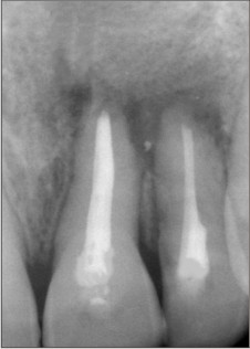
A 15 year old male patient reported to the Department of Conservative Dentistry and Endodontics, Panineeya Mahavidhyalaya Institute of Dental Sciences and Research Centre, Hyderabad, India with a chief complaint of spontaneous dull, throbbing, persistent pain in the upper front tooth region since 2 weeks. His medical history was unremarkable. Dental history revealed an incident of trauma to the upper front teeth region, 7 years ago. Maxillary left central #21 and lateral incisor #22 showed no response to palpation, percussion and vitality test. Radiographic examination revealed large periapical radiolucency of 1.5×2 cm in relation to 21 & 22 [figure 1] and diagnosed as phoenix abscess.
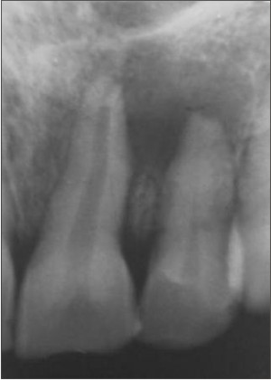 | Figure 1 : Preoperative Intraoral Periapical Radiograph Irt #21 And #22
 |
The initial phase of treatment included the administration of antibiotics (Amoxicillin 500 mg, 3 times a day for 5 days & Metrogyl 400mg 3 times a day for 5 days) and analgesics (ibuprofen 400 mg, 3 times a day for 3 days). The tooth #21, #22 were isolated with rubber dam, conventional root canal treatment was performed using step back technique till an apical size of #45 K-files( Sybron endo, Australia) [figure 2]. A5.25% sodium hypochlorite (Novo Dental Product Pvt Ltd, Mumbai, India) was used to irrigate the canals during rootcanal preparation.
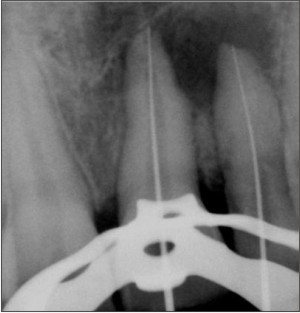 | Figure 2 : Working Length Radiograph
|
The treatment was planned in three visits and calcium hydroxide (Apexcal , ivoclor vivadent,Germany) was used as the intracanal medicament. The root canals were dried using paper points and obturated with gutta-percha points(Dentsply maillefer Ballaigues) and AH plus (Dentsply DeTrey GmbH, Philadelphia, USA) by lateral condensation technique [figure 3].
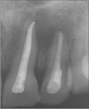 | Figure 3 : Post Obturation Radiograph
|
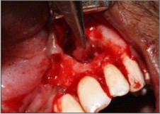
After three months follow up clinically and radiographically patient was having persistent pain and expected healing did not take place. Hence, a periapical endodontic surgery was planned using PRF as a regenerative material. The treatment procedure was explained to patient and before surgery a complete hemogram was advised and all the parameters were within the normal range. Under sterile, aseptic condition local anesthesia (1:200000 adrenaline, DJ Lab, India) was administered followed by the crevicular incision extending from mesial of #21 to distal side of #23 and full thickness mucoperiosteal flap was reflected.A large periapical defect was found extending till nasal floor [figure 4]. Debridement was carried out with through saline irrigation. Using tapered fissure bur (SS White burs), apicectomy was performed. The pathological tissue has been sent for histological examination, which revealed chronic inflammatory tissue.
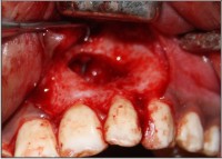 | Figure 4 : Mucoperiosteal Flap Reflected Showing Defect Extending Till Nasal Floor
|
For PRF preparation, 20 mL of blood was drawn from the patient's antecubital vein and centrifuged (REMI centrifuge machine Model R-8c with 12 × 15 mL swing out head Bench top centrifuge , Remi laboratory instruments, India) for 10 min under 3000 revolutions (approximately 400 g) per minute. It settles into the following layers: red lower fraction containing red blood cells, upper straw coloured cellular plasma and the middle fraction containing the fibrin clot. The upper straw coloured layer is then removed and middle fraction is collected, 2 mm below lower dividing line, which is the PRF [figure 5]. As the anticoagulant is not added, the blood begins to coagulate as soon as it comes in contact with the glass surface, therefore for successful outcome speedy blood collection and immediate centrifugation are the essential requirements.
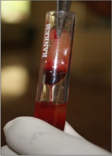 | Figure 5 : Prf Preparation
|
PRF was placed into the intrabony defect up to the surrounding bone level after thorough debridement [figure 6]. The mucoperiosteal flap was repositioned and figure of eight sutures were given using 3–0 nonabsorbable black silk suture (Sutures India Pvt. Ltd, Karnataka, India). A0.2% chlorhexidine gluconate solution as mouth rinse for a period of 5 days was instructed. Suture removal was done 1 week later and the healing was uneventful. Follow up at 6 months [figure 7] and one year [figure 8] showed no signs and symptoms of pain and discomfort both clinically and radiographically.
 | Figure 6 : Prf Placed Into The Intrabony Defect
|
 | Figure 7 : Intraoral Periapical Radiograph After Six Months
|
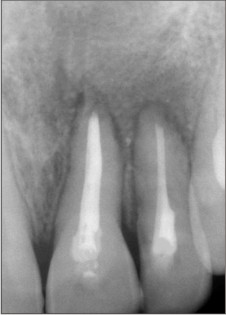 | Figure 8 : Intraoral Periapical Radiograph After One Year Follow Up
|
Discussion:
As understanding the disease process is the key to successfully treating the disease, it is important to understand the biological factors that are related to the failure of endodontically treated teeth that cause persistent periapical radiolucencies.[5]Traditional surgical procedures aimed to treat periapical lesions basically includes debridement of apical lesions, resection, retrograde preparation and retrograde filling of the apex, in which situations healing always and almost occurs by repair.[2]
The harmonized spurs of biology and mechanical regulators promoting cellular activities have critically enhanced the acceptance of regenerative therapy for dental tissues.[6]
Regeneration is defined as reproduction or reconstitution of a lost or injured part which fully restores the architecture or function of the part, it reconstitutes, although not completely, both the architecture and functions of the original components, lack of any of the components would result in repair rather than regeneration, they are : (1) recruitment of progenitor/stem cells to differentiate into committed osteoblasts, PDL cells, and cementoblasts (2) growth/differentiation factors as necessary signals for attachment, migration, proliferation, and differentiation of progenitor/stem cells; and (3) local micro environmental cues such as adhesion molecules, extracellular matrixand associated non-collagenous protein molecules.[2]
Prophylactic antibiotics were prescribed assome authorities feel it justifiable when the maxillary antrum or floor of the nose has been perforated or if there is through and through defect as seen in our present case.[7] According to the current concepts of evidence-based health care, selection between alternative treatments is based on the assessment of their respective benefits and risks. The most important decision whether or not to retreat the tooth is further influenced by technical quality of primary treatment. The praxis concept theory, a retreatment preference score was constructed to correspond to different cut off points and to vary between 0.0 to 1.0. The mean retreatment preference score was 0.38, representative of therapeutic intervention in cases of medium sized lesions which indicates that re-treatment is effective till medium sized lesions.[8] The macrophages and multinucleated gaint cells cannot destroy a great accumulation of cholesterol crystals “in a beneficial way to the host.”Because endodontic retreatment cannot resolve these bodies, periapical surgery was the treatment of choice in our present case.
In order to achieve optimal healing and bone regeneration in our case, where conventional endodontic therapy was failure, PRF (platelet rich fibrin) was used as the regenerative material of choice. Developed in France by Choukroun et al. in 2001, PRF is a second-generation platelet derivative because, unlike other platelet concentrates like PRP, this technique does not require anticoagulants nor bovine thrombin or any other gelifying agent. PRF is a strictly autologous fibrin matrix containing a large quantity of platelet and leukocyte cytokines It contains multitude of growth factors like platelet derived growth factor (PDGF), transforming growth factor β1 (TGF β1), insulin like growth factor (IGF), etc., exhibiting varied potent local properties such as cell migration, cell attachment, cell proliferation, and cell differentiation. It has been shown as an ideal biomaterial for pulp-dentin complex regeneration.[4],[9]
Conversion of fibrinogen to fibrin takes place slowly with small amounts of physiologically available thrombin present in the blood sample, because of this slow polymerization the fibrin network obtained is similar to natural architecture which is very favourable to the healing process and also protects growth factors from proteolysis, thus growth factors can keep their activity for relatively longer periods.A progressive polymerization causes increased incorporation of the circulating (intrinsic) cytokines in the fibrin meshes. This configuration increases the lifespan of these cytokines, as they are released and used only at the time of initial cicatricial remodelling.[10],[11],[12]
In the present case report, where surgical intervention was done using PRF showed a predictable clinical and radiographic findings which suggests that PRF is one of the most easy to obtain, inexpensive, promising biofuel in the field of bone regeneration.
References:
1. Schulz M, von Arx T,Altermatt HJ,Bosshardt D. Histology of periapical lesions obtained during apical surgery. J Endod 2009; 35: 634-42.
2. Jayalakshmi KB,Agarwal S,Singh MP,Vishwanath BT,Krishna A,Agarwal R. Platelet rich fibrin with β-tricalcium phosphate – A novel approach for bone augmentation in chronic periapical lesion: A case report. Case reports in dentistry 2012;Article ID 902858, pg 1-6.
3. Murray PE, Garcia-Godoy F, Hargreaves KM. Regenerative endodontics: a review of current status and a call for action. J Endod 2007;33:377–90.
4. Khiste SV, Tari RN. Platelet-Rich Fibrin as a Biofuel for Tissue Regeneration. ISRN Biomaterials Volume 2013 (2013), Article ID 627367, pp1-6.
5. Yan MT. The management of periapical lesions in endodontically treated teeth.Aust Endod J 2006;32: 2-15.
6. Hotwani K,Sharma K.Platelet rich fibrin - a novel acumen into regenerative endodontic therapy.Restor Dent Endod 2014; 39(1):1-6.
7. Carr GB, Bentkover SK. Surgical endodontics. In: Cohen S, Burns RC, eds. Pathways of the pulp. 7th ed. London: Mosby, 1998; 608 – 56.
8. Kvist T, Reit C, Esposito M, Mileman P, Bianchi S, Petterson K et al. Prescribing endodontic retreatment towards a theory of dentist behaviour. Int Endod J 1994;27:285-90.
9. Sunitha R V, Naidu ME. Platelet –Rich Fibrin: evolution of a second –generation platelet concentrate. Indian J Dent Res 2008;19(1):42-6.
10. He L, Lin Y, Hu X, Zhang Y, Wu H. A comparative study of platelet rich fibrin (PRF) and platelet rich plasma (PRP) on the effect of proliferation and differentiation of rat osteoblast in vitro. Oral Surg Oral Med Oral Pathol Oral Radiol Endod 2009;108: 707-13.
11. Dohan DM, Choukroun J, Diss A, Dohan Sl, Dohan AJ, Mouhyi J, et al. Platelet rich fibrin (PRF): A second generation platelet concentrate. Part III: A new feature for platelet concentrates. Oral Surg Oral Med Oral pathol Oral Radiol Endod 2006;101:e51-5.
12. Dohan DM, Choukroun J, Diss A, Dohan Sl, Dohan AJ, Mouhyi J, Gogly B. Platelet rich fibrin (PRF): A second generation platelet concentrate. Part I: Technological concepts and evolution.Oral Surg Oral Med Oral Pathol Oral Radiol Endod 2006;101:e37-44.
|