Routinely dental care professionals are at an increased risk of cross infection while treating patients. This occupational potential for diseasetransmission becomes evident initially when one realizes that most human microbial pathogens havebeen isolated from oral secretions. Because of repeated exposure to the microorganisms present in blood and saliva, the incidence of certain infectious diseases has been significantly higher among dental professionals than observed for the general population. Hepatitis B, HIV, Tuberculosis and Herpes Simplex Virus infections are well recognized and indicate the need for increased understanding of modes of disease transmission and infection control procedures by dental care providers. (1)
The general routes for transmission of microbial agents in dental clinics are as follows (2):
• Direct contact with infectious lesions or infected saliva or blood.
• Indirect transmission via transfer of microorganisms from a contaminated intermediate object.
• Splatter of blood, saliva / nasopharyngeal secretions directly into broken or intact skin or mucosa.
• Aerosolization, the airborne transfer of microorganisms.
Part of the problem lies in the fact that many practitioners and auxiliaries previously failed to comprehend or appreciate the infection potential presented by saliva and blood during treatment. The risk of potential infection was dismissed
because of the splatter coming from the patients mouth is not noticed readily. Organic debris may be transparent or translucent and dries as a clearfilm on skin, clothing and other surfaces. This article highlights various methods of infection control particularly in the field of Prosthodontics to prevent the transmission of diseases between patients, dentists and lab personnel.
GENERAL CONSIDERATIONS (3&4):
When the dental operatory is being prepared for treatment at the beginning of the day, the waterlines should be flushed for several minutes to remove bacterial growth that may have accumulated overnight. The equipments should
be disinfected. A hospital level tuberculocidal disinfectant that is registered with the environmental protection
agency should be used on hard surfaces in the dental office.
Protocol for universal precautions in dental clinic
• Staff protection measures: The wearing of gloves reduces contamination of hands with blood. They may be
disposable or sterilizable gloves. If re-sterilization is planned, the glowed hands should be washed with soap and rinsed again. The gloves should be checked for holes and discarded if defective. The gloves that pass the test can be
dried, powdered and packed for sterilization.
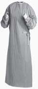 | Surgical Gown
 |
• Hands should be washed between patient contacts, after removing gloves and before wearing them again. Use of disinfectant scrub like chlorehexidine after washing will have a prolonged antibacterial effect against microbial
ingression through the gloves. Hands must also be washed after touching intimate objects likely to be contaminated by blood or saliva from patients and before leaving the dental treatment area.
• Clinic attire should be worn only in the dental environment and should be changed at the end of the treatment schedule.
• Use of mask is usually indicated especially during procedures that cause splashing or spattering of blood or saliva. It is recommended that facemasks should be changed once every hour or between each patient contact, whichever occurs first.
• Protective eye wear : It may be in the form of glasses and / or a face shield. It should prevent trauma to the eye tissue from flying droplets or aerosols. Protective glasses should be washed with soap first, rinsed with water and wiped with an appropriate surface disinfectant. Plastic safety lenses can also be immersed in alkaline glutaraldehyde solution
and should be thoroughly rinsed to avoid possible irritation to skin and eyes.
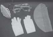 | Personal Protective Barriers
|
• Management of instruments (3 & 4): They should be cleaned and dried, lubricated if necessary and packaged before loading into the autoclave. Cleaning involves an initial presoaking with detergent solution containing disinfectants to soften organic debris and begin microbial kill. After cleaning the instruments should be dried.
• All moving parts of the instruments especially hand pieces should be lubricated prior to steam sterilization. The burs should be autoclaved or maintained in high level disinfection for not less than 3 hours. Thorough rinsing should
be followed to remove all traces of disinfectant. (5)
• Touch surfaces like unit handles, light handle, light switch, chair controls, head rest knob, trolley handle, trolley and 3-way syringescannot be disconnected and sterilized and therefore need to be treated with disinfectants or covered with a protective barrier. However instruments which enter oral cavity and are connected to some of the equipment e.g. air
rotor and surgical hand pieces, ultrasonic inserts or tips, air water syringe tips and light cure probes or tips should be disconnected, sterilized and rinsed before use(3).
• Disinfection of surfaces involves the cleaning of surfaces, after every patient and application of a disinfectant chemical. These chemicals include alcohol (spirit), iodophor products, synthetic phenols, glutaraldehyde, chlorines etc.
• The advantages of barriers include ease and speed of insertion, standard sizes and the protection of equipment from damage by chemicals, blood and fluids.
• Spittoons should be flushed with water, scrubbed and disinfected.
• Waste buckets should be used with disposable plastic bags as liners to be changed wherever
necessary.
• Reducing aerosols in the clinic: Preoperative mouth rinses with chlorhexidinegluconate or other suitable disinfectant mouth wash should help reduce infectious particles in aerosols. Rubber dam isolation is another method to
reduce potentially infective aerosols. High volume secretion during procedures using copious irrigation and even the routine use of saliva ejectors can restrict aerosolization. (6)
PROSTHODONTIC CONSIDERATIONS
• Disinfection of impressions (7):
A. Personal protective equipment: Protective eye wear, masks and gloves should be used when handling a
c o n t a m i n a t e d impression until it has been disinfected.
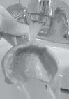 | Thorough rinsing of impression with water
|
B. Rinse the i m p r e s s i o n :
Immediately after an impression is taken in the dental operatory, rinse it under running water in order to remove any saliva or blood. This step is essential for allowing optimum disinfection of the impression.
C. Disinfection techniques: Once the impression is rinsed and shaken to remove excess water, it must be disinfected. This may be accomplished by immersing the impression in, or spraying it with, an acceptable disinfectant.
a. Disinfection of an impression by immersion: It is preferred over spraying. Spraying may not be effective because constant contact of the disinfectant with all surfaces of the impression cannot be assured.
i. Rinse the impression with running water and shake off excess water.
ii. Place rinsed impression into a zippered plastic bag containing appropriate disinfectant.
iii. Leave it immersed in disinfectant for 15 minutes. Polyethers and hydrocolloids may be adversely affected by disinfectants; therefore their immersion time is limitedto 10 minutes.
iv. Remove impression from disinfectant.
v. Rinse with running water and shake off excess water.
b. Disinfection of an impression by spraying:
Spray the cleaned impression and impression tray with an acceptable disinfectant. Seal the sprayed impression in a zippered plastic bag for 15 minutes. Remove the impression from the sealed bag.
 | Disinfection of Impression by spraying
|
• Hydrocolloid impressions (7&8): A number of investigators have evaluated disinfection of irreversible hydrocolloid (alginate) sometimes with contradictory results. Based on these findings, the ADA recommended disinfecting alginates by immersion in dilutedhy pochlorite, iodophor or glutaraldehyde with phenolic buffer. Investigators reported significant adverse effects of specific materials with disinfectants that are non-reactive with other alginates suggesting that caution should be exercised. Given the hydrophilic nature of the material, a minimal disinfection time should be used. Limited data are available on disinfection of reversible hydrocolloid, however research data suggest that there is no effect on
dimensional accuracy of impressions immersed in an iodophor diluted 1:213, 5.25% sodium hypochlorite with a dilution 1:10, 2% acid glutaraldehyde with dilution of 1:4, and glutaraldehyde with phenolic buffer diluted 1:16. Immersion in 2% alkaline glutaraldehyde has significant adverse effects on the impressions and resultant dies.
• Rubber base impression materials (7&8): They can be disinfected by immersion in iodophor, diluted chlorine solution, glutaraldehyde or complex phenols for the time required for tuberculocidal activity. It is important to review the method of disinfection with the manufacturers to prevent distortion of the impression or loosening of the adhesive bond between the impression tray and impression material. These impressions also should be rinsed with water before pouring. It is
important to inform the dental laboratory that the impression has been disinfected to prevent the laboratory personal from performing more disinfection procedures that might distort the impression. Studies by a number of investigators have
shown that polysulphides and silicones are relatively stable and can be disinfected without adverse effects by immersion in most disinfectants approved for use in dentistry. Although hydrophilic, polyether impressions also can be disinfected by immersion, but exposure times should be kept to minimum (10 minutes). Disinfectants requiring xposure
times greater than 10 minutes for tuberculocidal disinfection probably should be avoided with polyether. Immersion in acid glutaraldehyde actually improves the surface detail reproduction of elastomeric
impressions.
• Zinc Oxide Eugenol (ZOE) and compound impressions (7&8): Limited data are available on disinfection of ZOE and compound impressions. Adverse effect have been reported on ZOE immersed for 16 hours in diluted hypochlorite and on compound by all of the disinfectants tested (hypochlorite, formaldehyde and 2% alkaline glutaraldehyde).
• Once the impression has been disinfected it may be poured in the desired stone.
A. Disinfection of Dental prosthesis and appliances (1,3&4):
A. The ADA recommends disinfection by immersion in iodophor or chlorine compounds. Although both of these disinfectants are somewhat corrosive, studies have shown little effect on chrome cobalt alloy with short-term exposure (10 minutes) to iodophor or 1:10 hypochlorite. Damage of heat cured denture base resin has been shown to occur after only 10 minutes of immersion in a glutaraldehyde with phenol buffer, although immersion in 2% alkaline glutaraldehyde did not damage the acrylic surfaces. Given the tissue toxicity of glutaraldehyde and phenolic compounds, however iodophor or chlorine compounds are preferred for disinfection of acrylic appliances. Prostheses never should be stored in a disinfectant before insertion. After disinfection and thorough rinsing, acrylic items can be stored in diluted mouthwash until inserted.
b. Fixed metal/porcelain prosthesis may be disinfected by immersion in glutaraldehyde for the time recommended
for tuberculocidal inactivation by the disinfectant manufacturer. (5) In addition several clinical studies have confirmed
that fixed prosthesis can be disinfected by short immersion in diluted hypochlorite without apparent harm to the device. The higher the content of noble metal, the less the likelihood of adverse effects on the metal. Care should be taken to minimize the exposure times of metals to potentially corrosive chemicals. Iodophor probably could be used as well, but no data are available to substantiate this. Unglazed porcelain should not be exposed to any disinfectant and (porcelain firing/ glazing will suffice), fixed metal prostheses can be sterilized with ethylene oxide or even by autoclaving if desired. Any device that has been immersed in a disinfectant
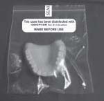 | Prosthesis should be disinfected and sealed before use
|
should be rinsed thoroughly before delivery to the patient.
• Prosthesis or appliances that have been worn by patients should be cleaned thoroughly before disinfection by scrubbing with a brush and an antiseptic hand wash or by cleaning in an ultrasonic unit.
· Dentures or other acrylic appliances that have been worn by patients and require repair should be disinfected, after cleaning and before handling should be handled (i.e. with gloves) as contaminated even after disinfection. The porous nature of acrylic makes such devices difficult to disinfect adequately.
DISINFECTION OF WAX BITES, OCCULUSION RIMS, STONE MODELS, CUSTOM IMPRESSION TRAYS & BITE REGISTRATIONS (1,3 & 4)
A. Wax rims and wax bites should be disinfected by the spray wipe spray method using an iodophor as recommended by the ADA. Rinse spray may be more appropriate for wax bites. For adequate disinfection these should remain in disinfectant for the time recommended for tuberculocidal disinfection. After the second spray, they can be enclosed in a sealed plastic bag for the recommended time. These items should be rinsed again after disinfection to remove any residual disinfectant.
B. Bite registrations made of various materials such as ZOE or compound can be handled in the same manner as impressions of the same materials. These registrations also can be disinfected, using the rinse spray rinse technique, with most EPA registered hospital level tuberculocidal disinfectants used as sprays. After disinfection, the registration should be rinsed again to remove residual disinfectant.
C. ADA recommends that stone casts be disinfected by the spraying until wet or immersing in a 1:10 dilution of sodium
hypochlorite or an iodophor. Casts to be disinfected should be fully set (i.e. stored for at least 24 hours). Investigators submerged die stone models in a variety of disinfectants and found that with 1:10 sodium hypochlorite and 1:213 iodophor, undesirable physical effects on set die stone ranged from none to minimal.
D. A disinfectant stone now is marketed and has been shown to have bactericidal property however this product is not yet EPA registered as a disinfectant. Several investigators have recommended adding disinfectants to gypsum during mixing (ie. As all or part of the liquid, when pouring casts). Although such products have potential for use in infection control, they do not solve the problem of the contaminated impression or tray as a source of infectious microorganisms during transit from the operatory to the laboratory.
E. Custom acrylic resin impression trays should be disinfected by spraying with surface disinfectants or immersing in either 1:213 iodophor or 1:10 sodium hypochlorite. They should be rinsed thoroughly to remove any residual disinfectant and allowed to dry fully before use. After use in the mouth custom trays should be discarded.
Other Prosthodontic items (5&6):
A. Heat stable items such as face bow forks orthodontic pliers and metal impression trays that come in contact with oral tissues should be heat sterilized rather than disinfected.
B. Articulators and face bows should be cleaned and disinfected. After manipulation at chair side wooden handled spatulas should be cleaned and disinfected. Other times such as Hanau torches should be disinfected after use.
The area to be touched should be covered with a barrier such as plastic wrap to prevent
 | Metal trays should be heat sterilized
|
contamination. Rubber bowls should be cleaned and disinfected after chair side use. (1,3&9)
C. Items such as shade guides should be cleaned and disinfected to avoid cross contamination. If iodophor is used on shade guides, they should be wiped with water or alcohol after the exposure time to remove any residual disinfectant. (9)
D. Ultraviolet light is a part of electromagnetic spectrum. It ranges from 400nm downwards to approximately 150nm. It is well established that greater germicidal effect is in the range of 240-280nm with the optimum being 253- 7nm. This is widely accepted as a near maximum for bactericidal and germicidal effect. Most investigators show that the rays are absorbed by the cellular DNA chain which is the initial event in the chain of events leading to cellular death.
Robert J. Boylan et al (1987) (10) under UV light with a wavelength of 254nm as a mode of sterilizing complete dentures, partial dentures and a rubber base impression contaminated with fine known species of microorganisms. The results showed that killing of microorganisms with greater than 98% within 15 seconds and 99% either 30 seconds and 100% in 2 minutes. They also concluded that UV light cannot be used as a sole means of disinfecting the impressions because of shadowing effect that allows the survival of microorganisms unexposed to UV light.
CONCLUSION
Dentists must use effective infection control procedures in their practices. A positive step by step approach should be used. One should determine and practice infection control and build upon them by adding new procedures to the dental
routine. The current knowledge in today’s society regarding infectious diseases in general and herpes, hepatitis and acquired immune deficiency syndrome (HIV) in particular dictates that all dental practices must incorporate acceptable
infection control techniques. Dental prostheses, impressions, models and items used in their fabrication are potential sources for cross infection and should be handled in a manner that prevents exposure of dental health care rofessionals and patients to infectious agents.
REFERENCES
1. Stewart k L, Rudd K D, Kuebker W A. Clinical Removable Prosthodontics. 2nd ed. AIPPD;2005. pp 126.
2. Samaranayake L P . Essential of Microbiology For dentistry 2nd Edition, Mosby , Pg 255-257.
3. CDC. Guidelines for Infection control in dental health-care settings – 2003. MMWR 2003; 52(No. RR-17):1–66.
4. USAF Guidelines for Infection control in Dentistry, September 2004
5. Nayar S, S. Bisnu. Prosthetic management of HIV/AIDS Patient. The Journal of Indian Prosthodontic society 2008:8;10-
16
6. Leggat PA, Kedjarune U. Bacterial aerosols in the dental clinic: a review. Int Dent J. 2001 Feb;51(1):39-44.
7. Anusavice Philips science of Dental Materials 11th edition pg 225-226.
8. Herrera S P and Merchant V A. Disinfection of Alginate , Polysulfide, Vinyl Polysiloxane and Polyether Dental
Impression J Dent Res 64, 194:1985.
9. Katberg JW. Cross contamination Via Prosthodontics laboratory J Prosthet Dent 1974; 32:412-419.
10. Robert J Boylan , Evaluation of an ultraviolet disinfection unit, Prosthet Dent 1987;58:650-654. |