Introduction
Surgical resection of the maxilla results in a communication between oral and nasal cavities that causes difficulty in swallowing, nasal reflux, unintelligible speech and unesthetic appearance. In addition, when the defect is associated with trismus, the complications of treatment procedure are exaggerated. Multiple-component prostheses have been advised to rehabilitate maxillectomy patients with severe trismus. A two-component prosthesis retained by magnets facilitates its easy placement and removal[1]. Advances in technology have made available a new family of magnetic alloys based on cobalt and other rare earth metals. They are small but strong and can be used in dental prostheses for retention. The mutual attraction of unlike poles has been utilized successfully to assemble multicomponent, maxillofacial prostheses[2]. This clinical report describes the prosthetic rehabilitation of a maxillectomy patient using a two-piece magnet retained hollow bulb obturator.
Case Report
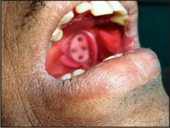
A 35 year old male patient reported to the Outpatient Department of Prosthodontics, Sardar Patel Post Graduate Institute of Dental And Medical Sciences, Lucknow, for the restoration of maxillary defect created after surgical intervention. The patient had been diagnosed with cancer of gingivo-buccal sulcus of maxillary left region, which was surgically resected. The resulting defect was classified as Aramany’s class II (Fig.1). Patient was undergoing radiotherapy and chemotherapy and had restricted mouth opening (15 mm) (Fig.2). Hence, an interim obturator with a separate closed hollow bulb was planned. For purpose of retention of the bulb, use of Cobalt-Samarium magnets was planned and written consent was obtained from the patient. The impression of the defect was made with Type II impression compound. The inferior surface of the impression was keyed for orientation and the remainder of the arch was recorded with alginate. The impression was assembled outside the mouth (Fig.3) and a master cast was poured in Type III gypsum. For preparation of the bulb, a sheet of modelling wax (wax shim) was adapted on the defect walls after blocking out the undercuts. The top of the sheet was keyed, to orient the lid (to be made later on). The wax shim was flasked and acrylized in heat cure polymethylmethacrylate. The acrylized shim was trial seated on the cast and a lid was prepared in wax (Fig.4). Four cobalt-samarium magnets of 3mm diameter were embedded in the wax, in accordance with the keys to create their housings, and then the lid was acrylized separately without the magnets. The magnets were then embedded in their respective housings in the acrylised lid and covered with a thin layer of autopolymerizing resin. The lid was then sealed to the hollow bulb with autopolymerizing resin. The closed hollow bulb was tried in the patient’s mouth, and verified for path of placement and fit (Fig.5). Next, the trial denture base was adapted on the cast and occlusal rim was prepared. Jaw relations were recorded. The casts were mounted and teeth arrangement was done for trial. The second molar was omitted, to reduce the load on the defect. Four magnets were embedded on the intaglio surface of the trial denture base, opposite to the previously placed magnets on the closed bulb lid, to prepare their housings, after which they were removed. After the denture was acrylized, the magnets were placed back in their housings and covered with a thin layer of autopolymerizing resin (Fig.6). The two-piece obturator was seated in the patient’s mouth (Fig.7), the bulb being placed first followed by the denture base. Necessary instructions were given on technique of placement, removal, and maintenance of the prosthesis. Post-insertion follow-up and patient care were carried out at the regular intervals of time. The patient was kept on physiotherapy (mouth opening exercises) and bite opener (Fig.8) to improve the mouth opening before making of the definitive prosthesis.
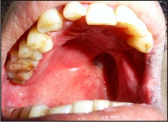 | Fig. 1. Intra Oral View Of The Defect
 |
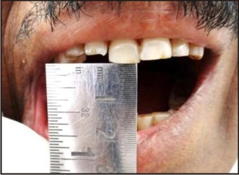 | Fig. 2. The Mouth Opening Was 15mm
|
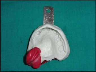 | Fig. 3. Impression Assembly Outside The Mouth
|
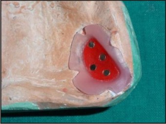 | Fig. 4. Acrylized Shim And Wax Lid With Magnets Embedded For Housing
|
 | Fig. 5. Trial Of Bulb In The Mouth
|
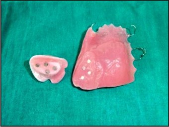 | Fig. 6. Acrylized Bulb And Denture Base With Magnets In Place
|
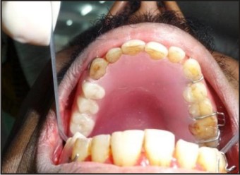 | Fig. 7. Prosthesis Seated
|
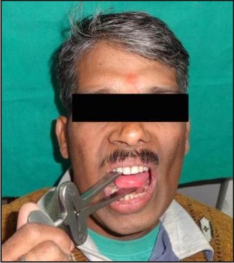 | Fig. 8. Bite Opener Given For Physiotherapy
|
Discussion
Maxillofacial defects can be due to surgical resection, infections, trauma or congenital reasons. As a result, such patients have compromised esthetics with decreased functional efficiency along with psychological disturbances as well. Often the situation is complicated with a decrease in mouth opening, due to ongoing radiotherapy, or fibrotic bands left after surgery[3].Obturator prosthesis play an important role in the recovery of oral function in postsurgical maxillectomy patients[4]. In this case, a closed hollow-bulb obturator was planned for the patient. A hollow bulb prosthesis (either one piece or two piece) is a better choice, as it is lighter in weight and is more hygienic. Light weight has also been considered for maxillary resection prosthesis, by Chalian and Barnett[5],[6]. One of the problems associated with oromaxillary obturators is insertion of the prosthesis due to compromised anatomic morphology in different planes[7]. Limited oral opening further worsens the problem, making it necessary to devise ways to easily seat and remove the prosthesis. In this particular case, a two-piece obturator was designed for ease of insertion and removal. There are several retentive devices available to secure the two sections in position[8]. Among the various retentive devices, magnetic attachments are more user friendly and cost effective compared to internal attachments which required extreme precision and good neuromuscular coordination from the patient to insert and use the prosthesis. In 1976, Federick[9] rehabilitated a patient with large orofacial defect using a two component obturator that was locked to each other with the help of magnets. Cobalt-Samarium magnets were used in this case for the 2-piece hollow-bulb prosthesis. Despite many advantages, magnets have poor corrosive resistance within oral fluids and therefore require encapsulation within a relatively inert alloy such as stainless steel or titanium. Coated magnets have been found to withstand the corrosiveness of saliva and thus no ill effects on the oral tissues[1]. In this particular case, the Cobalt-Samarium magnets were coated with a layer of acrylic resin though the resin is porous in nature. The magnets may lose their magnetism on heating, commonly encountered in processing of the acrylic denture. Hence, in this case, we seated the magnets, in their respective housings, only after the processing of the acrylic lid and the denture base. Patient was extremely happy with the masticatory efficiency and the improved quality of speech instantaneously.
References
1. V.Bhat. A close-up on obturators using magnets: Part II The Journal of Indian Prosthodontic Society.September 2006 ,Vol 6, Issue 3
2. V. Bhat. A Close-Up On Obturators Using Magnets: Part I - Magnets in Dentistry .The Journal of Indian Prosthodontic Society . July 2005 , Vol 5 , Issue 3
3. Ahmed B, Hussain M, Butt AM, Yazdanie N. Maxillofacial rehabilitation of a large cleft palate using fixed- removable prosthesis. J Coll Physicians Surg Pak 2011; 21:52-54.
4. Curtis TA, Beumer J, Rehabilitation of acquired hard palate defects. In: Beumer J, Curtis TA, Marunick MT (eds). Maxillofacial Rehabilitation, Prosthodontic and Surgical Considerations, ed 1. St. Louis: Ishiyaku Euro- America, 1996:225-284.
5. Chalian VA, Barnett MO. A new technique for constructing a one-piece hollow obturator after partial maxillectomy. J Prosthet Dent 1972;28:448-53.
6. Chalian VA, Drane JB, Standish M. Maxillofacial Prosthetics: Multidisciplinary Practice. The Williams and Wilkins Co: 1971. P. 133-48.
7. B. Rilo, J. L. Dasilva, I. Ferros, M. J. Mora, and U. Santana, “A hollow-bulb interim obturator for maxillary resection. A case report,” Journal of Oral Rehabilitation, vol. 32, no. 3, pp. 234– 236, 2005.
8. M. Fukuda, T. Takahashi, H. Nagai, and M. Iino, “Implantsupported edentulous maxillary obturators with milled bar attachments after maxillectomy,” Journal of Oral and Maxillofacial Surgery, vol. 62, no. 7, pp. 799–805, 2004.
9. Federick, David R. A magnetically retained interim maxillary obturator. J Prosthet Dent 1976;36:671-5.
|