Introduction
Autistic disorder (AD) is the third most common developmental disability and almost 400 000 people are affected. AD is categorized in the DSM-IV (Diagnostic and Statistical Manual of Mental Disorders, 4th Ed.) under the section Pervasive Developmental Disorders (PDD). [1]
Autism or autistic disorder is a neurodevelopment disorder characterized by impaired social interaction, communication, and restricted and repetitive behavior. These signs all begin before a child is three years old. It is a highly variable brain development disorder. [2], [3]
No specific oral manifestations of ASD have been described , though the oral hygiene of these subjects is known to be deficient .[4]
The first permanent molar (FPM) has been cited as being the most caries-prone tooth in the permanent dentition, probably as a termination of its early exposure to the oral environment.If FPMs are extracted during or after the eruption of the second permanent molars,occlusal forces encourage mesial tilting and the molar tilts lingually because the lingual plate is more fragile than the buccal plate of alveolar bone. Lingual rolling may result in the development of a scissor bite and non-working side interferences. Over-eruption of the opposing FPM. At this crucial juncture, treatment options available are removable partial denture , fixed partial denture, bonded prosthesis or an implant but all these different procedures or techniques have one or the other limitations.
In this case report, we discuss the dental management of early loss of first permanent molar in 14 year old autistic child with behavioral problems.
Case Report
A 14 year old male patient reported to the Department of Pedodontics and Preventive Dentistry with a chief complaints of painand food lodgment in left lower back region (tooth no. 36).
The patient was a known case of autism, mental retardation and hyperactive seizure disorder and delayed developmental milestones. He was inattentive, did not respond to verbal commands, made bubbling sounds and undefined gestures. His speech was not clear and was hyperactive with repetitive movements. His post-natal history was suggestive of birth asphyxia and delayed development milestones. Psychological testing report showed childhood autism rating scale (CARS) [4], which falls into a severe autistic category.[5] The family history was non-contributory. He was under the supervision of the concerned physician.
An intraoral periapicl radiograph revealed grossly carious 36, involving furcation area (Fig 1) and was a poor candidate for endodontic treatment . Treatment was planned extraction of nonrestorable first molar. The permanent second molar had not erupted completely (Fig 2) thus the crown height available was short, the various options for space maintenance were given to the patient. Since the patient’s sister was not ready for removable appliance, an economical fixed interim space maintainer was designed and delivered to the patient.
The appliance was intended to remain in place until the patient’s occlusion is enough to receive a permanent prosthetic replacement or an implant.
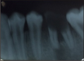 | Fig 1 : Iopa X-ray Of 36 Involving Furcation Area
 |
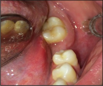 | Fig 2 : Intr Oral View After Extraction Of 36 & 37 Had Not Completely Erupted
|
Fabrication Of Appliance
Band adaptation was done for the adjacent mesialand distal abutment teeth i.e., left mandibular second premolar and left mandibular second molar using 0.004x0.150x2 inches and 0.006x0.180x2 inch band material respectively.(Fig 3 a)
Alginate impressions of both the arches were made, thus providing a working model.
A rectangular wire mesh was constructed by bending 23-gauge stainless steel orthodontic wire. The width of mesh was 2mm less than the bucco-lingual width of the crowns. The length and contour of mesh correspond to the edentulous space. The mesh was soldered to the bands contoured for the abutment teeth. The mesh served to hold the three units of bridge together. The gingival extension of the wire mesh was placed 1mm above the ridge to allow an adequate cleansing while not allowing food entrapment or gingival irritation. (Fig 3b)
The occlusal rest is prepared on distal fossa of premolar and mesial fossa of molar second molar for better retention . The solder joints were finished and polished.
A mandibular First pre molar resin tooth was is selected as pontic because there was space loss. (Fig 3c) The pontic was attached to the finished wire framework using auto polymer rising acrylic resin, matching the shade of the pontic. The acrylic attachment was finished and polished
The bridge was ready for a try in the patient’s mouth for final adjustments. The bridge was assessed for the gingival extension and soft tissue blanching. The occlusal and eccentric movements were adjusted. The bridge was cemented with Fuji I Glass ionomer luting cement. (Fig 4)
In a follow-up of 6 months, the bridge exhibited excellent soft tissue acceptance and has restored the masticatory function as well.
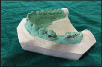 | Fig 3a : Band Adaptation On 35 & 37
|
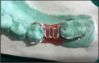 | Fig 3b : A Wire Frame Work
|
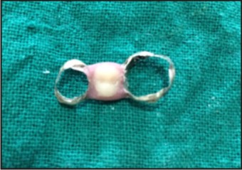 | Fig 3c : Fabricated Interim Space Maintainer
|
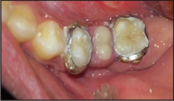 | Fig 4 : Post Operative Veiw , Cemented Space Maintainer
|
Discussion
Autism is an incapacitating disturbance of mental and emotional development characterized by severe deficits in social interaction and communication and the presence of repetitive, ritualistic behaviors.[6]
Autism was first described in 1943 by the American child psychiatrist Leo Kanner.[7]
He presented 11 children whose behavior were obviously different from others. Kanner suspected that they had an inborn feature which prevented their forming regular social contacts. Kanner suspected that they had an inborn feature which had prevented their regular social contacts . Autism Disorder is sometimes referred to as early infantile autism, childhood autism, or Kanner’s autism.
Autism is now recognized as an organic disorder characterized by abnormalities in the brain, especially the limbic system and cerebellum. Autism is not a disease but a syndrome with proposed etiological factors include post-encephalitic infection or sepsis, genetic, autoimmune factors and vitamin D deficiency, affecting boys 3-4 times more often than girls Family income, education, and lifestyle do not seem to affect the risk of autism.[8],[9] Children with autism have abnormal levels of serotonin or other neurotransmitters in the brain or have irregularities in several brain regions that affect normal development.[10]
Behaviour problems like hyperactivity and quick frustration can hamper the provision of oral health care, in patients with autism. Determining the presence of dental disease by means other than inspection often is difficult in patients with autism. The patient’s inability to communicate effectively often precludes obtaining an accurate dental history.Patients with autism have an increased threshold and that they may endure major pain without complaint.[11]
In general, children with autism prefer soft and sweetened foods and they tend to pouch food inside the mouth instead of swallowing it due to poor tongue coordination, thereby increasing the susceptibility to caries. Moreover, the risk for dental caries can be expected to be higher in these patients due to difficulties in brushing and flossing.Nevertheless, many authors have found that the prevalence of caries had no difference on comparing autistic and non-autistic individuals and in some cases, the prevalence of caries in children with AD even be comparatively lower.[12]
The first permanent molar is undeniably the most important unit of mastication and is essential in the development of functionally desirable occlusion. More than 50% of children over 11 years have some experience of caries in such teeth and may need to be extracted in their life. It is important to consider the following factors when planning extraction of FPMs: 1. the restorative state of the tooth; large occlusal or proximal restorations; irreversible pulpitis; periradicular infection; severe hypoplasia. 2. dental age of the patient; 3. degree of crowding in the buccal and labial segments; 4. the occlusal relationship; 5. presence and condition of the other teeth.[13]
The extraction event result in tipping of the molars and premolars may undergo greatest amount of distal drifting with all the teeth anterior to the space, including the central and lateral incisors on the side where the loss occurred, may show evidence of movement. When such disruptions do occur, appropriate corrective measures are needed to restore the normal process of occlusal establishment.
Such corrective procedures may involve some type of passive space maintenance, active tooth guidance, or a combination of both.
The space can be maintained, this may be accomplished in one of the several ways.[14], [15]
a. Cast overlay band and loop.
b. Band and loop maintainer with occlusal bar and rest.
c. Conventional fixed bridge work.
d. Etched casting, resin-bonded posterior bridge.
e. Single-unit implant prosthesis.
f. Auto-transplantation of third molars into the first molar position.
g. Stainless steel crown bridge.
The bridge provides us with an additional treatment modality in case of an early loss of a permanent tooth in young adolescent patients. It has soft tissue sparing as compared to a treatment partial denture.
The bridge is functional, readily acceptable, maintains the mesiodistal dimensions of the lost tooth, prevents supra eruption of opposing teeth and does not restrict normal growth and development, as is required of an ideal space maintainer.[16] The appliance has been shown to have an excellent patient compliance and is easy to clean and maintain. It has decreased likelihood of breaking or getting lost. Hygiene is easily maintained with no food entrapment. The bridge can be easily removed and recemented for fluoride application at recall appointments, if the need arises.
Children with autism have multiple medical and behavioral problems, which make their dental treatment extremely difficult. Long-term care includes frequent and effective oral hygiene measures with the help of the parents, application of fluoride gel or rinse, intake of healthy non-cariogenic foods such as cheese, nuts, plain milk and frequent recall appointments.
Our patient was totally caries-free except grossly carious 36; this could be ascribed to the absence of any retentive area Also, his sister was well educated and take charge of her brother’s oral hygiene care. Thus tooth extractionof grossly carious tooth 36 and the interim fixed prosthesis was planned.
Conclusion
AD is a heterogeneous disorder with a wide range of expressions. Therefore, treatment approaches that may yield a positive outcome in one patient may prove to be ineffective for another. Because of the limited attention span of AD patients short, well-organized appointments should be planned and the waiting time should not exceed 10-15 minutes, to avoid upsets . It is essential to adapt proper oral hygiene measures and also need for regular dental visit in future.
References
1. Ulrich Klein ,M S Arthur J.Nowak . Autistic disorder : A review for pediatric dentist . Pediatr Dent 1998 : 20:5,312-317
2. Amaral DG, Schumann CM, Nordahl CW. Neuroanatomy of autism. Trends Neurosci. 2008;31:137-45
3. Geschwind DH. Autism: many genes, common pathways? Cell. 2008;135:391-5
4. Lorena M. Orellana, Francisco J. Silvestre, Sonia Martínez-Sanchis,Victoria Martínez-Mihi, Daniel Bautista. Oral manifestations in a group of adults with autism spectrum disorder Med Oral Patol Oral Cir Bucal. 2012 May; 17(3)
5. Schopler E, Reichler R, Rochen Renner B. The childhood autism rating scale.Western Psychological Services; 1988.)
6. Gail Williams P, Sears LL, Allard A. Sleep problems in children with autism. J Sleep Res 2004;13:265-8
7. Kanner L : Autistic disturbances of affective contact . Nerv Child . 1943 ; 2:217-50,
8. Daniel R. Dental management of children with autism. Pediatric Dental Health. 2005
9. Muhle R, Trentacoste SV, Rapin I. The genetics of autism. Pediatrics. 2004;113:e472–86
10. National Institute of Neurological Disorders and Stroke, Autism fact sheet. NIH 2008; Publication No. 06-1877
11. Khatri A, Sugnani S, Kalra N, et al. Dental management of autistic child. Journal of Case Reports 2012;2(1):1-3.
12. Udhya J.,Varadharaja M.M, Parthiban J., Ila Srinivasan. Autism Disorder (AD): An Updated Review for Paediatric Dentists J Clin Diagn Res. 2014 Feb; 8(2): 275–279)
13. D.S. Gill, R.T. Lee C.J. Tredwin Treatment Planning for the Loss ofFirst Permanent Molars Dent Update 2001; 28: 304-308
14. McDonald RE, Avery DR. Dentistry for the child and adolescent. Missouri Elsevier 2004. p. 644-6.
15. Dimri M, Jain A. Stainlees Steel Crown Bridge Replacing Permanent Molar In The Adolescent Patient: A Case Report. J Indian Soc Pedo Prev Dent 2001;2:74-6.
16. Graber TM. Orthodontics: Principles and Practice. Philadephia W.B.: Saunders and Co; 1992. p. 640-1.
|