Introduction
Making an accurate impression of dental and dentoalveolar structures is important and is an essential requirement for the precise fit of the prosthesis. This is one of the important factors in determining the longevity of the restoration. The addition type silicon impression materials, polyvinyl siloxanes(PVS) have been reported to be most accurate and dimensionally stable.[1] Some authors claim that the extent of accuracy of dies is determined more with the technique than by the material itself,[2] and others reporting that the impression accuracy is governed more with material employed.[3] However with the proven accuracy of the material, the technique also has to be considered, especially in cases of fixed partial denture. Here inter-abutment relation is also equally important along with accuracy of the individual tooth/die.
Matrix impression system (MIS) developed by Gus J. Livaditis(1998) uses a precisely designed matrix which can provide a mean to better control the unpredictable dentogingival environment when making impressions which significantly improves the gingival displacement and sulcular cleansing phases.[4] The technique has shown to be beneficial in single crowns and its feasibility to use in FPD cases is still questioned.
Hence the present study was conducted to compare the accuracy of the matrix impression system with conventional technique for individual dies and also to compare the inter- abutment distance in master model using both impression methods.
Material & Methods
The materials used were polyvinyl siloxane putty material (3M ESPE, express STD, Germany) and Kalrock (class IV) die stone. All the measurements were made using profile projector. The samples were divided into two groups:
Group I-Impressions were made using Matrix impression system (MIS).
Group II-Impressions were made using multiple mix technique (MMT).
The typhodont teeth were embedded in the maxillary frasaco model base. The left canine and second premolar were prepared conservatively to receive a ceramometal fixed partial denture. The first premolar was removed and the socket was obliterated with wax to simulate a clinical case of 3-unit fixed partial denture (Fig 1). Four sharp hatch marks were made with a round bur on the finish lines of each prepare tooth. The hatch marks were placed diagonally opposite i.e., one each on labial, buccal, palatal, mesial and distal of each prepared tooth. Two more hatch marks were placed on incisal surface of canine and occlusal surface of premolar (Fig 2).The measurements of master model were made using profile projector.[5]
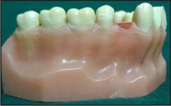 | Fig 1: Master Model With Typhodont Teeth
 |
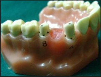 | Fig 2: Hatch Marks Placed On The Finish Lines Of The Prepared Teeth (23 And 24)
|
For Group-I, A matrix of the prepared teeth was made with polyvinyl siloxane putty material, which was extended to one tooth on either side. The matrix was relieved internally except for the incisal and occlusal portion which served as vertical stops. A definitive impression was made using the matrix of the preparations with a high viscosity elastomeric impression material (3M ESPE, Imprint II Garant), was seated over the remaining teeth to make an impression of the entire arch. Simultaneously, a stock tray filled with medium viscosity elastomeric impression material (3M ESPE, Germany) was seated over the remaining teeth to make an impression of the entire arch(Fig 3).[6]
For Group II, A mix of light body(3M ESPE, express, Germany) was injected over the prepared teeth on the master model and simultaneously a mix of heavy body was injected in to the custom tray which was seated on the master model to make an impression of the entire arch. The impressions for this group were made using custom trays for the prepared master casts(Fig 4).[7]
Fifteen impressions were made for each technique and the impressions were poured using die stone using standard W/P (water : powder) ratio and allowed to set completely to get the working casts. The master casts were recovered and the bases were formed by dental plaster. The working casts were checked carefully for any defects especially over the hatch marks. The mesiodistal, buccolingual, inciso-cervical dimensions of the canine and the second premolar and interabutment distance of the obtained casts (Group I and Group II) were measured using Profile projector and tabulated (Fig 5). Each measurement was repeated three times and the mean was recorded for a particular dimension. Thus obtained readings were then compared with master model and analysed.
 | Fig 3: Group - I Impressions
|
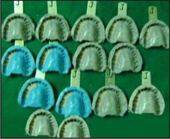 | Fig 4: Group - II Impressions
|
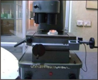 | Fig 5: Profile Projector With Sample
|
Results
Matrix Impression System: (Table 1 & 2)
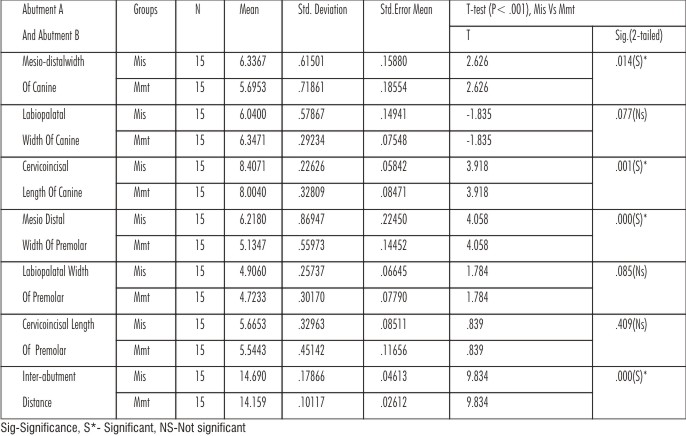
Mesio –distal width of canine and second premolar showed significant contraction from the master model i.e., -0.27mm and -0.18mm (p < 001). No significant differences were found in other dimensions.
 | Table I: Shows The Mean Differences Of Measurements Of Abutment A, Abutment B, Inter-abutment Distance Between The Master Model (Mm), Group I, Ii Working Casts
|
 | Table-II: Results Obtained From T Test Showing Difference Between The Groups I And II
|
Multiple Mix Technique: (Table 1 & 2)
Mesio – distal width of canine and second premolar showed significant contraction from the master model i.e., -0.92mm and -0.266mm(p< .001). Cervico – incisal width of canine showed significant contraction from the master model i.e., -0.816 mm (p< .001). Inter abutment distance showed significant contraction from the master model i.e., -0.48mm (p< .001).
Discussion
Fabrication of fixed prosthesis is an indirect technique, in which the prosthesis is to be fabricated in the laboratory and then it is placed in the oral cavity. For this purpose accurate replica of the dental and dentoalveolar structures are required. Making an accurate impression of individual tooth in their position is very vital in obtaining accurate working casts for the fabrication of FPD. Controlling the tissue fluids like gingival sulcular fluids, saliva and displacement of the gingival tissues around the abutments during impression procedure is a challenging task. The matrix impression system developed by Gus J. Livaditis (1998) requires a series of three impression procedures, using three types and /or viscosities of impression materials. This system effectively controls the four forces (relapsing, retraction, displacement, and collapsing) which impact over the gingiva during the critical phase of making an impression when attempting to register the subgingival margins.[6] The matrix impression system incorporates the attributes of traditional methods and overcomes important deficiencies in;
• Registration of subgingival margins
• Gingival retraction and relapse
• Hemostasis and sulcular cleansing
• Delivery of impression material subgingivally
• Strengthening the sulcular flange of the impression
• Simplification for making complex impression.[4]
The measurements of mesio-distal dimensions of canine and second premolar were 6.336 mm and 5.67 mm in group I, 5.693 mm and 5.13 mm in group II against the master model which was 6.61 mm and 6.42 mm. The contraction observed was more in group II when compared with group I. The results found in this study were similar to those found by Bomberg et al.,[8] which revealed the thickness of impression materials in custom and stock trays have reported a difference of less than 1.5mm and described that eccentric orientation of the tray on the arch was found in almost half of the impressions in both categories, and neither tray system confronted the non uniformity in thickness between prepared and prepared teeth. This may result in dies which are short mesio-distally.
The measurements of cervico-incisal dimensions of canine and second premolar were 8.40 mm and 5.66 mm in group I and 8.04 mm and 5.540 mm in group II against the master model which was 8.19 mm and 5.53 mm respectively. Group I showed a considerable amount of expansion in cervico-incisal dimensions of canine and second premolar. The results found in this study were similar to those found by Gordon et al.,[9] which revealed a slight increase in the vertical dimension of the dies when PVS impression material was used with stock trays. This may result in an elongated die in cervico-incisal direction.
The inter-abutment dimension of dies obtained from group I (14.69 mm) and group II (14.15 mm contraction) against the master model 14.63 mm. The contraction in the group II (14.15 mm) may be because of the uncontrolled wash bulk, which allows for differential contraction and results in uneven dimensional change. This may result in dies which are short mesio-distally, with decreased inter abutment distance. Increasing the thickness of the wash material increases the distortion of the impression because of greater polymerization shrinkage.
From the above mentioned results and discussion it can be concluded that group I impression (matrix impression system) produced less dimensional changes when compared to the dimensional changes shown in group II impression (multiple mix technique).The matrix impression system is more acceptable to obtain accurate dies with polyvinyl siloxane impressions.
Conclusion
Thus the present study concluded that the matrix impression system showed more accuracy of reproduction for individual dies and inter abutment distance when compared with multiple mix impression technique.
References
1. Nissan J, Laufer BZ, Brosh T, Assif D. Accuracy of three polyvinyl siloxane putty-wash impression techniques. J Prosthet Dent 2000; 83(2) : 161-5.
2. Winston W, Chee L, Terry ED. Polyvinyl siloxane impression materials : a review of properties and techniques. J Prosthet Dent 1992; 68: 728-32.
3. Hung SH, Purk JH, Tira DE, Eick JD. Accuracy of one step versus two step putty wash addition silicone impression technique. J Prosthet Dent 1992; 67(5) : 583-9.
4. Livaditis GJ. The matrix impression system for fixed prosthodontics. J Prosthet Dent 1998; 79: 208-16.
5. Gautam N, Sajjan S. Matrix impression system (MIS) versus conventional puttyreline technique (PRT): A comparative evaluation. JofIndProsthodontSoc.2003; 3(2): 44- 8.
6. Livaditis GJ. Comparison of the new matrix system with traditional fixed prosthodontic impression procedures . J Prosthet Dent.1998; 79(2):200-7.
7. Shillingburg HT, Hobo S, Whitsett LD, Jacobi R,Brackett SE. Fundamentals of fixed prosthodontics. 3rd ed.Illinois:Quintessence;1997.p.291-3.
8. Gordon GE, Johnson GH, Drennon GD. The effect of tray selection on the accuracy of elastomeric impression materials. J Prosthet Dent.1990; 63:12-5.
9. Goldfogel M, Harvey WL, Winter D. Dimensional changes of acrylic resin traymaterial. J Prosthet Dent.1985; 542:284-6.
|