|
|
|
| In Vitro Evaluation Of Effectiveness Of Two Different Endodontic Retreatment Systems In Removal Of Guttapercha. |
Mahendra Varma Nadimpalli 1 , Lekha Santhosh 2 , Srinivas Panchajanya 3 , Sudhakar Naidu 4 , Krishna Mohan 5 , Abitha Sheshadri 6
1 Senior Lecturer , Dept. Of Conservative Dentistry And Endodontics - Shree Sai Dental College And Research Center.
2 Professor , Dept. Of Conservative Dentistry And Endodontics - The Oxford Dental College, Bangalore,Karnataka
3 Reader , Dept. Of Conservative Dentistry And Endodontics - The Oxford Dental College, Bangalore,Karnataka
4 Senior Lecturer , Dept. Of Conservative Dentistry And Endodontics - Sree Sai Dental Colleege,Srikakulam,Ap
5 Senior Lecturer , Dept. Of Conservative Dentistry And Endodontics - Sree Sai Dental Colleege,Srikakulam,Ap
6 Reader , Dept. Of Conservative Dentistry And Endodontics - Sree Sai Dental Colleege,Srikakulam,Ap
|
| Address For Correspondence |
Dr. N.Mahendra Varma
D - No;3-3-8, Konna Street,
Srikakulam District, Andhra Pradesh.532001 |
| Abstract |
| Aim : To evaluate the effective removal of laterally compacted guttapercha by two different retreatment systems.
Methods and material : Forty-five freshly extracted human maxillary central incisors were used for the study. The teeth were instrumented with K-flex files, and obturated using lateral condensation technique with guttapercha and AH plus sealer. The teeth were divided into 3 retreatment groups, each group consisting of 15 teeth.
Group 1: D-RaCe desobturation files (D-RaCe);
Group 2: ProTaper retreatment files (PTUR);
Group 3: Hedstrom files (H-file). After removal of guttapercha, the teeth were split longitudinally and were divided into cervical, middle and apical third. The middle and apical thirds of all root halves were examined using SEM. The total surface area covered by the residual debris was evaluated using Motic Image plus 2.0 software and the data was subjected to statistical analysis.
Statistical analysis used: Statistical analysis was done by one way ANOVA test with a P value of X04;0.05 which was used to determine significance and Tukeys multiple post hoc test was used for comparison between the groups and ‘t’ test was done for comparison between the thirds within the same group.
Results: The amount of material remaining after using PTUR was significantly less when compared to D-RaCe and H-file at the middle third. PTUR and D-RaCe showed better efficacy than H-file at apical third.
Conclusions: ProTaper retreatment files cleaned the root canals more efficiently than D-RaCe and H-files during retreatment in middle third of the canal. |
|
| Keywords |
| Gutta-percha, ProTaper retreatment files, D-RaCe, H-files, SEM Key message: Protaper universal retreatment system showed better efficacy in removal of GP followed by D-RaCe and H-file. |
|
| Full Text |
Introduction
During the past two decades, there has been a tremendous improvement in the field of Endodontics. This includes better knowledge and understanding of the periapical pathology as well as the availability of various equipment and techniques to carry out a predictable root canal treatment. Although endodontic therapy has a very high degree of success today, failures can occur occasionally. These failures are attributed to the persistence of bacteria in the root canal system as a result of insufficient cleaning, inadequate obturation or poor coronal seal. Of late there is a growing interest in endodontic retreatment as it is a most conservative approach.
Removal of gutta-percha (GP) can be accomplished by various methods, that include H-files, GP solvent, Gates Glidden drills, heated pluggers, ultrasonic technique and lasers.[1],[2] Recently introduced specifically designed Ni-Ti retreatment rotary files have proven to be efficient and require less time when compared to hand instrumentation.[3],[4],[5]
D-RaCe desobturation files(D-RaCe) has been introduced which is specially designed for retreatment procedures. It consists of two retreatment files namely; DR1 and DR2. Protaper Universal Retreatment files(PTUR) consists of three files- D1, D2, and D3. Some studies have shown the efficacy of PTUR in comparison with Mtwo, D-RaCe and Profile.[6],[7],[8] Whereas few other studies [3],[9] comparing PTUR and D-RaCe have shown the latter to be more efficient and hence a need to do a study.
Scanning electron microscope(SEM) analysis generally enhance the inspection of surface remnants on the root canal wall, in comparison to stereomicroscope. [10] So, the aim of this study was to evaluate the efficiency of PTUR, D-RaCe and H-file in the removal of guttapercha from root canal walls and to evaluate the cleanliness of the canal wall in the middle and apical portions using SEM.
Materials & Methods
Forty-five freshly extracted, intact human maxillary central incisors with single canal and with fully developed apices were taken for the study. Access opening was made on each tooth and working length (WL) was established l mm short of root apex with No.15 stainless steel Kflex file. The crowns were sectioned horizontally such that working length was standardized to 15mm. The canals were prepared using step-back technique. Canal instrumentation was done using Kflex files with a master apical file size of 30, and canals were flared upto ISO No.60 file by reducing 1mm from each successive instrument. The canals were irrigated with 2ml of 3% sodium hypochlorite between the instrumentation. After the canal preparation the final sequence of irrigation was done with 5ml of 17% EDTA, and 5ml of saline.
Canal obturation was done by cold lateral compaction technique with a standardized GP cones with AH Plus sealer. The obturation was assessed radiographically to confirm the homogenous filling. All teeth were stored in an incubator with a temperature of 37°c and 100% humidity for 10 days to mimic clinical conditions and to allow complete set of the sealer. After this period, the temporary fillings were removed and the teeth were randomly divided into three retreatment groups.
Group 1: (n=15) D-RaCe, Group 2: (n=15) PTUR. Group 3: (n=15) H file.
To standardize the study, the coronal third of 5mm of GP, of all the samples were removed by using Gates Glidden drills #1, and #2.
Group 1: D-RaCe (FKG Dentaire):DR1 instrument of size 30, 10% taper, and DR2 instruments of size 25, 4% taper was used to remove root canal filling at middle third and apical third respectively with light pressure until the WL was reached. For standardization of the procedure, further instrumentation was done with size 30, 6% taper RaCe NiTi file.
Group 2: PTUR (DentsplyMaillefer) Retreatment files D1 (size 30, taper 0.09) and D2 (size 25, taper 0.08) were used to remove the filling from middle third and D3 (size 20, taper 0.07)was used to remove the filling from the apical third in a crown-down technique to remove the GP until the working length was reached. Apical preparation was performed with ProTaper instruments F2 (size 25, 0.08 taper) and F3 (size 30, 0.09 taper).
Group 3: H files (Mani): A drop of GP solvent (RC solve) was introduced into the canal for 2 minutes to soften the GP, and then the obturating material was removed with H-files of sizes 20, 25 and 30 in a circumferential quarter turn push and pull motion until working length was achieved.
For all the groups, instruments were discarded after removal of GP from five root canals. Preparation was considered complete when WL was reached and there was no GP/sealer covering the instruments. The total time required for the retreatment files to reach the WL was recorded using a stopwatch for all the groups. Longitudinal grooves were created on both the buccal and lingual surfaces which were used to split the teeth with a chisel. In each specimen; the half which contained the greatest amount of filling material was selected, divided into coronal, middle and apical thirds. Grooves were made in the root surface 2 and 6 mm from the anatomical apex specifying the area for investigation. All sample preparation procedures were performed by the same operator.
Preparation for SEM Evaluation
The specimens were dehydrated at 37°C for 7 days and sputtered with gold. The middle and apical thirds of all root halves were examined using a SEM at 20 kV and at a standard magnification of 1000 x, that corresponded to an area of 13,537sqµm. One image was made at the position of each groove prepared on the root surface for avoiding operator bias. The total surface area covered by the residual debris in the SEM images of middle and the apical third was evaluated using Motic Image plus 2.0 software. Debris free area was calculated by subtracting the total debris area from total surface area (13,537sqµm). Percentage of debris free area was calculated by mean score of the group divided by total surface area multiplied by 100. Statistical analysis was done by one way ANOVA test with a p value of <0.05 which was used to determine significance and Tukeys multiple post hoc test was used for comparison between the groups and ‘t’ test was done for comparison between the middle and apical third within the same group. Statistical software namely SAS 9.2, SPSS 15.0 version were used for the analysis of the data .
Results
All groups showed some amount of filling remnants inside the root canal, when the middle and apical third of the root canal was taken together. The two NiTi retreatment instruments performed better than hand instruments. Descriptive analyses of three techniques showing the mean and standard deviation(SD) values for middle and apical third and sum of them are presented in Table 1 and the p values in Table 2.
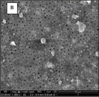
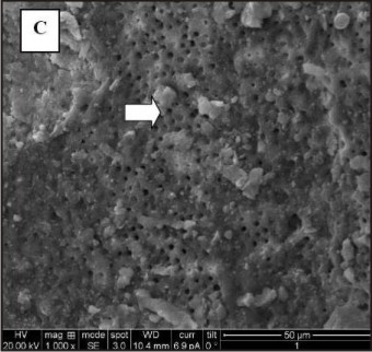
In the middle third of the retreatment technique Group 2 (Fig.1B) obtained better results than Group 1(Fig.1A)(p=0.0001) and Group 3(Fig.1C)(p=0.0002). In the apical third, Group 1(Fig.1D) and 2(Fig1E) obtained better results than Group 3(Fig.1F) (p=0.0071 and p=0.0078 respectively). There was no statistical difference between the D-RaCe and PTUR at apical third. In all groups middle third showed cleaner root canals than apical third (p=0.0454 in Group 1, p= 0.0000 in Group 2 and Group 3). Rotary instruments required less time than hand instruments to remove the GP as given in Table 3. In between the rotary systems PTUR was faster than D-RaCe.
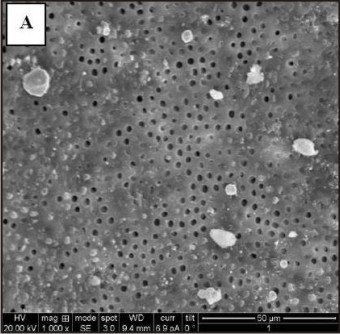 | Fig 1a : Group A, Middle Third
 |
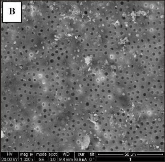 | Fig 1b : Group B, Middle Third
|
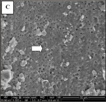 | Fig 1c : Group C, Middle Third
|
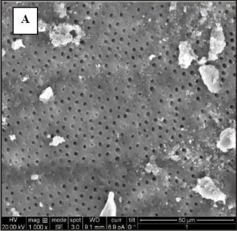 | Fig 1d : Group A, Apical Third
|
 | Fig 1e : Group B, Apical Third
|
 | Fig 1f : Group C, Apical Third
|
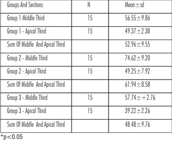 | Table 1 : Percentage Of Debris Free Area In Three Groups (Group 1, Group 2 And Group 3) At Middle And Apical Third.
|
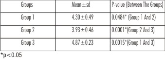
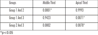 | Table 2 : Pair Wise Comparison Of Three Groups In Middle And Apical Third Of The Root Canal By Tukeys Multiple Post Hoc Procedures.
|
 | Table 3 : Mean And Sd Of Time Required For The Removal Of The Filling Material In Group 1, Group 2 And Group 3
|
Discussion
Despite being a difficult and time-consuming procedure, endodontic retreatment is undoubtedly the first choice for the management of endodontic failures when access to the root canal is possible. Advances in the endodontic field led to the use of NiTi rotary instruments which are not only effective in root canal shaping, but also proved to be efficient and require less time in removing gutta-percha/sealer during endodontic retreatment.[9]
D-RaCe Retreatment files have two retreatment instruments, DR1 and DR2, and were designed with alternating cutting edges and triangular cross section similar to the Race file. The first instrument DR1 has an active working tip to facilitate initial penetration of the filling material.
ProTaper Universal retreatment system is integrated with 3 Retreatment files D1, D2, D3 and two new ProTaper finishing files, F4, F5.The retreatment files remove large amounts of GP in spirals around the instruments. The active cutting tip and negative cutting angles and absence of radial lands permit more of a cutting rather than planing action. The nonactive tips of D2 and D3 reduce the incidence of ledging and perforation during the removal of filling material.
Traditionally, H-files have been used to remove the obturating material. They are made of stainless steel and can cut better than NiTi instruments in push and pull motion. Due to their positive rake angle they cut only in one motion that is the withdrawal stroke facilitating GP removal.
The use of H-files alone results in better cleanliness compared with hand instrumentation with solvent[11] but is tedious and time-consuming operation, especially in narrow and curved canals or when the filling material is well condensed.[12] Hence in this study solvent was used with H-file.
To standardize the apical preparation to size 30, instrumentation was further continued with Race NiTi file 30(6%) in Group 1 and finishing files of ProTaper Finishing file namely F2 (25, 8%) and F3(30, 9%) in Group 2.
Teeth were split longitudinally into two equal halves after the removal of filling material and the remaining debris in the canal was assessed by SEM.Operator bias was limited by grooves in the root surface prepared 2 and 6 mm from the anatomical apex specifying the area for investigation. Coronal third were prepared with GG drill for all the three groups for standardizing and thus coronal third assessment was not done.
It was impossible to completely remove all traces of gutta-percha/sealer from root canals with any of the retreatment files as revealed under SEM examination, which is consistent with previous studies.[6],[11],[16],[17] The reason for the presence of debris could be due to the universal inability of all instruments to contact 100% of the root canal wall. It may be due to the presence of residual paste within the irregularities or cul de sacs of the root canal system. Presence of debris also could be because of better adhesion of resin-based sealers to dentin walls, which makes their removal from root canals with rotary instrumentsand also H-file, more difficult. According to a study [13] AH26 was associated with the largest amount of remnant cement on the root canal walls when compared with other sealers. The manufacturer of the PTUR contraindicates its use in root canals filled with resin based sealers, which might help explain the presence of more filling debris.[15]
In all the groups; residual filling material was found to be more in apical third than the middle third. This may be due to the accumulation of more debris apically regardless of the protocol used.[19]
Results
The results of this study showed PTUR effectively removed GP from root canals in the middle third when compared to D-RaCe and H-files, which is in agreement with other studies.[4], [5], [6] D-RaCe and H File’s efficacy were comparable to each other. In case of the H-files the space created by GG drills at coronal third facilitated the removal of the remaining gutta-percha by serving as a reservoir for the solvent and improving the access for further instrumentation.
In the present study, in the apical third region D-RaCe and PTUR showed similar efficacy. The reason for this may be attributed to the fact that in Group 1 D-RaCe (DR1-30,10% and DR2-25,4%) followed by RaCe Ni-Ti files(30,4%) was used and in Group 2 the PTUR (D1-30,9% and D2-25,8%) followed by ProTaper finishing files (F2-25, 8% and F3 -30, 9%) was used, all of which possess greater taper. This resulted in more contact between the rotary instruments and the canal walls. Whereas, the H-files used were ISO sizes 20 to 30 of only 2% taper which may have resulted in less contact. The finding of the present study demonstrated that rotary NiTi systems reduced the retreatment time compared with hand instrumentation, and is in accordance with most previously published studies.[4],[7],[12],[14] Active tip and the cutting blades of both the NiTi rotary files that were used in this study might have influenced both the time required for retreatment and the safety of the instruments. Plasticization of gutta-percha during rotary instrumentation[12] may result in lower resistance to the action of the subsequent instrumentation and easier penetration and removal of the softened filling material. [18]
The results showed that the use of NiTi rotary files that are specifically designed for retreatment of root canal fillings appeared to be safe in retreatment procedures.[14] This may also be due to the fact that instruments were discarded after five uses, to the strict observance of manufacturers’ instructions.
Conclusion
To conclude, PTUR files were more effective in the removal of filling material from the middle thirds of the root canal walls as compared to the D-RaCe and H-files. In the apical third, PTUR files and D-RaCe systems performed fairly well as compared to H-files.
References
1. Ruddle CJ. Nonsurgical retreatment. In: Cohen S, Burns RC, eds. Pathways of the pulp(ed 8). St Louis, MO: CV Mosby; 2002:875–930.
2. ViducG1;ic´ D, Jukic´ S, Karlovic´ Z, Božic´ Ž, Miletic´ I, Anic´ I. Removal of gutta-percha from root canals using an Nd:YAG laser. IntEndod J 2003;36:670 –3.
3. Rödig T, Hausdörfer T, Konietschke F, Dullin C, Hahn W, Hülsmann M. Efficacy of D-RaCe and ProTaper Universal Retreatment NiTi instruments and handfiles in removing gutta-percha from curved root canals - a micro-computed tomography study.IntEndod J.2012 Jun;45(6):580-9.
4. Takahashi CM, Cunha RS, de Martin AS, Fontana CE, Silveira CF, da SilveiraBuenoCE.In vitro evaluation of the effectiveness of ProTaper universal rotary retreatment system for gutta-percha removal with or without a solvent. J Endod.2009 Nov;35(11):1580-3.
5. Kumar MS, Sajjan GS, Satish K, Varma KM. A comparative evaluation of efficacy of protaperuniversalrotaryretreatmentsystem for gutta-percha removal with or without a solvent. Contemp Clin Dent.2012Sep;3(Suppl 2):S160-3.
6. Marques da Silva B, Baratto-Filho F, Leonardi DP, Henrique Borges A, Volpato L, Branco Barletta F.Effectiveness of ProTaper, D-RaCe, and Mtwo retreatment files with and without supplementary instruments in the removal of root canal filling material.IntEndod J.2012 Oct;45(10):927-32
7. ValentinaGiuliani, RobertoCocchetti, Gabriella pagavino. Efficacy of ProTper universal Retreatment files in removing filling materials during root canal retreatment. JOE2008 NOV;34(11):1381-84.
8. Shrikanth V. Bhat, NithinSuvarna, K.Harish Kumar Shetty, K. Ravi Varma. Comparison of efficiency of guttapercha removal in retreatment using protaper retreatment files, RaCe instruments with and without ‘H’ files-An ex vivo evaluation. ENDODONTOLOGY
9. Marwan Z. Abdul-Jabbar . Abdul-Karim J. Al-azzawi .Comparison of the efficacy of three different techniques in the removal of gutta-percha and two types of sealers during endodontic retreatment. J BaghColl Dentistry2011;23(4):24-30.
10. Jayasenthil A, Sathish ES, Prakash P. Evaluation of manual and two-rotary NiTi retreatment systems in removing gutta-percha obturated with two rootcanal sealers. ISRN Dent.2012;2012:208241. Epub 2012 Sep 10.
11. Imura N, Kato AS, Hata GI, Uemura M, Toda T, Weine F.A comparison of the relative efficacies of fourhand and rotary instrumentation techniques during endodontic retreatment. IntEndod J.2000 Jul;33(4):361-6.
12. Hülsmann M, Bluhm V. Efficacy, cleaning ability and safety of different rotary NiTi instruments in root canal retreatment. IntEndod J.2004 Jul;37(7):468-76
13. Kosti E, Lambrianidis T, Economides N, Neofitou CEx vivo study of the efficacy of H-files and rotary Ni-Ti instruments to remove gutta-percha and four types of sealer. IntEndod J.2006 Jan;39(1):48-54.
14. Somma F, Cammarota G, Plotino G, Grande NM, Pameijer CHThe effectiveness of manual and mechanical instrumentation for the retreatment of three different root canal filling materials.J Endod.2008 Apr;34(4):466-9.
15. Dadresanfar B, Iranmanesh M, Mohebbi P, Mehrvarzfar P, VatanpourM.Efficacy of TwoRotaryNiTiInstruments in Removal of Resilon/Epiphany Obturants. Iran Endod J.2011;6(2):69-73
16. Kfir A, Tsesis I, Yakirevich E, Matalon S, Abramovitz I. The efficacy of five techniques for removing root filling material: microscopic versus radiographic evaluation. International Endodontic Journal, 45, 35–41, 2012.
17. HorvathSD, Altenburger MJ, Naumann M, Wolkewitz M, SchirrmeisterJF. Cleanliness of dentinal tubules following gutta-percha removal with and without solvents: a scanning electronmicroscopic study. IntEndod J.2009 Nov;42(11):1032-8.
18. Bramante CM, BettiLV.Efficacy of Quantec rotary instruments for gutta-percha removal. IntEndod J.2000Sep;33(5):463-7.
19. Hülsmann M, Stotz SEfficacy, cleaning ability and safety of different devices for gutta-percha removal in root canal retreatment. IntEndod J.1997 Jul;30(4):227-33.
|
|
|
|
|
|
|