Introduction
Eruption is a process of biological maturation which compromises the axial movement of tooth from the developmental position within the jaw towards the functional position in the the occlusal plane. Eruption is a multifactorial process whose biological mechanism remains unknown. The process of tooth eruption has been described in different stages (Pre-eruptive movements, intraosseous eruption, and pre-and post-occlusal eruption). Arrest in eruption may occur in any of these stages.[1] The mechanism that bring about tooth movement is till debatable and is likely to be a combination of a number of factors. Although many possible causes have been proposed, only four merit serious consideration: (1) bone remodelling, (2) root growth, (3) vascular pressure, and (4) ligament traction. Briefly stated, the bone remodelling theory supposes that selective deposition and resorption of bone brings about eruption. The root growth theory supposes that the proliferating root impinges on a fixed case, thus converting an apically directed force into occlusal movement. The vascular pressure theory supposes that a local increase in tissue fluid pressure in the peripapical region is sufficient to move the tooth. The ligament traction theory proposes that the cells and fibres of the ligament pull the tooth into occlusion.[2]
Case Report
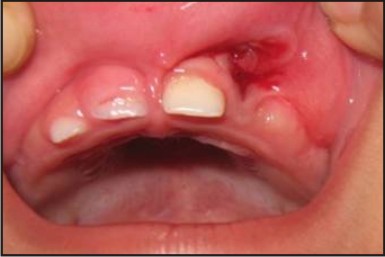
A 1 year old girl reported to our department of Paedodontics and Preventive Dentistry, Seema Dental College and Hospital with the chief complaint of inverted tooth in upper anterior teeth region (Fig.1). The history revealed by the parents that the tooth is causing trauma to the child especially the upper portion of the lip. The family and medical history were non-contributory. The parents could not recall any trauma to the oral cavity or the head and neck region.
An intraoral examination revealed that the primary maxillary primary central incisors are erupting and the maxillary lateral incisor on the right side is erupting normally whereas the primary maxillary lateral incisor 62 is inverted. The remaining primary dentition that are present are in normal shape and alignment. No abnormality was noted in the gingiva and alveolar bone but laceration in the upper lip was seen. An intraoral periapical radiograph revealed the presence of inverted primary maxillary lateral incisor 62. The involved tooth had well developed crowns and only partially developed roots (Fig 2).
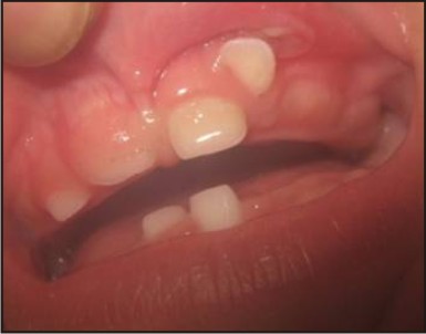 | Fig 1 : Inverted Primary Lateral Incisor Irt 62
 |
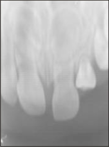 | Fig 2 : Iopa Xray Irt 62
|
Considering the age of the child, the position of primary maxillary lateral incisor 62, and the location and developmental stage of primary maxillary lateral incisor 62, it was decided to remove the inverted primary maxillary lateral incisor with normal extraction procedure as it was lacerating the upper lip. Informed consent was taken from the child’s parents. The inverted primary maxillary lateral incisor was removed under local anaesthesia (lidocaine with adrenaline 1:100,000) without disturbing the erupting permanent central incisors and lacerated lip was sutured (Fig 3, Fig 4 and Fig. 5). The child was very cooperative and tolerated the procedure well. Periodic recall visits were advised to monitor the developing dentition (Fig 6).
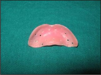 | Fig 3 : Custom Tray Fabrication For Maxillary Arch
|
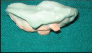 | Fig 4 : Cast Model Of Upper Arch
|
 | Fig 5 : Immediately After Extraction
|
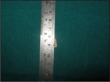 | Fig 6 : Inverted Primary Lateral Incisor 62
|
Discussion
An inverted tooth is usually associated with permanent dentition and is a rare entity during the development of primary dentition. Radiographic examination of the jaws of children would shows the extent of crown formation, amount of root development, all of which aid in a more correct assessment of the dental age. By far the greatest number of aberrations in eruption times are delayed eruptive movements, Premature eruption of teeth occurs infrequently, Sometimes infants are born with 'erupted' lower central incisors, but this is an example of gross maldevelopment. These teeth are termed Natal teeth or Neonatal teeth if they erupt during the neonatal period.[3] The failure of eruption has no identifiable systemic or local causes. There is no difference in incidence between maxillary or mandibular teeth or between sexes. It is suggested that defects in genes like CSF-1, c-fos may be responsible for this condition. It has been shown that the moment a tooth breaks through the oral epithelium, an acute inflammaory response occurs in the connective tissue adjacent to the tooth. This is een even in the germ-free animals and is seen in varying degrees around all teeth throughout life. Clinically as teeth break through the oral mucosa, there is often some pain, slight fever and general malaise, all signs of an inflammatory process. In infants these symptoms are populary called teething.[4]
Teeth which have erupted beyond the occlusal plane are reffered to as overerupted or supraerupted teeth. Lack of opposing teeth makes tooth to overerupt. The periodontal ligament and the bone develop together and therefore the gingival margin follows the tooth. In other case the gingival margin stays at the origingal level and the roots get exposed due to supraeruption. Treatment of supraerupted teeth and treatments on these teeth are challenging and require careful planning.[5]
Only a few cases of inverted lateral incisor have been reported earlier. In all these cases abnormal positioning of the primary tooth germ is mainly due to trauma. In the present case the abnormal positioning is not associated with trauma, since no trauma was reported by the parents. However many authors suggested that trauma cannot be ruled out as an etiological factor, as the parents may not even aware of the trauma which occur in young children prior to the eruption of the primary dentition.[6],[7]
The inverted primary maxillary lateral incisors are of concern to both the parents and dentist, because it poses problem with esthetics, speech and mastication. In the present case the inverted primary lateral incisor were extracted to allow the normal eruption of succedaneous tooth and to prevent the laceration of upper lip. Patient is recalled for follow up and intraoral periapical radiograph was taken and on viewing the intraoral periapical radiograph it is seen that the eruption of succedaneous tooth appears to be normal. The reason for inverted tooth is uknown in the present case but it may be due to some physical barrier in the eruption pathways (also known as primary failure or eruption).
References
1. Ten cate; oral histology – development, structure & function; 2005, 6th ed; Mosby.
2. Orban's oral histology and embryology; 2009, 12th ed; Mosby.
3. Pindborg JJ. Pathology of the dental hard tissues. WB Saunders Co: Philadelphia; 1970. p. 241.
4. Rasmussen P, Kotsaki A. Inherited primary failure of eruption in the primary dentition. ASDC J Dent Child 1997;64:43-7.
5. Miyanaga M, Takei K, Maeda T. Observation of a child with multiple submerged primary teeth. ASDC J Dent Child 1998;65:495-8
6. PH, Lindquist CC. Impactions should you bother them if they don’t bother you? J Dist Columbia Dent Soc 1977; 52: 55-8.
7. Kapur A, Goyal A, Jaffri S. Management of inverted impacted primary incisors: an unusual case. J Indian Soc Pedod Prev Dent. 2008 Mar; 26(1):26-8.
|