Introduction
Proper cleaning and shaping of the root canal system is one of the most important objectives for the success of endodontic therapy.[1] Canal preparation is adversely influenced by the highly variable root-canal anatomy and the relative inability of the operator to visualize this anatomy from radiograph.[2] Roots and root canals are rarely straight even when they appear so in a normal clinical radiographic projection.[3] Most root canals are curved and preparing a Bayonet or S-shaped canal presents additional problems, since they are; (1) not evident on a standard facial Intra-oral radiograph and (2) preparing such canals with stiff endodontic instruments may result in stripping, ledging and zipping.[4],[5] This report discusses the endodontic management of Bayonet-shaped canals occurring in a maxillary first premolar.
Case Report
A 18-year old male patient reported to the Department of Conservative Dentistry and Endodontics with chief complaint of pain in upper left back tooth since 2-weeks. History revealed previous restorative treatment in the same tooth at a private clinic 2-years back.
Intra-oral examination revealed moderate oral hygiene and marginal gingivitis. Hard tissue examination revealed a temporary restoration in relation to maxillary left first premolar (tooth no.24). The tooth was tender to percussion and gave delayed response to vitality test with thermal and electric method. A periapical radiograph reveled radiolucency beneath the restoration approximating the pulp space. The double curved anatomy of the root was also revealed [Fig -1].
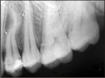 | Fig 1 : Pre-operative Iopar Showing Bayonet - Shaped Canal Curvature In Relation To 24 (Arrow)
 |
A diagnosis of irreversible pulpitis with acute apical periodontitis was established and root canal treatment was planned. Following administration of local anesthesia with 2% Lignocaine with 1:80,000 adrenaline (Lignox, Indoco Remedies Ltd, Mumbai, India), and placing rubber dam, standard access cavity was prepared with a no.2 round bur and safe-ended endo bur. The canals were irrigated with 3 % sodium hypochlorite (Parcan, Septodont India) and patency was established with a slightly pre-curved no.10 stainless steel K-file (Mani Inc Japan). A type III (Weine’s classification) canal configuration was revealed.
Keeping in mind the double curved anatomy, caution was exercised throughout the canal preparation. The modified double-flared technique was used to complete the canal preparation. This technique involves coronal flaring followed by working length determination and apical preparation.[6] Using 3 % Sodium hypochlorite and 17% EDTA as irrigant, coronal flaring was achieved with hand Nickel Titanium ProTaper S1 and SX instruments (Maillefer, Dentsply, Switzerland). Patency was again checked with a #10 K file (Mani Inc Japan) for both the canals and working length determined by Root ZX electronic apex locator. (J. Morita Mfg Corp, Kyoto, Japan) and confirmed radiographically [Fig-2].
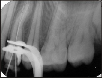 | Fig 2 : Working Length Radiograph
|
Both the canals were prepared with 0.02 taper Nickel titanium hand files (Maillefer, Dentsply, Switzerland) using a step-back technique to a master apical file # 25.
The hand files were used with minimal excursions, so as not to straighten the canal and avoid stripping and lateral perforation of the canal. It is preferable to use file and avoid reaming action. All the instrumentation was accompanied by lubrication with 17% EDTA (Glyde, Maillefer, Dentsply, Switzerland and copious irrigation with 3% sodium hypochlorite. The canals were then dried and intracanal Calcium hydroxide dressing (RC Cal, Prime Dental products, India) was placed and the access cavity sealed with a temporary filling material (Kalzinol DPI, India). One week later, the root canals were obturated by the cold lateral condensation technique with standardized gutta-percha points and zinc-oxide based sealer (Root canal sealer, Pulpdent Corporation, USA). [Fig- 3].
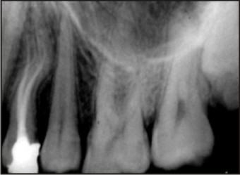 | Fig 3 : Post-obturation Radiograph
|
Discussion
Thorough cleaning and shaping of the complete root canal system are most important objectives of endodontic therapy.[1] Few root canals are straight, and even subtle curves introduce complexity into the instrumentation procedure.[7] A curvature may be a gradual curve of the entire canal or a sharp curvature near the apex. Double ‘S-shaped’ or Bayonet-shaped canal curvatures can also occur.[8] The bayonet or S- shaped curvatures are most often found in maxillary premolars; 20.6% in second premolars and 8% in buccal root of two rooted first premolars.[9]
Endodontic management of a tooth with bayonet-shaped canals is challenging, since they involve at least two curves, with the apical curve being more susceptible to deviations in canal anatomy and loss of working length. Hence to prevent problems like formation of a ledge, creating a zip, stripping canal wall or loss of working length, it is important to understand the three-dimensional anatomy of these canals.[5]
Various approaches have been suggested to prepare such canals.
Grossman recommends the straightening of the coronal curve by elimination of the middle third curve by filing with a Hedstrom file. A small Hedstrom file is introduced into the root canal until the junction of the middle and apical third is reached. The inner portion of this curve is then filed away. Recapitulate with No. 10 file to working length to maintain the patency. The root canal is irrigated, file cleaned, recurved, and the procedure is repeated until the curve is eliminated. Instrumentation of the apical part of the canal then follows with a curved instrument.[10]
Weine also recommends the straightening of the coronal curvature to prevent zipping in the apical third of the canal, which can be achieved by: precurving files, incremental instrumentation and flared preparation. Following negotiation and working length determination, flutes are removed from the file towards the outer side of the curve and minimal increments (0.5 mm) of file tip is clipped to create intermediate file size.[4]
To allow for more direct entry to the coronal curve, Gutmann recommends a divergent and skewed access preparation. This is followed by passive shaping of the coronal curve, to facilitate cleaning and shaping of apical curvature. To prevent stripping in coronal curve, anti-curvature or reverse filling is recommended, with primary pressure being applied away from the curve of coronal curvature.[5]
The Double-flared technique of preparing the straight portions of curved canal have being proposed by Fava, with advantage of reducing the forcing of materials through the apical foramen and early removal of microorganisms and decreasing the probability of carrying them to the apex.[11] The double flare technique was originally proposed for preparing the anterior teeth with hand instruments, however the benefits of this technique were more while preparing the teeth with curved canals, hence the modified double-flared technique was introduced which involves coronal flaring followed by working length determination and apical preparation with rotary or hand instruments.[7]
The advantage of initial Crown down (Coronal flaring) is reduction of periapically extruded debris and minimization of root canal straightening. Since during the stepdown there is less constraint to the files and better control of the file tip it has been expected that apical zipping is less likely to occur.[12],[13]
The introduction of hand and rotary nickel titanium files made the preparation of the curves of the middle third and in particular of the bayonet curves easier. Nickel titanium instruments maintain canal curvature and assure a better respect of the root canal anatomy.[14] One must be aware of the fact that although nickel-titanium files are flexible, nickel titanium metal, like any other metal, will eventually fatigue and fail when it becomes overstressed, especially during rotation in curved root canals or if improperly used or abused.[9]
Conclusion
Nature seldom draws a straight line. Nowhere this is more apparent than in the anatomy of roots and root canals system of human teeth. With careful clinical judgment the canals with multiple curvatures can be safely prepared by modified Double-flared technique, minimizing deviations, perforations or zipping of the canal. In the current case, we prepared the coronal part of the canals, by ProTaper hand files and apical part by using NiTi 0.02 taper hand instruments. The authors emphasize the use of hand instrumentation techniques in negotiation and preparation of Bayonet-shaped canals.
References
1. Schilder H. Cleaning and shaping the root canal. Dent Clin North Am 1974: 18: 269–296
2. Peters OA. Current challenges and concepts in the preparation of root canal systems: A Review.J Endod.2004; 30:559-567
3. Walker RT, Root canal morphology. In Stock C, Walker R, Gulabivala K, In:Endodontics. 3rd edn. Mosby. Pg. 125-134
4. Weine FS. Intracanal treatment procedures, Basic and Advanced Topics, In: Endodontic therapy. 6th edn. Mosby.pg 164-239
5. Glickman GN and Dumsha TC. Problems in canal cleaning and Shaping. In: James L.Gutmann ed. Problem solving in Endodontics 3rd edn. Elsevier Health Sciences. Pg 91-122
6. Saunders WP, Saunders EM. Effect of noncutting tipped instruments on the quality of root canal preparation using a modified double-flared technique. J Endod 1992: 18: 32–36.
7. Saunders EM and Saunders WP. Preparation of the root canal system. In: Pitt ford TR, ed. Harty’s Endodontics in Clinical Practice. 4th edn. Wright. Pg.81-105
8. Vertucci F J. Root canal morphology and its relationship to endodontic procedures. Endo topics 2005, 10, 3-29.
9. Ingle J I et al. Endodontic Cavity Preparation. In: Ingle and Bakland ed. Endodontics. 5th edn. B C Deker. Elsevier India. Pg. 405-570
10. Louis I Grossman, Seymour Olie and Carlos Del Rio .Preparation of the Root canal:Equipment and Technique for Cleaning, Shaping, and Irrigation. In: Endodontic practice. 11th edition. Pg. 179-227. Varghese Publishing House. India
11. Fava LR. The double-flared technique: an alternative for biomechanical preparation. J Endod 1983 feb;9(2): 76-80
12. Hulsmann M, Peters O A, Dummer MH. Mechanical preparation of root canals: shaping goals, techniques and means. Endo Topics 2005;10:30-76
13. Saunders E M. Hand instrumentation in root canal preparation. Endo Topics 2005;10:163-167
14. Castellucci A. Curved canals. In Arnaldo Castellucci ed. Endodontics Volume II .IL tridente. Pg.502-517.
|