Introduction
The aim of the periodontal aesthetic procedure is based on the coverage of exposed root surfaces. The main indication for root coverage procedures are establishment of aesthetics, to eliminate root hypersensitivity, management and prevention of root caries and cervical abrasion, enhancement of restorative outcomes, and facilitation of plaque control efforts. The main causative factor for gingival recession are traumatic tooth brushing and tooth mal-positioning which have been considered as the main etiologic factor for gingival recession.[1] The rationale of periodontal therapy is to protect and maintain the patient’s natural dentition over his or her lifetime for optimal comfort, function and esthetic appearance.[2] Multiple gingival recession-type defects present a challenge because of several recessions must be treated at a single surgical session to minimize patient discomfort.
Since 20th century, various techniques have been introduced to cover denuded roots. Among them are the free autogenous grafts and pedicle grafts including rotational flaps, coronally advanced flaps (CAF), and semilunar flaps have been advocated. Combination grafts with either autogenous grafts or allograft and with GTR membranes were developed later to correct mucogingival defects.[3] Many materials have been proposed to improve clinical outcomes. Fibrin glue (FG) has been tested in conjunction with tetracycline root conditioning, but the addition of FG may not enhance the outcome of the CAF procedure.[4] The homogeneous @257;brin network that is obtained and considered by the promoters of the technique to be a healing biomaterial and is commonly used in implant and plastic periodontal surgery procedures to enhance bone regeneration and soft tissue wound healing.[5] Platelet concentrate contains PDGF, TGF and many other unidentified growth factors that modulate and up regulate one growth factors function in the presence of second or third growth factor.[6] This particular advantage influenced the decision to use platelet concentrates as the material of choice in this case report. In the case report, platelet rich derivative (PRF membrane) was combined with coronally advanced flap for root coverage.
Case Report
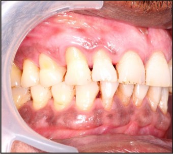
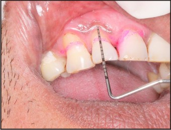
A 28-year-old male patient reported to the Department Of Periodontics, Institute Of dental Sciences, Bareilly with a chief complaint of sensitivity to hot and cold beverages since 6 to 7 months. On evaluation, multiple recession defects were noticed in maxillary right anterior teeth region. At the time of presentation, clinical examination revealed 3mm, 5mm, 6mm of clinical attachment loss in 11, 12, and 13 respectively (Fig. No.:- 1). The distance between the cemento-enamel junction and gingival margin was 2mm, 4mm, 5mm in 11, 12, and 13 respectively. (Fig. No.:- 2).
 | Figure No.:- 1
 |
 | Figure No.:- 2
|
Pre - Surgical Therapy:
The surgical procedure was explained to the patient and the informed consent was obtained. Preparation of the patient included scaling and root planing of the entire dentition and oral hygiene instructions 1 month prior to the surgery. The following parameters were recorded before (baseline) and after surgery (after 6 months). Keratinized gingival width (KGW) was measured from the mucogingival junction to the gingival margin, recession width (RW) was measured at the CEJ, probing depth (PD), clinical attachment level (CAL), and gingival/mucosal thickness (GTH).
Platelet Rich Fibrin Membrane Preparation
For the preparation of platelet rich fibrin membrane, intravenous blood was collected from the anti – cubital vein in two 10-ml centrifugation test tubes without anticoagulant and immediately centrifuged at 3,000 revolutions per minute for 10 minutes. The resultant product consists of the following three layers:
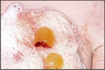
Top most layer consisting of acellular poor platelet plasma (PPP), platelet rich fibrin (PRF) clot in the middle and Red blood cells at the bottom. The @257;brin clot was easily separated from the lower part of the centrifuged blood and spread on a sterile gauze. The obtained PRF is in the gel form and the PRF membrane is prepared by pressing the gel between two dry gauze and they are pressed to obtain a PRF membrane.
Surgical Procedure
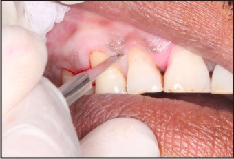
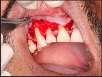
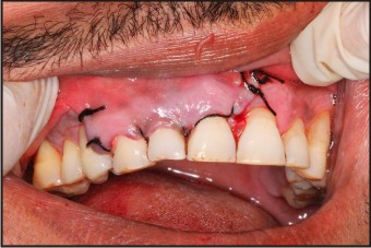
Recession defects were thoroughly scaled using Gracey curettes. No root conditioning was used. A Coronally advanced flap technique was undertaken using a continuous sling suturing technique. The first incision involving the submarginal incisions (Fig. No.:- 3) were made in the interdental areas, and intrasulcular incisions were made around those teeth with recession defects. Partial-full- partial @258;ap incisions were performed in a coronal–apical direction. Gingival tissue adjacent to the root defect and the interproximal bone was elevated with blunt dissection with the help of periosteal elevator in the full thickness manner, whereas the most apical portion of the @258;ap was partial thickness obtained by the sharp dissection with the help of scalpel. To allow coronal repositioning of the @258;ap without tension two vertical incisions were incorporated on mesial and distal aspect of the recession defect which were diverging in nature (Fig. No.:- 4). The involved interdental papillae were deepithelialized to create a connective tissue bed. At the recession defect site, the prepared @257;brin clot (Fig. No.:- 5) was positioned over the recession defects, just below the CEJ (Fig. No.:- 6). The gingival @258;ap was coronally advanced, with its margin located on the enamel. It was held in that position with horizontal suspensory sutures around the contact points (Fig. No.:- 7). Stabilization of the blood clot was achieved by the application of gentle pressure for 3 minutes.
 | Figure No.:- 3
|
 | Figure No.:- 4
|
[Image 5]
 | Figure No.:- 5
|
 | Figure No.:- 7
|
Post-Surgical Protocol
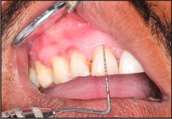
Patient was advised not to brush their teeth in the operated areas until after suture removal 2 weeks later, and also was instructed to rinse their mouth with a 0.12% chlorhexidine solution, three times a day for 1 minute, for 3 weeks. Fifteen days after surgical treatment, patient was reviewed and the sutures were removed. There was no clinical signs of inflammation present at the operated site and the healing was satisfactory without any complications. The patient was motivated for mechanical tooth cleaning in the operated areas using a soft toothbrush and a roll technique. Patient was recalled for prophylaxis 1 month after suture removal and at 3 and 6 months post – operatively. Complete root coverage was achieved six months after the procedure, with excellent tissue contour and colour (Fig. No.:- 8).
 | Figure No.:- 8
|
Discussion
Multiple gingival recessions may be a concern for patients with a high lip smile line and severe root hypersensitivity. The ultimate aim of any therapeutic intervention aimed at root coverage is to restore the tissue margin at the cemento - enamel junction (CEJ) and to achieve an attachment of the tissues to the root surface so that a normal healthy gingival sulcus with no bleeding on probing and a minimal probing depth is present.[7] Different surgical procedures have been introduced to treat gingival recessions, but these have been demonstrated to heal with a long junctional epithelium, and regeneration has been observed only in the most apical portion of the lesion. The use of PRF membrane in our case report to attain root coverage may alleviate the need for donor site procurement of connective tissue. A recent 6-month study evaluated the use of PRF in the treatment of multiple gingival recessions with coronally advanced flap procedure and found the significant improvement during the early periodontal healing phase with a thick and stable final remodelled gingiva.[8]
A clinical study on Clinical Evaluation of a Modi@257;ed Coronally Advanced Flap Alone (MCAF) or in Combination with a Platelet-Rich Fibrin Membrane for the Treatment of Adjacent Multiple Gingival Recessions was done by Sofia Aroca et. al in 2009 and they concluded that MCAF is a predictable treatment for multiple adjacent Miller Class I or II recession-type defects. The addition of a PRF membrane positioned under the MCAF provided inferior root coverage but an additional gain in GTH at 6 months compared to conventional therapy.[9] PRF containing the growth factors has been shown to accelerate bone repair and promote fibroblast proliferation, and increase tissue vascularity, rate of collagen formation, mitosis of mesenchymal stem cells and endothelial cells, as well as osteoblasts, playing key roles in the rate and extent of bone formation. This activity, together with increased vessel ingrowth, is mediated by PDGF and TGF. Because of all of these potential effects on tissue regeneration, a growing number of human clinical studies have detailed the use of growth factors in reconstructive oral and maxillofacial surgery, periodontal surgery, implants, and sinus grafting.[10]
Conclusion
The use of autologous platelet preparations like PRF allows the clinician to optimize tissue remodelling, wound healing and angiogenesis by the local delivery of growth factors and proteins. This case report reflects the success of this biomaterial for coverage of multiple recession defects and the ability to increase the thickness of the keratinised gingival tissue.
References
1. Gorman WJ. Prevalence and etiology of gingival recession. J Periodontol 1976;38:316-22
2. Zander HA, Polson AM, Heijl LC. Goals of periodontal therapy. J Periodontol 1976;47:261-6. ,
3. Pin Prato GP, Tinti C, Vincenz G, Magnani C, Cortellini P, Clauser C. Guided tissue regeneration versus mucogingival surgery in the treatment of human buccal gingival recession. J Periodontol 1992;63:919-28
4. Trombelli L, Scabbia A, Wikesjo ¨ UM, Calura G. Fibrin glue application in conjunction with tetracycline root conditioning and coronally positioned @258;ap procedure in the treatment of human gingival recession defects. J Clin Periodontol 1996;23:861-867.
5. Choukroun J, Diss A, Simonpieri A, et al. Platelet-rich @257;brin (PRF): A second-generation platelet concentrate. Part IV: Clinical effects on tissue healing. Oral Surg Oral Med Oral Pathol Oral Radiol Endod 2006;101:e56-e60.
6. Grif n TJ, Cheung WS. Treatment of gingival recession with a platelet concentrate graft: A report of two cases. Int J Periodont Restorat Dent 2004;24:589-95
7. Guptha R, Pandit N, Sharma M. Clinical evaluation of a bioabsorbable membrane (polyglactin 910) in the treatment of miller type II gingival recession. Int J Periodont Restorat Dent 2006;26:271-7.
8. Del Corso M, Sammartino G, Dohan Ehrenfest DM. Clinical evaluation of a modified coronally advanced flap alone or in combination with a platelet-rich fibrin membrane for the treatment of adjacent multiple gingival recessions: a 6- month study. J Periodontol 2009; 80: 1694–1697.
9. So@257;a Aroca, Tibor Keglevich, Bruno Barbieri, Istvan Gera, and Daniel Etienne Clinical Evaluation of a Modi@257;ed Coronally Advanced Flap Alone or in Combination With a Platelet-Rich Fibrin Membrane for the Treatment of Adjacent Multiple Gingival Recessions: A 6-Month Study JOP 2009;80:244-252.
10. Lozada JL, Caplanis N, Proussaefs P, Willardsen J, Kammeyer G. Platelet-rich plasma application in sinus graft surgery: Part I -Background and processing techniques. J Oral Implantol 2001;27:38-42.
|