Introduction
Natal teeth are teeth that are already present at the time of birth. They are different from neonatal teeth, which grow in during the first 30 days after birth.
Terms such as congenital teeth, fetal teeth, predeciduous teeth, and precocious dentition (Mayhall and Bodenhoff), as well as dentitia praecox and dens connatalis, have been used to describe these teeth. More recently, it was been found that these terms were too broad to accurately describe teeth that erupted at birth or shortly thereafter. Currently the terminologies ‘natal’ and ‘neonatal,’ used by Massler and Savara, have been adopted. Even these terms only define the time of eruption and give no consideration to anatomy and histology or whether the tooth is a component of primary dentition or whether it is supernumerary.[1],[2]
Natal teeth are uncommon. They vary in occurrence from 1:1000 to 1:30000 and 85% of natal and/or of Neonatal teeth are mandibular incisors.[3] Natal teeth most often develop on the lower gum, where the central incisor teeth will appear. They have little root structure. They are attached to the end of the gum by soft tissue and are often wobbly.
Natal teeth are usually not well-formed, but they may cause irritation and injury to the infant's tongue when nursing. Natal teeth may also be uncomfortable for a nursing mother.
Natal teeth are often removed shortly after birth while the newborn infant is still in the hospital or may be removed as early as possible. Tooth extraction is indicated if the tooth is supernumerary or if the tooth is poorly implanted and excessively mobile, which is associated with a risk of aspiration.[4] This is very often done if the tooth is loose and the child runs a risk "breathing in" the tooth.[5]
Case Report
The case report presents the 4 cases of natal teeth who reported to the Department of Pedodontics, JCD Dental College, Sirsa, Haryana.
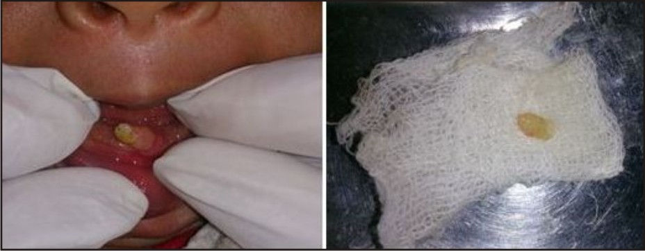
Case 1: A 15 days old female child reported to the department with complaint of presence of tooth since birth. Oral examination revealed single tooth whitish opaque in colorin mandibular anterior region with grade II mobility and edema of gingival tissue around the neck of the tooth. (Image Case 1).
 | Case 1
 |
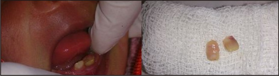
Case 2: A 21 days old female child reported department with complaint of presence of tooth like structures since birth and refusal to suck milk. Oral examination revealed two teeth in mandibular anterior region, with normal size and exhibiting grade II mobility. (Image Case 2).
 | Case 2
|
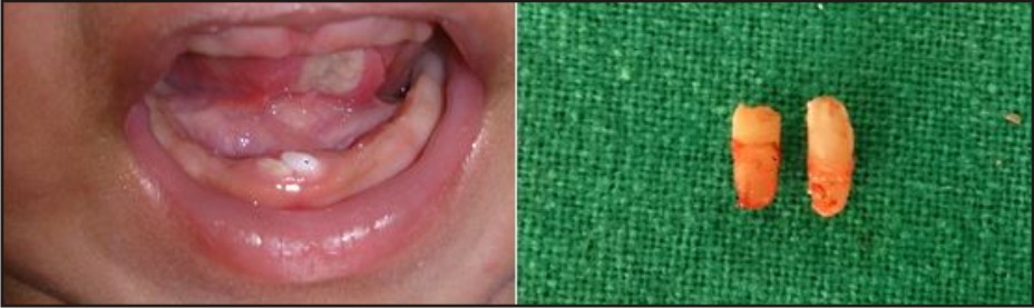
Case 3: A 13 days old male child reported to the department with mother complaining of difficulty in feeding process. Oral examination showed a presence of two teeth in mandibular anterior region which was normal in size with no mobility. Tip of the tongue & floor of mouth showed massive ulceration which was a Riga-Fede disease presentation. (Image Case 3).
 | Case 3
|
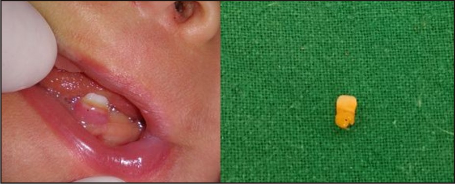
Case 4: A 5 days old male child reported to the department with complaint of presence of a tooth since birth. Oral examination revealed single tooth in mandibular anterior region with grade II mobility and small ulceration on tip of the tongue. (Image Case 4).
 | Case 4
|
Immediate extraction was the treatment of choice for Case 1, 2 & 3. The teeth were extracted under topical local anaesthesia in Case 1 and 2 which was well tolerated by both the patients. In case 3, 0.9 ml local anaesthesia was administered as the two teeth present were firm. The patients were re-evaluated after 2 days, and recovery was found to be uneventful in all three cases. The sharp edge of the tooth were trimmed and made smooth with finishing bur for Case 4. Patient was reviewed after three days, the recovery was uneventful and the ulcer disappeared, as there was grade I mobility and mother was not comfortable in breast feeding, the tooth was also extracted after consultation with the paediatrician and vitamin K (0.5 = 1.0mg) was administered intra muscularly as a part of immediate medical care to prevent haemorrhage. The extracted teeth had a thin shelled crown and were devoid of roots in Case 1, 2 and 4 but Case 3 showed some degree of calcified root structure.
Discussion
The etiology of natal and neonatal teeth remains undetermined; however it was suggested to be related to various factors, including superficial position of the tooth germ, increased eruption rate due to pyretic incidents, hormonal stimulation, developmental abnormalities, syndromes, heredity, and osteoblastic activity within the germ zone related to the remodeling phenomenon [6], [7], [8].
Natal and neonatal teeth could be either conical or of normal shape and size. They usually have an opaque yellow brownish color. The dimensions of the crowns of these teeth are smaller compared to primary teeth that have erupted normally [6], [7], [8], [9], [10].
Clinically, natal and neonatal teeth can be classified according to their degree of maturity: (1) a mature natal or neonatal tooth is nearly or fully developed and has moderately good prognosis and (2) an immature natal or neonatal tooth is incomplete or having a substandard structure with a poor prognosis [8],[9], [10], [11]. Hebling et al.[12] suggested another clinical classification in their case report according to tooth morphology during eruption into the oral cavity: (1) shell-shaped crown that is poorly fixed to the alveolus by gingival tissue with root absence, (2) solid crown that is poorly fixed to the alveolus by gingival tissue with little or no root, (3) eruption of the incisal margin of the crown through the gingival tissues, and (4) gingival edema with palpable but unerupted tooth.
Natal/neonatal teeth that show mobility of more than 1mm are indicated for extraction; this is due to the probability of aspirating or ingesting natal teeth. Another reason for the removal of the natal/neonatal tooth is to alleviate feeding difficulties or complications like Riga- Fede disease. If extraction is the treatment of choice, it can be deferred till the child is 10 days of age or more and has appropriate blood levels of vitamin K. This ten-day waiting period is to allow the normal flora of the intestine to become established to produce vitamin K, an essential factor for prothrombin production in the liver [6], [8], [13]. Since parenteral vitamin K prevents a life threatening haemorrhagic disease of the newborn, the American Academy of Pediatrics recommends that all newborns be given a single intramuscular dose of 0.5 to 1mg of vitamin K [14]. If it is not possible to delay the extraction, a consultation with the pediatrician should be initiated, so they can assess if there is a need to administer vitamin K, if the newborn did not receive vitamin K immediately after birth.
Once extraction is performed, it is essential to remove the underlying dental papilla and Hertwig’s epithelial root sheath during the extraction of natal tooth/teeth to prevent the development of root structure that could continue if these structures are left in situ.
In the cases reported above the first three patients were in age of 15, 21 and 13 days respectively and all of them presented with either great deal of mobility or chronic ulceration on ventral surface of the tongue and immediate extraction was the treatment of choice and the patients also responded satisfactorily to the treatment. Taking into consideration the age of the patient in case 4 prophylactic administration of vitamin K (0.5 - 1.0 mg, i.m) which was the base line treatment management discussed above.
Conclusion
Natal teeth are rare occurrences in the oral cavity and proper evaluation and diagnosis are crucial to provide the best treatment option. Pediatricians are usually the first to detect these teeth and early consultation with the dentist can prevent complications. The decision to maintain or remove these teeth should be assessed in each case independently. Thus far, no studies confirmed the cause and effect relationship with any of the proposed theories so far. The etiology of natal and neonatal teeth still required further investigations.
References
1. Alvarez MP, Crespi PV, Shanske AL. Natal molars in Pfeiffer syndrome type 3: A case report. J Clin Pediatr Dent. 1993;18:21–4.
2. Anderson RA. Natal and neonatal teeth: Histologic investigation of two black females. ASDC J Dent Child. 1982;49:300–3.
3. Bodenhoff J, Gorlin RJ. Natal and neonatal teeth: Folklore and fact. Pediat 1963; 32: 1087-1093.
4. Alexander K.C. Leung,William Lane M. Robson. Natal Teeth: A Review. Journal of the national medical association.2006; 98: 226-228.
5. Smith JB. Initial evaluation. In: Gleason CA, Devaskar SU. Avery's Diseases of the Newborn.
6. A. K. C. Leung and W. L. M. Robson, "Natal teeth: a review," Journal of the National Medical Association, vol. 98, no. 2, pp. 226–228, 2006
7. M. H. Chow, "Natal andneonatal teeth," Journal of the AmericanDental Association, vol. 100, no. 2, pp. 215–216, 1980.
8. R. F. Cunha, F. A. C. Boer,D.D.Torriani, andW. T. G. Frossard, "Natal and neonatal teeth: review of the literature," Pediatric Dentistry, vol. 23, no. 2, pp. 158–162, 2001.
9. S. Mhaske, M. B. Yuwanati, A. Mhaske, R. Ragavendra, K. Kamath, and S. Saawarn, "Natal and neonatal teeth: an overview of the literature," ISRN Pediatrics, vol. 2013, Article ID 956269, 11 pages, 2013.
10. R. S. Rao and S. V.Mathad, "Natal teeth: case report and review of literature," Journal of Oral and Maxillofacial Pathology, vol. 13, no. 1, pp. 41–46, 2009.
11. J.D. Spouge andW. H. Feasby, "Erupted teeth in the newborn," Oral Surgery, Oral Medicine, Oral Pathology, vol. 22, no. 2, pp. 198–208, 1966.
12. J. Hebling, A. Zuanon, and D. Vianna, "Dente Natal—a case of natal teeth," Odontologia Clinica, vol. 7, pp. 37–40, 1997.
13. M. Rusmah, "Natal and neonatal teeth: a clinical and histological study," The Journal of Clinical Pediatric Dentistry, vol. 15, no.4, pp. 251–253, 1991.
14. Z. A. Bhutta, G. L. Darmstadt, B. S. Hasan, and R. A. Haws "Community-based interventions for improving perinatal and neonatal health outcomes in developing countries: a review of the evidence," Pediatrics, vol. 115, no. 2, supplement, pp. 519–617, 2005.
|