Introduction
Hemimandibulectomy is a procedure that is used to eradicate disease that involves the lower jaw of mandible. Depending on the thickness of the native mandible and the specific disease process, the manibulectomy can either be full thickness in which both cortices in addition to the upper and lower surfaces of the mandible are removed, or partial thickness, in which either the inner or outer cortex of the mandible is spared in order to maintain some mandibular continuity.
Indications of hemimandibulectomy:[2]
1. Malignant tumor invading the mandible, either a primary alveolar ridge tumor or from an adjacent site such as the floor of mouth.
2. Involvement of the mandible with a benign tumor,cyst of the jaw that has destroyed much of the mandibular integrity such that tumor would leave mandible so unstable that it can lead to pathological fracture.
3. Significant osteomyelitis that involves a significant portion of the mandible.
4. Significant osteonecrosis related to bis phosphonate use that has led to significant mandibular deterioration.
5. Osteoradionecrosis of the jaw following head and neck radiotherapy.
6. Severe mandibular trauma that has devitalised a significant portion of the mandibular bone and whose debridement will lead a full thickness segmental defect in the mandible.
Contraindications of hemimandi bulectomy:
The most common contraindications to hemimandibulectomy are related to medical complications. Cardiovascular hemodynamic instability or other metabolic complications may prohibit the surgical procedure.
Post operative complications include incisional dehiscence, infection, injury to salivary ducts, emphysema, mandibular instability, abnormal salivation, lingual dysfunction, anorexia, cosmetic defects and local tumor recurrence.[3]
The most common problem with patients affected with such lesions is late diagnosis, generally at stage III and IV[4] and due to this surgeon not only do excision of soft tissues but also resects part of mandible adjoining the neoplasm[5]. Resection of mandibular portion and excision of soft tissues leads to alterations in mandibular function related to mastication, deglutition, phonetics and facial aesthetics. This generates the need of rehabilitation requirements for these patients[6]. In patients where rehabilitation is not done after surgery, stiffening of tissue occurs due to scar formation and further rehabilitation becomes difficult[7].Cantor R, Curtis TA[8] grouped patients subjected to mandibulectomies into six classes, according to the anatomical characteristic of the remaining mandible as well as the alterations in its function.
CLASS I patients are those subjected to radical alveolar resection without loss of mandibular continuity. This class does not include de insertion of masticatory muscles.
CLASS II patients are those subjected to unilateral mandibular resection comprising from the distal section of canine up to the condyle. As the insertion of masticatory muscles is lost it results in deviation of mandible towards the affected side.
CLASS III patients are those subjected to unilateral resection spanning from midline up to the condyle. This class has greater muscle insertion loss causing increased instability to the mandible
CLASS IV Those patients that are treated for unilateral mandibular resection and rehabilitated with bone and soft tissue grafts. These patients have more support for placement of prosthesis than class II and III.
CLASS V Those resection patients where condyles are not affected and there is reestablishment of mandibular continuity.
CLASS VI These patients are similar to class V but lacks bone continuity.
Both interim and final surgical prosthesis are used to rehabilitate patients subjected to hemimandibulectomy[9]. Various reconstruction chains and k wires[10] are used for stabilization transitionally along with radiotherapy. For final rehabilitation palatal ramp prosthesis or guided flange prosthesis are used. They guide mandibular teeth to an inter cuspation position during mandibular closure. The aim of this article is to present the case of a patient who was rehabilitated with occlusally modified conventional prosthesis after hemimandibulectomy surgery.
Case Report
A 62 year old female reported to our clinic with chief complaint of pain and swelling near angle of mandible. Patient noticed this swelling 4 months back, with gradual increase in size and complaint of pain since 15 days. General physical examination ruled out existence of a basal cell nevus syndrome. Intra oral examination showed no alteration in sensation over the mental nerve however complaint of tenderness on palpation in the angle of mandible region. The labiobuccal sulcus was obliterated from 33 to 37 region, 33 and 37 were grade II mobile and 34, 35 and 36 were grade III mobile. The OPG was taken which revealed a multi locular swelling extending from 33 to 37 region along with root resorption in relation to 34, 35 and 36. FNAC was done and the electrophoresis of the cystic fluid demonstrated low soluble protein content (< 3.5 g/100). Provisional diagnosis of Odontogenic Keratocyst was made and was confirmed by histopathological examination of the specimen. Block resection was planned under general anaesthesia as the CT scan showed multiple areas of bone perforation on the buccal and lingual cortical plates. After surgery there was deviation of mandible to left side, asymmetrical face and convex profile. On basis of clinical examination the patient was classified as class III mandibular defect according to Cantor and Curtis[8]. Patient was recalled one month after surgery for prosthodontic rehabilitation but she reported after 6 months. We planned for guided flange prosthesis but because of scar tissue formation and tissues stiffening desirable deviation was not achieved, so we decided to continue with conventional prosthesis with double row of teeth on unresected side of maxillary arch. On intraoral examination teeth present in maxilla were 13,14,24,25. and in mandible all anterior teeth were present along with 37 and 44. There was no occlusion on right (unresected) side. (Figure-1).
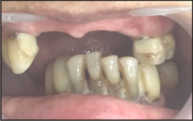 | Figure 1. Showing Deviated Mandible With No Occlusion On The Right Side
 |
Clinical Procedures
Upper and lower primary impression were made with irreversible hydrocolloid (Zelgan) using stock trays and casts were poured with type III dental stone. Custom trays were fabricated on both the primary casts with self cure acrylic resin (DPI) following Boucher’s selective pressure impression technique. Border moulding was done using green stick compound and impressions of edentulous areas were recorded with zinc oxide eugenol impression paste (septodont) followed by dual impressions with alginate using stock trays. Final impressions were poured with type III dental stone to obtain master casts. Denture bases were fabricated with shellac base plates and occlusal rims were made. Maxillary cast was articulated using face bow record on a semi adjustable articulator and maxillo mandibular relations were recorded[1]. The patient was advised to move his mandible as far as possible on the unaffected side and then close to record functional maxilla mandibular relationship. After articulation of rims, two rows of teeth were arranged on right (unaffected) side of maxilla. First row of teeth was arranged according to ridge and second row of teeth was arranged palatal to first row. (Figure-2) Lower posterior teeth occlude with second row of teeth. (Figure-3). Try in was done and record was verified. Dentures were cured using heat cure acrylic resin (DPI) using long curing cycle. Dentures were finished, polished and delivered to the patient[1].
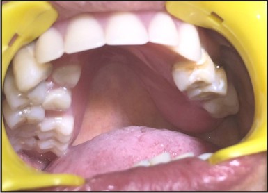 | Figure 2. Showing Double Row Of Teeth On Right Side Of Maxillary Denture
|
 | Figure 3. Denture In Occlusion
|
Discussion
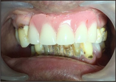
Rehablitation is an essential part of hemimandibulectomy patients and it should be planed from the time of diagnosis. The main objective of rehablitation is the restoration of function and esthetics. In this article, we fabricated conventional maxillary and mandibular removable partial denture with two rows of teeth in order to facilitate proper intercuspation and mastication. The palatal row of teeth intercuspated with the remaining mandibular teeth and the buccal row of teeth supported the cheeks. The patient reported with increase in masticatory efficiency. Guided flange prosthesis was not possible in this case because of increase deviation and scar tissue formation. Patient was recalled after one week, there was improvement not only in mastication but also in facial appearance and patient was quite satisfied. Patient was then kept on 6 months recall appointments. Earlier the mandibular positioning therapy is initiate, more successful is the final occlusal relationship[11]. Hemimandibulectony causes facial asymmetry and malocclusion so restoration is indicated as soon as possible to limit any further complication and to restore mandibular function. We can use grafts or other definitive surgical modes of treatment, cast metal prosthesis which are also quite effective but most patients subjected to hemimandibulectomy come from low socio-economic status so cheaper alternatives like acrylic based prosthesis are quite commonly used.
Conclusion
Present article explain prosthodontic rehabilitation of hemimandibulectomy patients with a conventional prosthesis but with a slight modification of occlusion. This is not only the cost effective treatment but also prevent patient from undergoing surgical treatment like implants, grafts etc. Prosthesis like guided flange or palatal ramp are also good options but not in the present case. The patient was able to achieve good functional intercuspal position and was able to masticate properly.
References
1 Zarb G A, Bolender C L, Carlsson G E. In: Boucher’s prosthodontic treatment for edentulous patients. Mosby; St Louis 2004; 425-26.
2 Mark A Varvares, Arrlen D Myers; Mandibulectomy; Indications and contraindications emedicine.medscape.com/article/1890889.
3 Matthiesen D T, etal. : Results and complications associated with partial mandibulectomy techniques. Probl Vet Med 1990.
4 Shafer, Hine, Levy; Textbook of Oral Pathology; Elsevier.
5 Shah JP Patel SG. Head and neck surgery and oncology. 3rdEd. New York; Mosby; 2003:589-92,614-31.
6 Schneider R, Taylor T D. Mandibular resection guidance prosthesis: J Prosthet Dent 1986; 55: 84-86.
7 Beumer J, Curtis T A, Marunick M T. Maxillofacial rehabilitation; Prosthodontic and surgical considerations. Ishiyaku Euro America: St. Louis. 1996: 184-188
8 Cantor R, Curtis TA. Prosthodontic management of edentulous mandibulectomy patients. Part 1. Anatomoic, physiological and psychological considerations.j Prosthet Dent 1971;25: 446.
9 Taylor TD. Clinical maxillofacial prosthetics. Chicago: Quintessence publishing Co, Inc; 2000:205-213.
10 Lee KY, Kore JM, Perry CJ. Use of Kirschner wire for mandibular reconstruction. Arch Otolaryngol Head Neck Surg 1988; 114 (1): 68-72.
11 Thomas D Taylor, Clinical maxillofacial prosthetics. Prosthodontic Rehablitation of mandibulectomy patients. 2000; 171-188.
|