Introduction
Extra-oral cutaneous odontogenic sinus tracts pose a diagnostic dilemma and are often misdiagnosed. It is well documented that cutaneous sinus tract of dental origin is rare entity[1] therefore, patients seeks treatment from dermatological or general surgeon and often undergo multiple antibiotic regimens, surgical interventions or biopsies before being referred to the dental surgeon.[2],[3] Misdiagnosis count up to the chronicity of the lesion which further compromise the beauty of face by scarring and dimpling. Therefore complete dental evaluation is mandatory for all chronic draining sinus tracts of face and neck region.
These cutaneous lesions usually arise as sequel of bacterial invasion of dental pulp through a rupture by trauma or carious defect.[4] The vicinity of lesions does not always arise from the infected tooth and have been commonly reported on the chin or jaw line as it follows the path of least resistance.[5],[6] Microorganisms along with their by-products present in the necrotic and infected pulp spread beyond the confines of the tooth into the periradicular area, might perforate the cortical plate through inflammatory and immunological processes with the infection draining onto the intraoral mucosa or cutaneous surface resulting in scarring or pimple of skin.[7] This article presents a case of extraoral sinus in sub mental region, managed non-surgically by endodontic treatment with the application of triple antibiotic paste.
Case Report
A 16-year old girl had reported to the Department of Conservative Dentistry and Endodontics, Gian Sagar Dental College & Hospital with chief complaint of pain in lower front tooth region since 3 days. Patient gave history of trauma two years back which resulted in pus discharge inside the mouth with persistence of mild symptoms, but she did not get any dental treatment. No relevant medical history was found. Extra oral clinical examination showed cutaneous draining sinus in the sub-mental region (Figure 1). Swelling was tender on palpation, had raised temperature and was hard in consistency. Intraoral examination revealed no vestibular swelling or any carious teeth but all mandibular anterior teeth were tender to percussion. Pulp sensibility test was done using EPT (Electronic Pulp Tester) in mandibular four anterior teeth (32, 31, 41, and 42) and negative response was observed. An OPG showed well-defined radiolucency with respect to mandibular anteriors in the periapical region (Figure 2). A diagnosis of chronical apical abscess causing extra-oral draining sinus in submental region was concluded.
Root canal treatment was initiated for the offending teeth (32, 31, 41, and 42) after local anesthesia and rubber dam placement. Working length was determined with Root ZX electronic apex locator (J Morita, Kyoto, Japan) and confirmed with an intraoral periapical radiograph. Biomechanical preparation was done with hand instrumentation using step back technique. Irrigation was done with chlorhexidine solution and final preparartion was performed upto size #35 2% hand K-file (Mani Dental mart, INC, Japan).Canals were dried using paper points. Triple antibiotic paste (ciprofloxacin, minocycline and metronidazole) was placed in root canals for 2 weeks. Access cavities were then temporarily sealed with glass-ionomer cement.
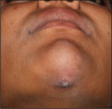 | Figure 1 : Cutaneous Sinus Tract In The Submental Region Before Treatment.
 |
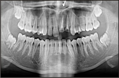 | Figure 2 : Preoperative Panoramic View Of The Lesion.
|
After 2 weeks healing of extra-oral sinus was in progress and patient was asymptomatic. Triple antibiotic paste was given again for 4 weeks. At the following appointment, rubber dam was placed, irrigation was performed, canals were dried and obturation was done using cold lateral compaction technique. The post operative periapical radiograph was taken (Figure 3) and teeth were restored with composite resin material.
 | Figure 3 : Periapical Radiograph Of Final Obturation.
|
The patient was recalled after 3, 6, 9 and 12 months for follow-up. Patient was evaluated at each visit with no recurrence of symptoms and radiograph showed signs of healing with radiolucency grew progressively smaller (Figure 4). The clinical appearance of skin had returned to a healthy status with no scare or dimple (Figure 5).
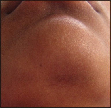 | Figure 4 : Post Operative View Of Healed Cutaneous Lesion After 12 Months.
|
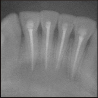 | Figure 5 : Post Operative View Of Periapical Radiograph After 12 Months.
|
Discussion
The cutaneous draining odontogenic sinus tract is uncommon and can present a diagnostic challenge as offending tooth is usually asymptomatic and lesion may be misdiagnosed as dermatological pathology.[8] The differential diagnosis of an extra-oral cutaneous sinus includes endodontic lesion, suppurative apical periodontitis, salivary gland fistula, osteomyelitis, congenital fistula, pyogenic granuloma, infected cyst and deep mycotic infection.[9] Winstock documented cutaneous lesion of dental origin[10] and Kaban defined the path of spread of chronic odontogenic infections.[11] Approximately 80% of the reported cases of extra-oral sinus tract with purulent drainage in the chin or submental area are associated with mandibular teeth whereas 20% are associated with maxillary teeth.[12]
For correct diagnosis, a thorough history of patient may reveal the history of trauma to the face which could lead to injury of dental pulp and then necrosis. Clinical examination along with periapical radiographs and pulp vitality tests provide invaluable help for diagnosis. Once definitive diagnosis is made, the first line of treatment for extra-oral sinus tracts of endodontic origin is non-surgical endodontic therapy.[8],[13] Prognosis of the disease depends on the successful elimination of microorganism within the root canal system. Thoroug instrumentation with chlorhexidine irrigant is considered ideal in this case as chlorhexidine shows substantivity and action against microorganism even Enterococcus.
Modern concept of medicine emphasizes prevention and reversal of the diseases. Lesion asepsis and tissue repair requires use of a combination of antibiotics to promote healing of pulp, peri-radicular tissue and done due to their synergistic effect.[14] TAP (Triple Antibiotic Paste) has been found to be successful in promoting the healing and hence used as intracanal medicament in this case.[15] Single empirical antibiotic is insufficient in disinfection of the root canal due to polymicrobial nature of root canal. Therefore it is essential to use combination of antibiotics to act against all endodontic pathogens and to prevent resistance. The use of antibiotic in endodontics was first reported in 1951 by Grossman which was known as polyantibiotic paste (PBSC). Penicillin was used for targeting against Gram-positive organisms; Bacitracin for penicillin-resistant strains; Streptomycin for Gram-negative organisms and Caprylate sodium to target yeasts.[16] Tri-antibiotic paste containing metronidazole, ciprofloxacin and minocycline was used in this case as metronidazole exhibits broad spectrum of activity against protozoa and anaerobic bacteria; Ciprofloxacin is a synthetic floroquinolone with rapid bactericidal action, and minocycline is primarily bacteriostatic, exhibit broad spectrum of activity against gram positive and gram negative microorganisms. For successful outcome of treatment, ratio of triple antibiotic paste is important,according to Hoshino et al (1996) antibiotic (3Mix) ratio is 1:1:1 (Ciprofloxacin 200mg, Metronidazole 500mg and Minocycline 100mg) with Macrogol ointment and Propylene glycol Carrier (MP ratio 1:1) [17] and according to Takushige et al (2004)drugs are powdered and mixed in a ratio of 1:3:3 (Ciprofloxacin, Metronidazole and Minocycline respectively) and added with macrogol-propylene glycol (3 Mix-MP).[18] This approach of synergistic action may help to reduce the chances of even resistant microbes. It has also been shown to be a great success in the management of large periapical lesions and promoting regeneration of lost tissue by providing antimicrobials at the foci of infection.[19],[20] Also spontaneous closure of the sinus tract should be expected in 5-14 days after root canal therapy.[21]
Conclusion
Elimination of primary cause of infection of extra-oral sinus is mandatory for complete healing of cutaneous lesion without the need of surgical intervention. The presented case report demonstrates endodontic treatment with triple antibiotic paste placement as intracanal medicament resulted in complete healing and repairs of cutaneous sinus tract without any surgical intervention.
References
1. Cioffi GA, Terezhalmy GT, Parlette HL. Cutaneous sinus tract: an odontogenic etiology. J Am Acad Dermatol 1986; 14: 94.
2. Cantatore JL, Klein PA, Lieblich LM. Cutaneous dental sinus tract, a common misdiagnosis: A case report and review of the literature. Cutis 2002; 70: 264-67.
3. Mittal N, Gupta P. Management of extra oral sinus cases: a clinical dilemma. J Endodon 2004; 30: 541-47.
4. Stoll HL, Slolmon HA. Cutaneous sinuses of dental origin. JAMA 1963; 184: 120-24.
5. Donohue WB, Maisonneuve C. Sinus tract of odontogenic origin in the face and neck. J Can Dent Assoc 1984; 50: 99-102.
6. Sammut S, Malden N, Lopes V. Facial cutaneous sinuses of dental origin- a diagnostic challenge. Br Dent J 2013; 21: 555-58.
7. Kotecha M, Browne MK. Mandibular sinuses of dental origin. Practitioner 1981; 225: 910-15.
8. Pasternak-Junior B, Teixeira CS, Silva-Sousa YT, Sousa-Neto MD. Diagnosis and treatment of odontogenic cutaneous tract of endodontic origin: Three case reports. Int Endod J 2009; 42: 271-76.
9. Wilson SW, Ward DJ, Burns A. Dental infections masquerading as skin lesion. Br J Plast Surg 2001; 54: 358-60.
10. Winstock D. Four cases of external facial sinuses of dental origin. Proc R Soc Med 1959; 52: 749-51.
11. Kaban LB. Draining skin lesions of dental origin: The path of spread of chronic odontogenic infection. Plast Reconster Surg 1980; 66: 771-77.
12. Foster FH, Primack PD, Kulild JC. Odontogenic cutaneous sinus tract. J Endod 1992; 18: 304-06.
13. Witherow H, Washan P, Blenkinsopp P. Midline odontogenic infections: A continuing diagnostic problem. Br J Plast Surg 2003; 56: 173-75.
14. Hoshino E, Takushige T. LSTR 3Mix-MP method- better and efficient clinical procedures of lesion sterilization and tissue repair (LSTR) therapy. Dent Rev 1998; 666: 57-106.
15. Vijayaraghavan R, Mathian VM, Sundaram AM, Karunakaran R, andVinodhS.Triple antibiotic paste in root canal therapy. J Pharm Bioallied Sci 2012; 4(Suppl 2):230–33.
16. Parasuraman VR, Muljibhai BS. “3Mix- MP in Endodontics – An overview” J Dent Med Sci 2012; 3(1): 36-45.
17. Hoshino E, Kurihara-ando N, Sato I, Umetsu H, Sato M, Kota K. In vitro antibacterial susceptibility of bacteria from infected root dentin to mixture of ciprofloxacin, metronidazole and minocycline. Int Endod J 1996; 29: 125-30.
18. Takushige T, Cruz EV, Moral AA, Hoshino E. Endodontic treatment of primary teeth using a combination of antibacterial drugs. Int Endod J 2004; 37: 132-38.
19. Sisodia N, Manjunath MK. Chronic cutaneous draning sinus of dental origin. Annals of Medical and Health Sciences Research 2015; 4: 962-64.
20. Jung IY, Lee SJ, Hargreaves KM. Boilogically based treatment of immature permanent teeth with pulpal necrosis: A case series. J Endod 2008; 34: 876-87.
21. Spear KL, Sheridan PJ, Perry HO. Sinus tracts to the chin and jaw of dental origin. J Am Acad Dermatol 1983; 8: 486-92.
|