INTRODUCTION:
Missing teeth and supporting oral tissues have traditionally been replaced with dentures or bridges permitting restoration of chewing function, speech, and aesthetics. Dental implants offer an alternative. These implants are inserted into the jawbones to support a dental prosthesis and are retained because of the intimacy of bone growth
on to their surface. This direct structural and functional connection between living bone and implant surface, termed osseointegration, was first described by Branemark 1977(2) and has undoubtedly been one of the most significant
scientific breakthroughs in dentistry over the past 30 years. Teeth may have been lost through dental disease or trauma or they may be congenitally absent. However in many clinical situations compromised teeth or roots may still be present
in the patient’s mouth. Traditionally, before placing dental implants, compromised teeth were removed and the extraction sockets were left to heal for between several months and 1 year. However, the great majority of patients are interested in
shortening the treatment time between tooth extraction and implant placement or even better in having the implants inserted during the same session as the teeth are extracted (immediate implants). This would result Interventions for
replacing missing teeth: dental implants in fresh extraction sockets. Implant placement in fresh extraction sockets is well documented. Animal and human studies have demonstrated attainment of osseointegration following immediate placement of implant in freshly extracted tooth/teeth at a light microscopic level. In addition, numerous human
clinical studies have documented high levels of success of implant placed at the time of tooth extraction and subsequently restored and in function (6).
CASE REPORT
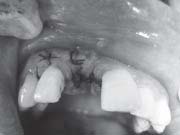
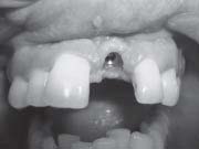
25 year old boy reported in the department of Oral Medicine, Diagnosis and Radiology in Himachal Dental College, Sunder Nagar, with the chief complaint of root stump in upper right front region and gave history of trauma to maxillary
Left Central incisor due to accidental fall on floor during Cricket game three years back in which he had fractured crown and patient went for root canal treatment but left it incomplete due to negligence and slowly tooth crown was worn out
(Fig 1).
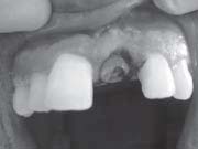 | Figure - 1 (Pre Operative Photograph)
 |
He was referred to department of Periodontics and Implantology for needful. Patient’s general and medical history was taken and was non significant. Patient was examined clinically and Orthopantomograph was taken. After thorough
analysis clinically and radiographically it was evaluated that there is no underlying pathology and tooth root was unrestorable but was surrounded by healthy bone. It was there and then decided to do extraction and place the implant
immediately and so was desired by the patient. Tooth root was extracted atraumatically under local anaesthesia after carefully raising the flap (Fig.2).
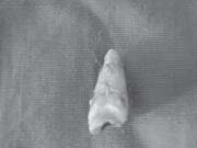 | Figure - 2 (Root Stump)
|
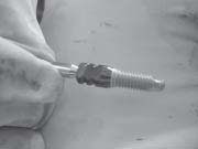
The pilot drill (D-2.O mm) was put to use for creating a osteotomy site of the appropriate depth i.e. 13mm for implant placement (LEADER, ITALIA-implant system) (Fig.3).
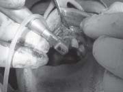 | Figure - 3 (Pilot Drill)
|
It is indexed with various markings corresponding to the desired implant lengths. When appropriate depth was reached with the pilot drill, the implant depth probe was used for tactile perception of intact bony plates & or any perforations & the confirmation of desired osteotomy depth. Once desired depth was confirmed, paralleling pins were placed to check the proper alignment of the implant with adjacent teeth & opposing occlusion. After confirmation of depth & angulation, the osteotomy site (fig.4)
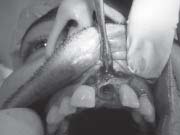 | Figure - 4 (Implant Site Prepared)
|
was prepared by a series of gradually larger drills (D2.6, D2.8, D3.2 and D3.8) to the requisite width with a speed of 1400-1600 r.p.m at 1: 16 reduction torque as per manufacturer instructions. The Implant site was generously irrigated with sterile saline to remove any residual bone chips/other residue following preparation. The depth of the osteotomy site was ascertained with Implant depth probe.
 | Figure - 5 (Implant before Insertion)
|
The implant was removed from the sterile vial using the insertion tool and delivered directly into the osteotomy site. Contamination by touching the implant with instruments made of a dissimilar metal or by contact with soft tissue, cloth or even surgical gloves may affect the degree of osseointegration. The implant was then pressed into the prepared site with manual pressure aided by the insertion mount & insertion tool attached to the implant head. Following which, the
insertion mount was removed and hex driver was placed into the implant internal hex & ratcheted with torque-controlled implant ratchet. Implant was never forced into the socket with excessive force as this might lead to micro cracks in the surface bone resulting in improper osseointegration. Implant that was placed was checked for stability by applying gentle pressure to determine if it could be depressed or rotated. Also, primary implant stability was assessed with
the torque controlled ratchet. The cover screw, provided with the implant package was then placed using the hex-driver using finger pressure (Fig.6). At this point, implant was confirmed to be immobile, which re-affirmed primary implant
stability.
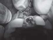 | Figure - 6 (Implant placement with cover screw placed)
|
The flap margins were then repositioned & sutured tension free using 3-0 mersilk in interrupted fashion (Fig.7). A radiograph was taken post operatively to evaluate the implantangulation & position (7).]
 | Figure - 7 (Closure of Socket)
|
The patient was on regular recall and under strict oral hygine measures.
The patient was recalled after 12 weeks radiograph was taken and prosthetic phase was carried out under the opinion & supervision of a prosthodontist. After soft tissue healing (fig.8), Impressions were made with the impression post
attached to the implant using the closed tray
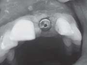 | Figure - 8 (Post 3 months 2nd stage surgery - Gingival Former Placed)
|
impression technique. Shade selection was also done during this appointment. Casts with impression post-implant analogue complex, abutment, lab drivers and selected shade were sent to laboratory for preparation of cement retained
porcelain fused to metal crown. Healing abutment/ gingival former was replaced till the time taken for laboratory manufacture of prosthesis. After approx. 4-7 days, the healing abutments were removed and replaced with final abutment. The PFM crown was checked for its passive fitting to abutment and non-interference with adjacent teeth.
Crown was then cemented with glass ionomer cement (Fig.9 & 10). The patient was dispatched
 | Figure - 9 (Shape of Gingiva after 7 days of Gingival Former Removal)
|
with a reminder of oral hygiene instructions & the recall programme.
DISCUSSION
Many clinical reports and experimental studies in the animal model demonstrated the favourable outcome of dental implants immediately inserted in freshly extraction socket, without the use of any regenerative materials (3, 5, 10). Our data shows
 | Figure - 10 (Abutment in Situ)
|
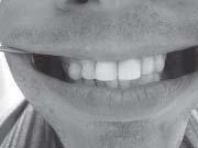 | Figure - 11 (Prosthesis in Situ)
|
a survival rate of immediately implantation carried out on this patient at one year after immediate implantation and does not differ from the cases in which implant was placed in healed sites. These data agree with those from other authors who evaluated the clinical success rate of immediate implantation without use of any membrane or graft material in both humans and animals. It must be kept in mind that the present study is related to immediate implant not subjected to functional loading and therefore not fully comparable with the results from loaded implants. However, it has been demonstrated that functional loading does not impair, but rather enhances, bone maturation.
REFERENCES
1. Lindeboom JA, Tjiook Y, Kroon FH. Immediate placement of implants in periapical infected sites: a prospective
randomized study in patients. Oral Surgery, Oral Medicine, Oral Pathology, Oral Radiology, and Endodontics
2006;101(6):705–10.
2. Branemark et al. Osseointegrated implants in the treatment of the edentulous jaw.Experience from a 10-year period.
Stockholm: Almqvist & Wiksell Int, 1977.
3. Fugazzotto PA. Treatment options following single-rooted tooth removal: A literature review and proposed hierarchy of
treatment selection. Journal of Periodontology 2005;76(5):821–31.
4. Gomez-Roman G, Schulte W, d’Hoedt B, Axman-Krcmar D. The Frialit-2 implant system: five-year clinical experience
in single-tooth and immediately postextraction applications The International Journal of Oral and Maxillofacial Implants
1997;12(3):299–309.
5. Yukna RA. Clinical comparison of hydroxyapatite-coated titanium dental implants placed in fresh extraction sockets and healed sites. Journal of Periodontology 1991;62(7):468–72.
6. Michele Paolantonio, Marco Dolci, Domenico D’ Archivio, et al. Immediate implantation into fresh extraction sockets. A
controlled clinical trial and histological study in man. J of Periodontol 2001; 72:1560-1571.
7. Schwartz-Arad, D,Chaushu G. The ways and wherefore of immediate placement of implants into fresh extraction sites.
A literature review. J of Periodontol 1997; 68:915-923.
8. Esposito M, GrusovinMG,Worthington H, Coulthard P. Intervention for replacing missing teeth: bone augmentation
techniques for dental implant treatment. Cochrane Database of Systematic Reviews.2006; issue 1.
9. Esposito M, Hirsch JM, Lekholm U, Thomsen P. Biological factors contributing to failures of osseointegrated oral implants. (II) Etiopathogenesis. European Journal of Oral Sciences 1998;106(3):721-764
10. Schwartz-Arad, D, Chaushu G. Placement of implants into fresh extraction sites:4-7 years’ retrospective evaluation of
95 immediate implants. J of Periodontol 1997; 68:1110-1116. |