|
|
|
| Short Communication : A Simplified Localization Technique |
Abdulla Mufeed 1 , Johson K Issac 2 , Abdul Shameem 3 , Vadi Vazhagan 4
1 Asst. Professor , Dept. of Oral Medicine & Radiology - MES Dental College
2 Professor & Head , Dept. of Oral Medicine & Radiology - MES Dental College
3 Professor , Dept. of Conservative & Endodontics - MES Dental College
4 Reader , Dept. of Oral Medicine & Radiology - MES Dental College
|
| Address For Correspondence |
Dr. Abdulla Mufeed
Department of Oral Medicine & Radiology
MES Dental College
Perinthalmanna
Malappuram - Kerala
Pin: 679338 |
| Abstract |
| This article is intended to introduce and familiarize an easy and effective modality for localizing impacted tooth or objects in the jaws; a technique that is quiet simple and economic for the general dental practioners. |
|
| Keywords |
| Localization Techniques, Object Localization |
|
| Full Text |
Introduction
This article is intended to introduce and familiarize an easy and effective modality for localizing impacted tooth or objects in the jaws; a technique that is quiet simple and economic for the general dental practioners.
The conventional radiographs have an inherent limitation that they are of limited use in exactly locating the impacted tooth, retained roots, or foreign bodies.
The common techniques for localization include clark’s tube shift method (parallax) and Miller’s right angle technique. A recent survey among dentists concluded that, Clark is the best known technique. However, 44% of the general dentists did not know how to apply or interpret it. Moreover, it was shown that the other localization techniques are hardly known to them[1].
We would like to familiarize a simplified localization technique utilizing two IOPAR films with the principles of Millers right angle technique.
After obtaining a regular IOPA radiograph and evaluating the position of the object of interest, a second radiograph is taken by placing the film parallel to the occlusal plane and asking the patient to bite gently on it, ensuring that the area of interest is centered in the film. (the same fashion how an occlusal radiograph is taken) Exposure to be done with the X ray beam oriented perpendular to the film. The first film gives the frontal view where as the second image provides the axial view. Having both these projections, the clinician can easily identify the position of the desired object in vertical and horizontal planes.
Two cases featured below depicts the usefulness of this technique.
Case 1 : Impacted super numerary tooth
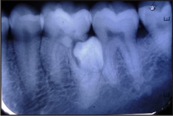 | Fig. 1A
 |
Figure 1A : regular IOPA projection showing the presence of impacted supernumerary tooth.
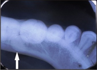 | Fig. 1B
|
Figure 1B : Occlusal view of the same case with IOPA film showing the lingual postioning of the impacted tooth.
Case 2 : A bony swelling of alveolus
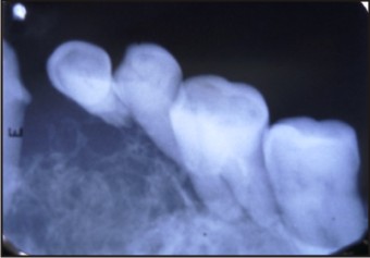 | Fig. 2A
|
Figure 2A : Regular IOPA projection showing a destructive bony lesion.
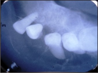 | Fig. 2B
|
Figure 2B : Occlusal view taken using IOPA film showing bucco-lingual extension of the lesion and the tipping of roots.
Advantages:
1. General dentists are comfortable and familiar with IOPAR radiographs than occlusal radiographs/ films
2. In terms of availability, ease of handling, and cost of IOPA films are favorable than occlusal films.
3. Easier to interpret as compared to SLOB or Tubeshift technique
References:
1. LP Cruz et al. Survey of dentists of knowledge about radiographic localization methods.Revista Gaucha de Odontolgia, 57(3) 269-272,2009.
|
|
|
|
|
|
|