Introduction
Restoring the primary maxillary incisors that are severely destroyed by trauma or caries is a common problem faced by any pediatric dentist, especially in developing and underdeveloped countries. Most cases are observed on children with nursing bottle caries.[1],[2],[3] In early childhood caries there is early carious involvement of the maxillary anterior teeth.[4] A child with premature loss of carious maxillary incisors, mainly suffers from speech difficulty. The lost maxillary incisors affect the change in the pattern of speech by interfering with the pronunciation of tongue tip consonants and labial sounds. Also it decreases the masticator efficiency, brings forth abnormal tongue habits which subsequently leads to potential malocclusion. The child may also suffer from psychological problems if esthetics are compromised.[5] The restoration of primary incisors is often a difficult procedure that presents a special challenge to dentists.[6]
For decades, esthetic restoration of severely mutilated primary anterior teeth has been a great challenge for the pediatric dentists due to limited availability of cost effective materials and techniques besides the children who require such restorations are usually among the youngest and least manageable group of patients. Added to this, these teeth usually have short and narrow crowns, only a small surface for bonding; a pulp chamber that is relatively large and enamel that is inherently difficult to acid etch due to its aprismatic nature. In many cases, destruction of whole crown occurs leaving only dentine in root for bonding. So, in the past and even now, many of these teeth are extracted.
A restorative technique that is able to provide efficient, durable and functional restorations, and that is simple to perform would enhance the management of patients presenting with carious maxillary primary incisors. Such a technique could help to ensure the child’s cooperation and reduce the anxieties associated with restorative treatment [6] because of the reduced coronal structure, direct restorative procedure do not always give satisfactory results. Shape, function and esthetics can be better restored by means of prosthodontics techniques. The child’s growth and development may be improved.[8]
This case report describes the challenging task of treating a four year old ECC patient with mutilated maxillary incisors with composite resin using an Omega loop post made with 0.8 mm wire to increase the potential surface area for attachment of the restorative material and consequently increase the long term stability of an esthetic restoration.
Case Report
A 4 year old female child reported to the Department of Pedodontics and Preventive Dentistry, MES Dental College, Perinthalmanna with complaint of severely decayed teeth. The child was shy and withdrawn.
On Examination
Intraoral examination revealed teeth present 55, 54, 53, 52, 51, 61, 62, 63, 64, 65, 85, 84, 83, 82, 81, 71, 72, 73, 74, 75.
Dental caries in respect to 54, 63, 64, & 74 Grossly destructed and decayed 52, 51, 61, 62.[Fig 1].
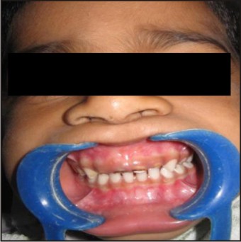 | Figure 1 : A 4yr Old Girl Child With Mutilated Primary Anterior Teeth
 |
A treatment plan was drawn up which was as follows:
i Diet analysis and counseling
ii Fluoride application
iii Oral prophylaxis
iv Restoration i.r.t 54,63,64&74 with GIC.
v Pulpectomy with composite restoration using custom made posts i.r.t. 52,51,61,62.
The pulpectomy of 52,51,61,62. with zinc oxide eugenol was carried out along with other required treatments. About 4mm of cement was removed from the coronal end of the root canal and 1mm of zinc polycarboxylate cement was placed. A 0.8mm stainless steel orthodontic wire was bent using no .130 orthodontic plier into a Omega loop in such a way as to allow the ends to be hooked in the entrance of the root canal.[7] The incisal end of loop of the wire projected 2-3mm above the remaining root structure. [Fig 2].
 | Figure 2 : Placement Of Omega Loop Post
|
This provided better mechanical retention and support for the restorative material. Shade selection of composite was made in the daylight. After the polycarboxylate cement set, the canal was prepared to get a space of about 3mm. The root canal and remaining coronal structure was etched with 35% phosphoric acid for 20 seconds. Composite restorative material of the selected shade was placed in the canal. The loop was inserted into the canal with composite. The composite was light cured for 40 seconds. A strip crown was used and the crown was reconstructed. The occlusion was checked and after removal of any interference, final finishing and polishing of the restoration was performed using soflex tips. [Fig 3] Patient was asked to come for regular checkup. [Fig 4].
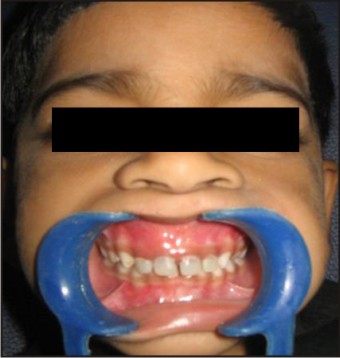 | Figure 3 : After Composite Resin Restoration
|
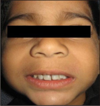 | Figure 4 : 6 Months After Restoration
|
Discussion
Restoring primary anterior teeth that are grossly destructed due to caries is very challenging for the Pediatric Dentists. There is high rate of failure not only because of absence of tooth structure, poor adhesion of bonding agent to primary teeth, limited availability of material and techniques, but also because children who require such restoration are among the youngest and least.
To provide shape, function and esthetics in such teeth, use of intracanal retainers is necessary. After endodontic treatment and placement of intracanal retainers, the remaining coronal structure can be restored with direct or indirect technique or with single tooth prosthesis such as celluloid strip crowns, stainless steel crowns, porcelain veneers, polycarbo nate crowns and acrylic resin crowns.[8]
Rifkin[9] described restoring primary anterior teeth with post and crown. But it was not widely accepted because of potential for interference with physiological root resorption if the wire extends along way into the root. In addition, it can increase internal stresses within the root leading to fracture if the post is forcibly fitted into a narrow canal.
Threaded posts used in permanent teeth are expensive for a pediatric dentist because it is bought as a kit but the whole kit is never utilized. Furthermore, apical tensions may be created, which may lead to root fracture during installation.[8]
Preformed and cast metal posts have been utilized; however, they are expensive and require an additional lab stage. The use of metal posts needs the use of opaque resin to mask the post and could pose addition problems, during the course of natural exfoliation.[6]
More esthetic option may be the use of biological post. The disadvantage of this technique include the need of tooth bank, donor and recipient acceptance and stringent cross infection control policies.[6]
Studies have shown that intracanal retention in primary teeth can be obtained by directly building resin composite posts or preparing an ‘inverted mushroom shaped ‘undercut in the root canal prior to the buildup of resin. However, resin composite posts have low strength of loading.[8]
Motisuki et al[6] have restored severely decayed primary teeth using an indirect composite resin restoration using fibre glass post. His technique was expensive and required lab work.
Conclusion
The direct composite resin restoration using a Omega loop post with orthodontic wire used in this case report had demonstrated good retention and esthetics, further more the post can be manipulated by gently bending on the bow of loop which makes it a novel technique. It was easy to perform and benefited the child immensely.
References
1 Johnson D C. Characteristics and Background of Children with “nursing caries”. Pediatric dentistry 1982: 4:218-224
2 Ripa L W. Nursing caries: A Comprehensive review: Pediatric Dentistry 1988; 10(4): 268-282.
3 Yui CKY, Wei SH. Management of rampant caries in children. Quintessence Int 1992; 23:159-168.
4 Mcdonald, Avery and Dean. Dental caries in the Child and Adolescent. Int: Dentistry for the child and Adolscent.8th ed. Mosby 2005. pg209-10
5 Ngan p, Fields H. Orthodontic diagnosis and treatment planning in the primary dentition. ASDC Journal of Dentistry for Children1995; 62:25-33.
6 Motisuki C; Santos-Pinto L. and Giro EMA. Restoration of severely decayed primary incisors using indirect composite resin restoration technique. International journal of Pediatric Dentistry 2005;15:282-286.
7 Usha Mohandas. Restorative Challenges In Severe Early Childhood Caries. Indian Dentist Research and Review, September 2007: 52-54
8 Mortada A and King NM. A simplified technique for the restoration of severely mutilated primary anterior teeth . The Journal of Clinical Pediatric Dentistry 2004;28(3):187-192
9 Rifkin A. Composite post crowns in anterior teeth. Journal of the dental association of South Africa 1983;38:25-227.
|