Introduction
Fusion is defined as union between the dentine and / or enamel of two or more separate developing teeth. It can be complete with the formation of one abnormally large tooth or incomplete with the union of crown or union of roots only. The prevalence of this anomaly is estimated as 0.5%-2.5% in primary dentition with a lower prevalence in permanent dentition[1]. Fusion of primary teeth can appear in several ways, usually involving two teeth but occasionally three[2].
Triplication of primary teeth is a rarer phenomenon with a prevalence of 0.02 %[2]. Triplication is defined as a lateral supernumerary tooth doubly joined to two teeth of normal series on the mesial side and lateral incisor on the distal side[3].
Primary triple teeth suggest an idiopathic abnormality in the distribution of the dental material originated very early in the dental development. It can be an early double fusion between three tooth germs, initially separate but in close proximity and developing synchronically[3].
Rarely supernumerary teeth may be fused to normal teeth in the arch, usually by medial or lateral fusion. Supernumerary teeth are seen infrequently in the primary dentition with prevalence rate of 0.2% to 0.8%[4].
Problems associated with fused teeth includes esthetics, caries, periodontal disease, malocclusion, delayed exfoliation and anomalies in permanent dentition.
This article reports the case of unusual fusion of three teeth in a 10 year old boy involving one supernumerary and maxillary right primary central and lateral incisors.
Case Report
A 10 year old boy came to the department of Pedodontics and Preventive Dentistry, I.T.S Dental College, Muradnagar, Ghaziabad, with the chief complaint of large tooth in the maxillary anterior region of the jaw. No other member of the family was affected with similar dental anomaly. Patient was mentally challenged.
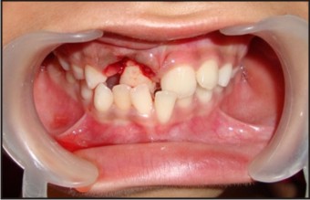
Intra-oral examination revealed that the patient was in mixed dentition phase along with fusion of the maxillary right primary central incisor and primary lateral incisor with an additional tooth. (Figure 1) Dental caries was evident on the incisal edge and proximal surface of supernumerary tooth. The child’s dentition revealed Angle’s class 1 molar relation on both right and left side. Maxillary right permanent central incisor was erupted but it was in cross-bite relationship. A midline diastema was also noted. Clinical examination revealed missing right permanent lateral incisor.
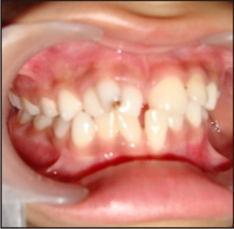 | Figure : 1 Pre-operative Intra Oral Photograph.
 |
Further investigation included Intra-oral periapical radiograph of maxillary anterior region and OPG (Orthopantomogram) (Figure 2-3). Based on clinical and radiographical findings, the diagnosis of fused triple teeth was confirmed.
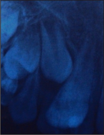 | Figure : 2 Intra-oral Periapical X-ray Showing Fused Teeth.
|
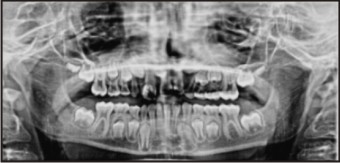 | Figure : 3 Orthopantomogram Showing Missing Permanent Maxillary Lateral Incisor.
|
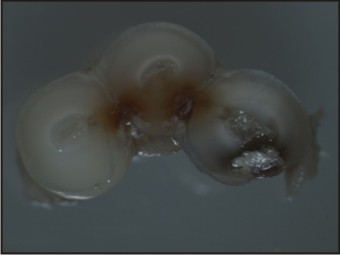
The treatment plan was aimed at removal of fused teeth. Under local anesthesia fused teeth were carefully extracted.(Figure 4 - 5) Macroscopically root resorption was evident on apical portion of maxillary right primary lateral incisor.
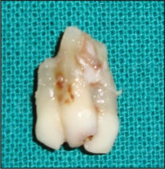 | Figure : 4 Extracted Fused Teeth
|
 | Figure : 5 Post-operative
|
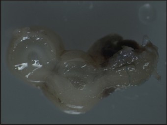
For histological examination, the extracted teeth were sectioned at three levels: the coronal, middle root and apical levels, respectively. These sections were visualized under a stereomicroscope at magnification of 50x. Based on the findings, a final diagnosis of an incomplete fusion was confirmed.
Histological view of the coronal section revealed three teeth fused with each other with confluent dentin without intervening cementum and with separate pulp chambers. (Figure 6).
 | Figure : 6 Coronal Section Of Fused Teeth Showing Confluent Dentin Without Intervening Cementum And With Separate Pulp Chambers.
|
The middle third of the roots of three fused teeth also showed confluent dentin without intervening cementum. (Figure 7) The apical third showing merging of root canals of the fused teeth. (Figure 8).
 | Figure : 7 Middle Third Of The Roots Of Three Fused Teeth Also Showed Confluent Dentin Without Intervening Cementum.
|
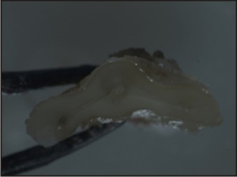 | Figure : 8 The Apical Third Showing Merging Of Root Canals Of The Fused Teeth.
|
The treatment plan involved thorough oral prophylaxis and correction of anterior cross bite. After oral prophylaxis and pit and fissure sealant application, a removable Hawley’s appliance with Z-spring was fabricated for the correction of the anterior tooth cross bite, but as patient was mentally challenged co-operation from him was poor as the child was not ready to wear the appliance. Again Catalan’s appliance was made and cemented on the lower incisors but the attempt for the correction of cross-bite failed because of insufficient compliance of the patient as he was not occluding his teeth.
Discussion
Triple fusion is rarely encountered. Fusion is thought to be caused by pressure or force during development of adjacent roots. If the fusion begins before calcification, then the union will involve all components of the tooth, including enamel, dentin, cementum and pulp. If the union begins at later stage of tooth development, then the affected teeth may have separate crowns and fusion may be limited to the roots. The pulp canals may be either fused or separate. It can occur between normal teeth or between normal and supernumerary teeth[5].
In the present case fusion was seen between maxillary right primary incisors and supernumerary teeth and teeth were incompletely fused involving dentin in coronal, middle and apical portion of the teeth with separate pulp canals.
The anomalies of permanent dentition are strongly associated with anomalies in the primary dentition[6]. The most common problem related to fused teeth is hypodontia of the permanent dentition[7]. In the present case, the maxillary permanent lateral incisor on the affected side was missing. Fused teeth can also cause delayed resorption of roots which may lead to delayed or ectopic eruption of permanent successor. In the present case fused teeth showed minimal resorption of primary lateral incisor at the apical portion of the root. Maxillary right permanent central incisor was palatally erupted and was in cross bite relationship.
The treatment of fused teeth depends on clinical situation. Esthetic is usually the determining factor regarding the decision to retain or extract these teeth. In the present case, we decided to extract the fused teeth since the permanent central incisor of that side was already erupted. As in this case the child is mentally challenged fixed orthodontic treatment can be anticipated for the desirable occlusion relationship.
Importance of anomalies in primary dentition is usually underestimated especially when they are asymptomatic. But their presence may have marked effect on the permanent dentition.
References:
1 Mohapatra A,Prabhakar AR, Raju OS. An unusual triplication of primary teeth: A rare case report. Quintessence Int 41(10):815-20, 2010.
2. Erdem GB, Uzami#1; M, Olmez S, Sargon MF Primary incisor triplication defect. J Dent Child. 68(5-6):322-5, 301, 2001.
3. Aguilo L, Catala M, Peydro A Primary triple teeth: histological and CT morphological study of two case reports. J Clin Pediatr Dent. 26(1):87-92,2001.
4. Shah A,Gill DS, Tredwin C,Naini FB. Diagnosis and management of supernumerary teeth.Dent Update 35:510-520,2008.
5. Scheid RC, Woelfel J.B, Woeltel’s Dental Anatomy –Its relevance to Dentistry 7th Ed Lippincott Williams and Wilkins 403-431 2007.
6. Nik-Hussien N .N and Majod Z.A .Dental anomalies in the primary dentition distribution and co-relation with the permanent dentition. J Clin Pediatr 2(1):15- 19, 1996.
7. Gellin ME .The distribution of anomalies of primary anterior teeth and their effect on the permanent successor. Dent Clin North Am.28:69-80, 1984.
|