|
|
|
| Comparative Analysis Of Sealing Ability Of A New Resin Based Obturation System Using Two Different Techniques - An In Vitro Study |
Kishan K V 1 , Vani Hegde 2
1 Reader , Dept. Of Conservative Dentistry And Endodontics - Coorg Institute Of Dental Sciences
2 Professor , Dept. Of Conservative Dentistry And Endodontics - Ame's Dental College,Raichur
|
| Address For Correspondence |
Dr. Kishan K.V. MDS, Reader,
Dept. of Conservative Dentistry & Endodontics
Coorg Institute Of Dental Sciences, Virajpet
Karnataka, India. Pincode: 571218
Phone No: (91)9902522937
Fax No: (91)8274256479
E-Mail: drkishankv@yahoo.co.in |
| Abstract |
| Aim:This study has been designed to evaluate, in vitro, the apical sealing ability of a new thermoplastic synthetic polymer resin based obturation system (Resilon) with AH26 – gutta-percha obturation system using two different techniques. Methodology A total of sixty single rooted extracted human teeth are subjected for the study. The roots are divided randomly into four groups of fifteen each. All the groups are subjected to root canal instrumentation while maintaining the patency of the apical foramen.
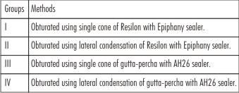
Group 1: Obturated using single cone of Resilon with Epiphany sealer.
Group 2: Obturated using lateral condensation of Resilon with Epiphany sealer.
Group 3: Obturated using single cone of gutta-percha with AH26 sealer.
Group 4: Obturated using lateral condensation of gutta-percha with AH26 sealer. All samples from each group are subjected for dye leakage study. Teeth are sectioned and observed under stereomicroscope for apical linear dye penetration. The results are evaluated statistically.
Results: There was no significant difference between both the materials but Lateral condensation technique was significantly better than single cone technique with both Resilon/Epiphany and AH 26/Gutta-percha groups.
Conclusion: Under the conditions of present study AH 26/Gutta-percha groups were comparatively better than the Resilon/Epiphany groups and lateral condensation technique sealed root canals significantly better than single cone technique. However, the clinical relevance of the results of this in vitro study should be interpreted with caution. |
|
| Keywords |
| Apical sealing, AH26, Epiphany, Gutta-percha, Resilon, Single cone |
|
| Full Text |
Introduction
The preliminary objective of operative Endodontics is total debridement of the pulpal space, development of a fluid tight seal at the apical foramen and total obliteration of the root canal.To achieve this,over the years there are many different filling materials and sealers have been introduced. Currently the material used most often as a solid core filling is Gutta-percha. Despite having most of the ideal characteristics, Gutta-percha has failed to provide effective fluid tight apical seal[1].
For complete sealing of the canal, the root canal sealer/resin is required to have the capability of producing a bond with the dentin of the canal. Improvements in adhesive technology have fostered attempts to reduce apical and coronal leakage by bonding to root canal walls. Total-etch adhesives have been tested with resin cements as alternative root filling materials. Those results showed that dentin adhesives significantly reduced apical leakage. Self-etching primers have also been used for bonding to root canal dentin[2]. As epoxy resin sealers do not copolymerize with methacrylate resin-based adhesives, a methacrylate resin sealer was developed with a self-etching primer that resulted in improvements in the apical seal and adhesion to root dentin[3].
A new thermoplastic, synthetic, polymer based, root canal filling material Resilon, has been introduced in an attempt to replace and potentially to challenge Gutta-percha as a root filling material. It has been claimed that Resilon obturated with resin sealer Epiphany in combination with self etching primer, allow creation of a solid `Monoblock` (a material which is contiguous from its resin tags in cleared dentinal tubules through sealer to the core canal filler). Thermoplasticity of Resilon is because of polycaprolactone, biodegradable polyester with a moderately low melting point, while its bondability is derived from the inclusion of resin with methacryloxy groups. This filling material also contains bioactive glass fillers and barium chloride as fillers, and is capable of coupling to resin sealers, an example of which is Epiphany (Pentron Clinical Technologies, Wallingford CT). It performs like gutta-percha, has the same handling properties and for re-treatment purposes may be softened with heat or dissolved with solvents like chloroform. Epiphany Root Canal Sealant is a dual-curable resin com¬posite containing a new redox catalyst that enables optimal auto-polymerization under acidic environments. The promising Monoblock concept of Resilon, filled with Epiphany sealer claims to eliminate or superior to conventional gutta-percha obturation technique[3].
The objective of the study is to compare the apical sealing ability of a new thermoplastic synthetic polymer resin based obturation system (Resilon) with AH26 - gutta-percha obturation system using both single cone technique and lateral condensation technique in vitro.
Materials and Methods
A total of 60 single rooted extracted human teeth were subjected for the study.
Preparation of the sample:
The length of all the roots were prepared approximately 16mm from the coronal surface to the apex of the root. The working length were established with size 15 k-file, 0.5 to 1mm short of the minor foramen. The instrumentations were performed using K3, 0.06 taper nickel titanium rotary instruments to size 35 up to the working length.
A total of 15ml of 5.25 % sodium hypochlorite were used for irrigation during instrumentation, 5 ml of 17% EDTA rinses were used during and after instrumentation to remove the smear layer and a final rinse with 5ml of 0.2% chlorhexidine. The root canals were dried with sterile paper points before filling.
The roots were divided randomly into four groups of fifteen each.
 | Table 1 : Experimental Groups
 |
Table 1: Experimental groups
Group 1: Single cone Resilon with Epiphany sealer.
After instrumentation, a self etching primer (Epiphany primer, Pentron clinical technologies) were placed in to the canal with a micro brush. Excess primers were removed with paper points. Roots were filled with 0.06 taper, 35 size, single cone of Resilon and Epiphany sealer.
Group 2: Lateral condensation of Resilon with Epiphany sealer.
After the placement of primer, the canal is coated with the epiphany sealer. The Resilon master cone of 35 size, 0.06 taper, coated with the sealer is placed in the canal to the working length. Lateral condensation was performed using a spreader and placing medium-fine Resilon accessory points dipped in resin sealer until the root canal was filled.
Group 3: Single cone of gutta-percha with AH26 sealer.
The sealer was mixed according to manufacturers instructions and is coated into the canal with finger spreader. Obturation was performed using 35 size 0.06 taper master gutta-percha cone till the working length.
Group 4: Lateral condensation of gutta-percha with AH26 sealer.
After coating the canal with the sealer, a master gutta-percha cone of size 35 and 0.06 taper placed till the working length. Lateral condensation was performed using finger spreader and medium fine accessory gutta-percha cones until the root canal was filled.
After obturation, the excess gutta percha was removed with the hot instrument and condensed vertically. Resilon Groups were cured at the coronal orifice using visible light for 30 seconds. Coronal orifice was sealed with Fuji IX GP GIC.
All the samples were stored in relative humidity under anaerobic condition in an incubator for 7 days. The samples were dried and the root surface coated with two layers of nail varnish leaving the apical 1-2 mm.
Samples were placed in India ink and stored at room temperature for 24 hrs after which they were thoroughly washed under tap water and dried.
The roots were split longitudinally with diamond discs and were checked for apical linear dye penetration using stereomicroscope. The dye penetration was measured from the apex to the most coronal extent of the dye visible on the filling material or root canal walls.
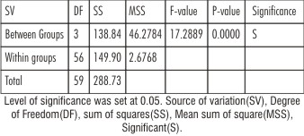
Results were analyzed statistically using one way analysis of variance test at 5% level of significance.
 | Table 2 :One Way ANOVA
|
Group IV - Lateral condensation of AH 26/ GP
Results
Table 2: One Way ANOVA
Result of one-way ANOVA for leakage, which was used for the comparison of the mean values among the groups. P<0.001 indicates that the F- statistical value (F = 17.2889, Degree of Freedom = 3, 56) is highly significant.
 | Table 3: Pair Wise Comparison Between Four Groups By Student’s Unpaired T-test
|
Table 3: Student’s t test
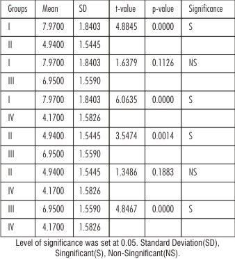
For categorical data comparison, amount of dye penetration between four groups was done by student’s t-test. For all the test a P-value of 0.05 or less was considered for statistical significant
Discussion
The preliminary objective of operative endodontics is total debridement of the pulpal space, development of a fluid tight seal at the apical foramen and total obliteration of the root canal[4]. There is wide variation in the sealing capacity of different endodontic materials and achieving an adequate apical seal is an important goal in endodontics. When evaluating a new root canal filling material analysis of its sealing ability is therefore very important and relavant.
Conventional epoxy resin based sealer AH26 was selected as a sealer due to its better apical seal and good adhesion, thus providing optimal conditions for all methods of obturation[5]. The physical properties of AH 26 are well known, so in the present study this material has been chosen for comparison with the new material.
Resilon is a thermoplastic synthetic resin material that is based on polymers of polyester and contains a bifunctional methacrylate resin, bioactive glass and radio opaque fillers. The resin sealer, Epiphany Root Canal Sealant contains BisGMA, ethoxylated Bis-GMA, urethane dimethacrylate, hydrophilic difunctional methacrylates, silane-treated barium borosilicate glasses, barium sulfate, silica, calcium hydroxide, bismuth oxychloride with amines, peroxide, photo initiator, stabilizers and pigment. The primer is an aqueous solution of an acidic monomer[6].
With the tendency to preparation techniques of greater taper, gutta-percha points of matching taper may be used. These fit the prepared canal so well that some manufacturers recommend using single gutta-percha point and sealer. The only advantage of technique is its simplicity. The disadvantage is that the majority of sealers are soluble. As the canal will not be fully filled in three dimensions, tissue fluids may leach out the sealer over time[1]. But in the present study single cone technique was considered because the manufacturers claimed that the Resilon cone bonds with the Epiphany sealer, which in turn bonds to the root dentine with the help of a self etch primer acting as a ‘monoblock’[3].
Under the conditions of this study, neither material produced an effective apical seal but there was significant difference (p<0.05) between the two techniques used for obturation i.e., the lateral condensation technique provided significantly better results than single cone technique.
But within the materials gutta-percha with AH 26 proved to be comparatively better than the Resilon/Epiphany group with both lateral condensation and single cone techniques.
The possible reason for the poor seal of Resilon/Epiphany in both the groups, could be the shrinkage forces acting on the methacrylate based sealer during polymerization.
Shrinkage stresses associated with polymerization of methacrylate based resins are higher in low filled lower viscosity resin cements and root canal sealers than highly filled resin composites. A major problem associated with endodontic bonding is the lack of relief of shrinkage stresses created in deep narrow canals[7].
The possible reason for, GP/AH 26 shown better results when compared with RES in both the techniques could be because GP expands in the presence of humidity and closes microgaps. Expansion of GP may have occurred in this experiment because of the moisture present during the setting and testing period. This may have enhanced the sealing ability of Both the GP groups[8].
In the present study single cone technique was considered because the manufacturers claimed that the Resilon cone bonds with the Epiphany sealer, which in turn bonds to the root dentine with the help of a self etch primer acting as a ‘monoblock’[3].
Both Group I and Group III showed significant leakage (P<0.05) when compared with Group II and Group IV. This result could be compared with the earlier study which states that the single cone methods of obturation have been shown to be inferior in providing an effective apical seal. The immediate reason for this may be because of the existence of substantiate amount of interfacial gap between dentin and sealer and also in the body of the sealer[9].
According to a study most leakage studies have been performed in vitro and the clinical significance of such results is questionable. Most of the results of the in vitro dye penetration after root canal obturation with lateral condensation technique are rather high ranging from 4.16 to 9.25mm which also supports the present study[10].
It is unlikely, however that the sealing failure observed in both the materials resulted solely from problems inherent in the sealers or the obturating technique per se. Other factors such as the possible presence of entrapped air at the interface, accessory canals, fins or oval shaped canals that are difficult to prepare and fill adequately as we have standardized the preparation with 0.06 taper, may also responsible for sealing failure.
Conclusion
Under the conditions of this study, neither material produced an effective apical seal but there was significant difference (p<0.05) between the two techniques used for obturation i.e., the lateral condensation technique provided significantly better results than single cone technique.
But within the materials, gutta-percha with AH 26 proved to be comparatively better than the Resilon/Epiphany group with both lateral condensation and single cone techniques.
However, the clinical relevance of the results of this in vitro study should be interpreted with caution.
References
1. Grossman L I, Oliet S, Dee Rio L E. Endodontic practice. Eleventh Ed. Varghese publishing house.
2. Imai Y, Komabayashi T. Properties of a new injectable type of root canal filling resin with adhesiveness to dentin.J.Endod.2002;29(1):20-23.
3. Tay F R.,Loushine R J,Weller R N, Kimbrough W F, Pashley D H, Mak Y-F, Lai C-N S, Raina R, Willum M C. Ultrastructural evaluation of the apical seal in roots filled with a polycaprolactone-based root canal filling material. J.Endod.2005;34(7):514-519.
4. Ingle J I, Backland L K. Endodontics; Fifth Ed. B.C.Decker. Elsevier.
5. Limkangwalmongkol L, Burtscher P, Abbott P V, sandler A B, Bishop B M, Comparative study of the apical leakage of four root canal sealers and laterally condensed gutta-percha. J.Endod. 1991; 17(10):495-499.
6. Shipper G, Ostavik D, Teixeira F B , Trope M:“An Evaluation Of Microbial Leakage In Roots Filled With A Thermoplastic Synthetic Polymer Based Root Canal Filling Material (Resilon)”. J Endod 2004; 30(5):342-347.
7. Tay F R, Loushine R J, Lambrechts P, Weller R N, Pashley D H. Geometric factors affecting dentin bonding in root canals - a theoretical modelling approach. J.Endod. 2005;31(8):584-588.
8. Wu M K, Fan B, Wesselink P R, Diminished leakage along root canals filled with gutta percha without sealer over time: a laboratory study. Int Endod J 2000;33:121-5.
9. Pommel L, Jacquot B, Camps J. Lack of correlation among three methods for evaluation for apical leakage. J. Endod. 2001;27:347-350.
10. Wu M.-K, Wesselink P R. Endodontic leakage studies reconsidered. Part I. Methodology, application and relevance. Int. Endod J.1993; 26:37-43.
|
|
|
|
|
|
|