Introduction:
Congenital epulis (CE) is a rare benign soft-tissue tumor of uncertain histogenesis, which is also known as “gingival granular cell tumor of the new-born”[1]. It occurs on the alveolar ridge in the anterior region of the maxilla or mandible of newborn infants.It is also termed ascongenital granular cell tumor, congenital granular epulis,congenital granular cell myoblastoma and congenital granular cellfibroblastoma[2]. The Greek word epulis means “swelling on the gingiva”.The tumour may be sessile or pedunculated. The exactpathogenesis of CE is still uncertain, as is its growthand progression[3]. It can range from normal to reddish pink in color. It is seen three times more frequently on theanterior alveolar ridge of the maxilla than from the mandible with a female to maleratio of 8 to 10:1[4]. Congenital epulis usually occurs as a single mass, although 10% of the cases occur as multiple masses[5].
The diagnosis is made clinically and tissue biopsy is not usually indicated before removal of the lesion, since the excises surgical specimen is usually sent for histopathologic examination and the diagnosis is confirmed retrospectively. The excessive tissue is composed of cellular, inflamed fibrous connective tissue. The appearance of an epulisfissuratum microscopically is an overgrowth of cells from the fibrous connective tissue. The epithelial cells are usually hyperkeratotic and irregular, hyperplastic rete ridges are often seen.
Treatment is by surgical excision (complete removal) of the fibrous tissue overgrowth and addressing the causative factor to prevent recurrence of the lesion. Other sources suggest that surgical excision may not be required in allcases. Common techniques for removal of the excess tissue include traditional removal with a surgical scalpel, electrical scalpel, or laser excision with a laser scalpel, e.g. a carbon dioxide laser, erbium:YAG laser, Neodynium-YAG laser, or diode laser.
In this report we describe the case of a one year old male infant with a growth on his mandibular alveolar ridge.
Case Report:
A 1-year old male born after 39 weeks of gestation weighing 22 pounds was referred to the Outpatient Department of Periodontology and Oral Implantology, Himachal Dental College, Sundernagar, H.P with the chief complaint of a localizedgrowth on theanteriorregion of mandibular alveolar ridge. The growth did not interfere with feeding and respiration, but with mouth closure. This growth was present since the birth of the patient as the history given by his parents. There was no family history of this type of a case.
Intraoral examination revealed a painless, reddish pink, pedunculated soft tissue mass measuring 2.5×1.5×1.5 cm, attached to the anterior mandibular ridge. The mass prevented normal closure of oral cavity. After taking the complete medical and dental history, the mass was excised with electrocautary under local anesthesia and fixed in 10% formalin solutionfor histological analysis.Thepostoperative period was uneventful.
Histological analysis revealed it to be similar to granular cell tumour composed of polygonal cells with abundant granular cytoplasm and small eccentric nuclei with occasional small nucleoli. There was a prominent vascular stroma with perivascular lymphocytes and histiocytes. There was no pseudoepitheliomatous hyperplasia of the overlying squamous mucosa and no nerve bundles were seen within these lesions. The findings were consistent with congenital epulis (Image 1-4).
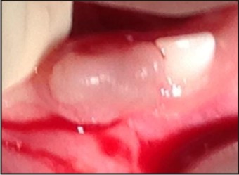 | Fig 1 : Pre - Operative Photograph
 |
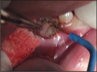 | Fig 2 : Excision Of Epulis
|
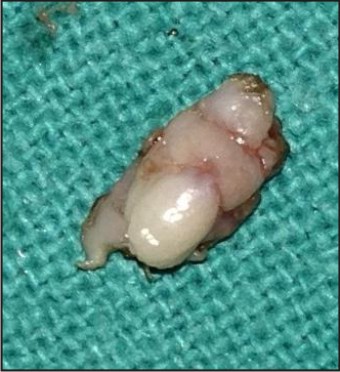 | Fig 3 : Excised Lesion
|
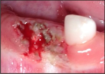 | Fig 4 : Post - Operative Photograph
|
Discussion:
The congenital epulis (congenital granular cell myoblastoma, Abrikossoff’s tumor, granular cell epulis of infancy, granular cell fibroblastoma) is a unique and rare congenital tumor of the alveolar mucosa of the jaws.The case presented in this article is noteworthybecause the lesion was present in themandible, which is reported to have a lowerfrequency of occurrence than the maxilla[6]. Congenital Epulis is an overgrowth with unclear histogenesis. It usually presents at birth with an obvious mass arising from the gingival mucosa of the maxilla or mandible[9]. Congenital epulis has an 8:1 incidence in females and a 3:1 incidence of occurring at maxillary alveolar sites[10]. The lesions do not have familial origin.The lesion develops late in utero as it often is not detected on antenatal ultrasound. If there is any fear of airway obstruction or difficulty with feeding, then prompt surgical treatment is necessary. Congenital epulis is often misdiagnosed before surgery because of its rarity and a lack of awareness of the condition by clinicians. The differential diagnosis of a large mass in the fetal or neonatal oral cavity includes hemangioma, lymphatic malformations, teratoma,rhabdomyoma and rhadomyosarcoma[8]. Treatment consists of CE removal by surgery underlocal or general anesthesia. A “watchful waiting”procedure can be followed because small lesions pontaneous involution can occur, although this is rare[7]. Dentists may be consulted initially regarding such cases and should be aware of the potential for airway compromise and familiar with the differential diagnosis.
References
1. Hiradfar M, Zabolinejad N, Gharavi M, Sebt S. Multiple Congenital Epulis of a mandibular ridge: A Case Report.Iranian Journal of Otorhinolaryngology 2012;4(24): 193-196.
2. Lee JM, Kim UK, Shin SH. Multiple congenital epulis of the newborn: A case report and literature review. J Ped. Surg. Case Reports 2013;1: 32-33.
3. Merrett SJ, Crawford PJ. Congenital epulis of the newborn: Acase report. Int J Paediatr Dent 2003;13:127-9.
4. Gokhale UA, Malhotra CJ. Congenital epulis of the newborn. IJPM 2009;52(3): 436-437.
5. Sahu S, Maurya RK, RaoYaswant, Agarwal A. Multiple congenital epulis in newborn – A rare presentation. JOMFP 2009; 13(2): 78-80.
6. Soft tissue tumors. In: Neville BW, Damm DD, Allen CM, Bouquot JE (eds). Oral and Maxillofacial Pathology,ed 2. Philadelphia: Saunders, 2002:466–467.
7. Ruschel HC, Beilke LP, Beilke RP, Kramer PF. Congenital epulis of newborn: Report of a spontaneous regression case. J ClinPediatr Dent 2008;33:167-9.
8. Hasanov A, Musayev J, Onal B, Rahimov C, Farzaliyev I. Gingival granular cell tumor of the newborn: A case report and review of literature. Turkish J Pathol 2011; 27: 161-3.
9. Lapid O, Shaco LR, Krieger Y, Kachko L, and Sagi A. Congenital epulis. Pediatrics 2001; 107(2): 22–24.
10. Dash JK, Sahoo PK, Das SN. Congenital granular cell lesion "congenital epulis" - Report of a case. J Indian SocPedo. Prev. Dent. 2004; 22(2): 63-7.
|