Introduction
Stents are required in various regions of the body to counteract stenosis, to maintain the patency of a lumen, to minimize scar contracture, and sometimes to better perform the radiotherapy.
Vaginal stents are used primarily to counteract the stenosis and are also modified to perform brachytherapy for vaginal cancers. They are now also used widely to support the newly formed vagina during healing stages following sex reassignment surgery (SRS).
In literature, the use of vaginal dilators, preformed vaginal stents and regular sexual intercourse has been advised to prevent vaginal stenosis. However it was seen that vaginal dilators are ineffective in preventing the stenosis of the upper third of vaginal canal as their design doesn’t take into the account the fact that the vagina is most distensible in its upper third [1]. As far as the sexual intercourse is concerned for preventing the stenosis, it is not effective because it makes a medical necessity out of what should be a spontaneous act.
Many prefabricated vaginal stents are available in the market but as the severity of stenosis and the positions of the fibrotic bands differ in different patients, prefabricated stents may need a lot of modifications to conform to the internal vaginal anatomy of the patient concerned.
This article thus describes a technique, through a case report, for the fabrication of custom made vaginal stent to counteract the developed vaginal stenosis and to prevent the development of stenosis ( e.g. after brachytherapy). Along with this, the technique can be modified to fabricate radiation stent for the brachytherapy in the vaginal cancers.
Case Report
A 23 year old female from rural background was referred to the department of Prosthodontics and Dental material sciences, K.G.D.C Lucknow, India, from the department of Urology, K.G.M.C, C.S.M.M.U, .Lucknow, India, as a case of vaginal stenosis with bladder and vaginal calculi, along with longstanding (3 years) recto-vaginal and vesico-vaginal fistulae. The patient was operated at the Urology department for the removal of the bladder and vaginal calculi under spinal anesthesia. Sigmoid colostomy was done to prevent irritation for the healing fistulae. Surgery for the repair of fistulae and simultaneous correction of stenosis by release of fibrotic bands in vaginal canal was planned after 3 months. Till that period the patient was prescribed a vaginal stent to maintain the patency of the vaginal canal. Such a vaginal stent is required (1) to have a surface topography according to the internal surface anatomy of vagina of the patient concerned, (2) to have highly polished surface, (3) to have sufficient strength, (4) not to encroach the fistulae (in this particular patient), (5) to be easy to use and comfortable to the patient, (6) to be easy to maintain the vaginal hygiene, and finally, (7) to be retentive.
Technique Of Fabrication
(I) Examination Of The Vaginal Canal
The dimension of the vaginal canal was tentatively examined by digital palpation. The tentative position of the fibrotic band was assessed, which was found to be at approximately 10 o’clock to 2 o’clock position. The maximum length, up to which prosthesis can be inserted to achieve sufficient retention and still be comfortable to the patient, was also determined.
(II) Fabrication Of Special Tray.
A special tray was fabricated, about 2 mm smaller than the canal width and relieved further at the approximate portion of the fibrotic
band. Light body poly vinyl siloxane impression material (3M ESPE, Dental products, St. Paul, MN, U.S.A.) was chosen because of sufficient tear strength and flow properties. (Image 1).
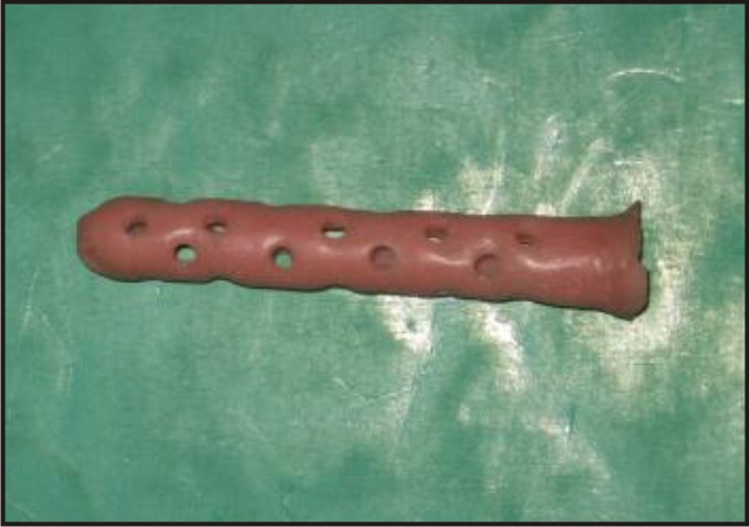 | Figure 1 - Impression Tray For Vaginal Canal.
 |
Special tray fabricated was made highly polished and cylindrical in shape having a central canal running along its whole length. Holes were drilled into the special tray. An opening was made at the end of the tray which projected out of the vaginal canal, through which impression material was injected.
(III) Impession Making
(a) Special tray was checked for its fit into the canal .Necessary adjustments were made. Special tray was coated with rubber base adhesive.
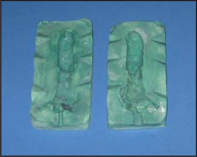
(b) Impression was then taken using light body addition silicone with an automixing delivery gun. (Image 2, 3).
(c) The impression was examined for any tear of material. Vaginal canal was also examined for any teared piece of impression material.
(d) In the impression a notch corresponding to the fibrotic band can easily be appreciated at the 10 o’clock to 2 o’clock positions in the impression.
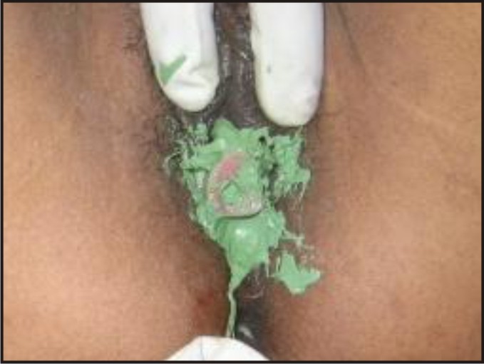 | Figure 2 - Impression Making Of Vaginal Canal.
|
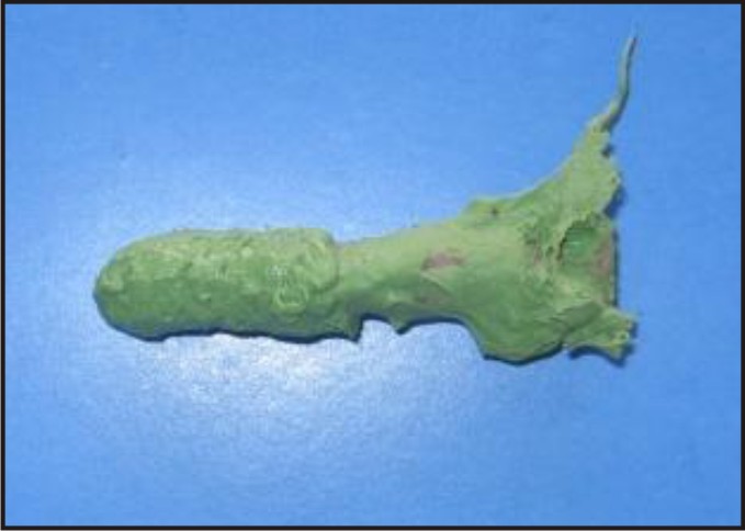 | Figure 3 - Vaginal Canal Impression.
|
(IV) Lab. Procedures
(a) The impression was half invested with the dental stone and key indexing was done (Image 4).
(b) Tin foil substitute was applied as separating media before pouring the second half.
(c) The halves of the mold were separated (after the dental stone has achieved sufficient strength) along the separation line and the impression was slowly eased out. Necessary blockout was done with the help of base plate wax (Image 5).
(d) The prosthesis was then fabricated using clear heat cure acrylic resin ( Densply India pvt. Ltd, Plot no. 9, Phase 1, Udyog Vihar, Gurgaon 122016 , Haryana, India) and finished and polished (Image 6).
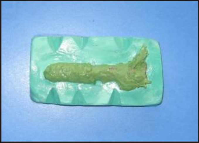 | Figure 4 - Poured First Part Of Cast With Indexing.
|
 | Figure 5 - Two Halves Of The Cast.
|
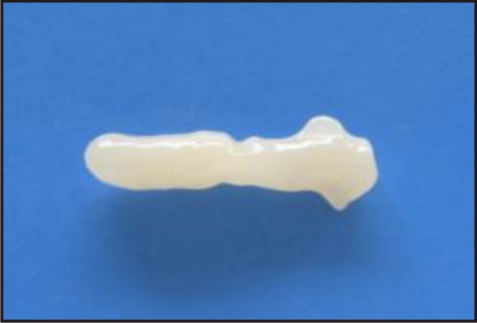 | Figure 6 - Cured, Finished And Polished Vaginal Prosthesis.
|
(V) Delivery Of Prosthesis
Before delivery of the prosthesis necessary arrangements were done to provide required retention to the prosthesis.
Elastic bands were used to secure the stent into vagina. The bands were kept passive or with little tension and used only to keep the prosthesis into its place and no force was applied. Sanitary napkins were advised for assisting the elastic bands in providing the necessary retention (Image 7).
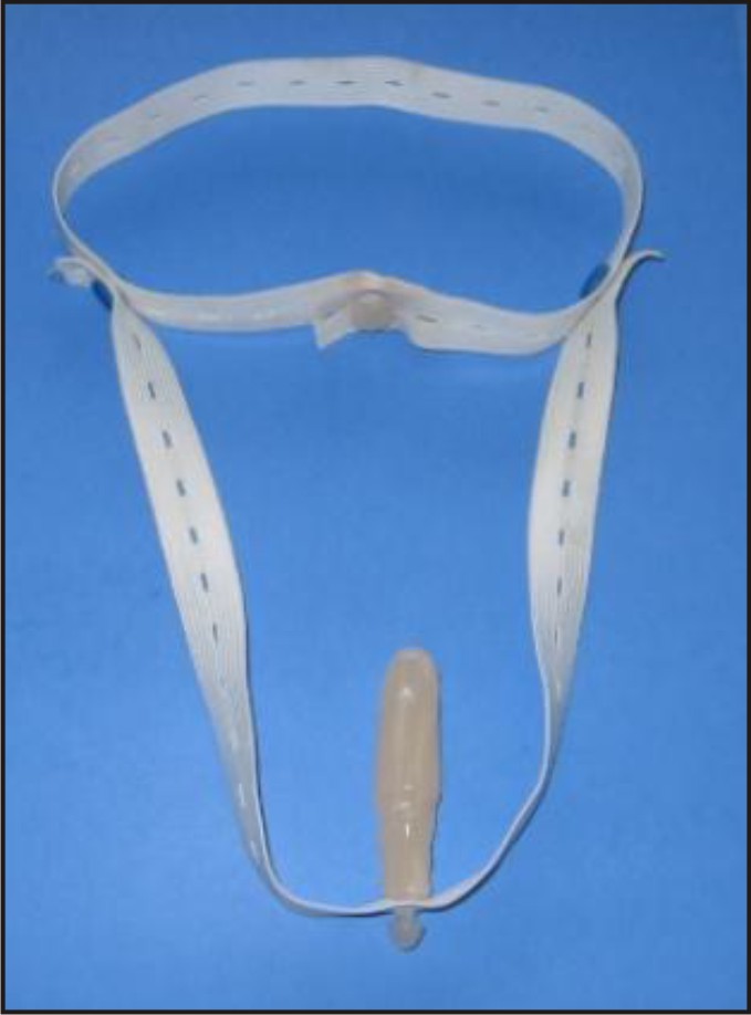 | Figure 7 - Final Prosthesis Ready For Delivery.
|
At the time of delivery
(i) Prosthesis was checked whether it was comfortable to the patient or not.
(ii) Patient was advised to wear the prosthesis with both the retentive accessories i.e. elastic bands and sanitary napkins.
(iii) Patient was advised to keep the prosthesis clean and wash it with soap water or Betadine, at least 3-4 times a day.
(iv) Patient was advised to wear the prosthesis, at least 4- 5 hours per day initially, and then 8 – 10 hours per day.
(v) Patient was explained the importance of vaginal hygiene, and advised to wash the vagina at least twice .
(vi) Patient was instructed to avoid sexual intercourse till the repair of fistulae can be undertaken.
(vii) Finally as per the instructions from the Urology department, the patient was advised to attend urology clinic after 2 weeks and recalled after 3 month for repair of vesico- vaginal and recto- vaginal fistulae.
Discussion
In literature many treatment alternatives for vaginal stenosis are mentioned such as prefabricated vaginal stents, vaginal dilators, regular sexual intercourse etc [2].
Vaginal stents have been shown to be better than vaginal dilators as latter’s design don’t take into the account the fact that vagina is most distensible in its upper third part, and vaginal dilators are ineffective in that region for preventing stenosis [1].
Vaginal stents have also been proved to be better than regular sexual intercourse because of compliance difficulties on emotional grounds[1]. The described technique of fabrication of custom made vaginal stent offer definite advantage over prefabricated vaginal stents. Custom made stent conform more intimately to altered internal vaginal anatomy than prefabricated one and thus more effective.
Moreover, as already mentioned in the case report, the patient also had fistulae. Hence, custom made stent would be far more suited than a prefabricated one in preventing any encroachment toward the fistulae.
Irreversible hydrocolloid has been used in the literature for impression making [3]. We have chosen poly vinyl siloxane impression material because of its much important high tear strength, better flow, better detailing, and adequate working time.
Elastic bands have been used to achieve retention of vaginal stent[4]. We have used elastic bands only to secure the stent into the vagina and any tension or force was avoided, to prevent any undue pressure over the fistulae. Sanitary napkins were used to assist in achieving the retentionSexual intercourse has been advised in the past to prevent the stenosis, but we have asked the patient to avoid it on two important grounds. Firstly sexual intercourse is ineffective in preventing stenosis[1]. Secondly intercourse may hurt the existing fistulae.Vaginal stenosis is a very common side effect of any brachytherapy received in the vaginal region e.g. vaginal cancers[5]. In these patients the preoperative impression can be used to form two stents one for carrying out the brachytherapy and other one to be used as stent between intertherapy period. This way the preoperative dimensions of the vaginal canal be maintained and stenosis can be prevented.
Conclusion
The patient well tolerated the custom made vaginal stent, after she was made to understand all procedures of use and maintenance of hygiene. The stent helped to maintain patency of the lumen till the repair of the fistulae was undertaken without encroaching the same.
References
1. Decruze SB, Guthrie D, Magnani R. Prevention of vaginal stenosis in patients following vaginal brachytherapy. Clinical Oncology 1999; 11: 46-48.
2. Lancaster L. Preventing vaginal stenosis after brachytherapy for gynaecological cancer: an overview of Australian practices. European Journal of Oncology Nursing 2004; 8: 30-39.
3. Grisius R, Moore DJ. Miscellaneous prosthesis In: Livenwood W (ed-in-chief). Maxillofacial rehabilitation: Prosthodontic and surgical considerations. Missouri: Ishiyaku EuroAmerica,Inc.1996: 520
4. Lacy J, Correll GR, Walmer DK, Price TM. Simple vaginal mold for use in postoperative care of patients with a transverse vaginal septum. Fertility and Sterility vol.xx, no.x, month, 2007, article in press
5. Brand AH, Bull CA, Cakir B. International Journal of Gynaecology Cancer, and 2006 Jan-Feb; 16 (1): 288-93.
|