Introduction
Forensic odontology plays an important role in establishing the gender of victims when bodies are mutilated beyond recognition in mass disasters[1].
Teeth are known for being the most resistant mineralized tissue against different agents of destruction. They are therefore often used as a way of reconstructive identification. They are particularly useful in determination of gender by using different odontometric techniques of real interest in the case of major catastrophes where bodies are often damaged beyond recognition[2].
The need for identification of a person can be of social, emotional or legal importance. Confirmation regarding identity of the deceased is needed for obtaining a death certificate, claims of insurance or to carry out investigation of offenses like murder, abuse[3].
Teeth are extremely durable during high temperature and may be identified in the rest of the body after it has often undergone decomposition. Of all the teeth in human dentition, the canines are the least frequently extracted teeth. Also canines are reported to withstand extreme conditions and have been recovered from human remains even in air disasters and hurricane[4].
Identification of gender in damaged dead bodies (non living population) is an essential step for medico legal purposes. Teeth are excellent material in living and non living population for anthropological, genetic, odontological and forensic investigations. Their extreme durability in the face of fire and bacterial decomposition makes them invaluable for identification. Being the hardest and chemically the most stable tissue in the body, they are preserved[5].
Sex assessment constitutes an important step in constructing a postmortem profile and is useful in identifying skeletal remains. The accuracy of sexing using diverse parameters of the body such as craniofacial morphology and measurements on the pubis ranges from 96 to 100%. In forensic context, it is uncommon to recover full remains, with fragmentary skull and pelvic bones. Therefore, the dentition is considered a useful adjunct in sex determination[6].
Correct sex determination limits the pool of missing persons to just one half of population[7]. Teeth may be used for sex determination with aid of odontometric analysis. The mandibular canine is found to exhibit the “greatest sex dimorphism” amongst all teeth[5].
The sexual dimorphism specific to canines has been explained by Eimalad Derne on the basis of their function which forms an evolutionary point of view and is different from other teeth. During the evolution of primates the aggressive function of the canines in apes is transformed to the fingers in man[8].
Sexual dimorphism refers to those differences in size, stature and appearance between male and female that can be applied to dental identification because no two mouths are alike. The study of permanent mandibular and maxillary canine teeth offers certain advantages in that they are the least extracted, are less affected by periodontal diseases, are exposed to less plaque, calculus, abrasion from brushing and are the last teeth to be extracted with respect to age. The findings indicate that mandibular canine can be considered as key teeth for personal identification[5].
The existence of sexual dimorphism in permanent teeth is a known phenomenon as observed in several investigations. This behavior morphogenetically determines that the shape and dimensions of the tooth are fairly stable and has been seen as a determining factor in providing sexual dimorphism in skeletal remains, which is required for forensic identification purposes[9].
Mandibular canine index was employed in numerous studies in large populations as it is simple, reliable, inexpensive and easy to perform. This is of definite significance, as tooth morphology is known to be influenced by cultural, environmental and racial factors. Studies performed on the lower canines using the ratio between the maximum crown width and intercanine width, resulting in a mandibular canine index (MCI), have shown an ability to determine gender with an accuracy of 84.3% in males and 87.5% in females, and 83.3% in males and 81% in females by comparing the observed MCI with a standard MCI value respectively[10].
The purpose of this study is to investigate the accuracy with which gender can be differentiated by using the mandibular canine index in the present study group.
Materials and Methods
Sample
The sample comprised of dentitions from 1000 individuals of Indian origin (500 males and 500females), all young adults in the age group of 17 – 24 years. Most of the participants were undergraduate and postgraduate students of our institute, Bangalore. The participants were from different states of India and the sample was the composite of ethnic groups, castes and religions since the aim of the study was to assess sexual dimorphism in Indian population. Following an informed consent, impressions of the teeth were made using alginate and the cast were poured in dental stone.
Measurements
The mesiodistal (MD) measurements of the mandibular canines and the intercanine distance were measured on the casts using a digital caliper calibrated to 0.01mm (Mitsoyo, Japan). The mesiodistal dimension was defined as the greatest distance between contact points on the approximate surfaces of the tooth crown and was measured with the caliper beaks perpendicular to the long axis of the tooth. The intercanine distance was measured between the cusp tips of the right canine to the cusp tip of the left canine using the caliper beaks placed occlusally along the cast.
Statistical Analysis
The measurements were evaluated using the student‘t’ test (two tailed, independent) to find the significance of the study parameter on continuous scale between two groups (inter-group analysis). Discriminant analysis has been performed to predict the correct classification by study variables for discriminating gender. All statistical analysis were performed using statistical software namely SAS 9.2, SPSS 15.0, Stata 10.1, Med calc 9.01, Systat 12.0 and R environment 2.11.1.
Results
An observational study with 1000 subjects with equal male and females 500 each (table 1) was undertaken to study and establish that the dimorphism of canines can be used for sex determination, to establish that the mandibular canine as a quick and reproducible method of sex determination.
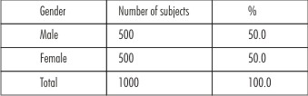 | Table 1: Gender distribution
 |
The descriptive statistics and the degree of sexual dimorphism for mesiodistal measurements of the mandibular canine and the mandibular intercanine arch width are depicted in the Table 2. The mesiodistal width of right canine in males was 6.74 ± 0.54 where as in female it was 6.34 ± 0.52. The p value was < 0.001 which was statistically significant. In case of the mesio distal width of left mandibular canine, in males, it was 6.73 ± 0.59 and in females it was, 6.39 ± 0.55 with a p value of <0.001. This was again statistically significant. The inter-canine distance was found to be 27.56 ± 1.98 in males and was 27.06 ± 1.86 in females and p value was < 0.001 which was statistically significant.
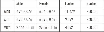 | Table 2: Discrimination of MDR, MDL and MICD between male and female
|
Discriminant analysis of the mandibular mesio- distal canine width and canine arch width using, discriminant analysis was done (table 3). Wilk’s lambda denotes how useful a given variable is in the step wise analysis and determines the order in which the variables enter the analysis. The mesio- distal width of the right canine has a Wilk’s lambda value of 0.883, which is statistically significant with 63% predicted value for correct classification. The mesio- distal width of left canine has a Wilk’s lambda value of 0.092, with 62.9% predicted value for correct classification and was not statistically significant. The inter-canine width has a Wilk’s lambda value of 0.983 and 56.2% predicted value for correct classification.
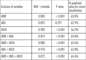 | Table 3: Discriminant analysis to predict the sex determination
|
The combined Mesiodistal right [MDR] and Mesiodistal left [MDL] Wilk’s lambda value is at 0.874 with 64 % predicted value for correct classification and is statistically significant. The MDR and MICD Wilk’s lambda value is 0.880 with 63.6% predicted value for correct classification, while the MDL + MICD Wilk’s lambda is 0.910 with 62.8% prediction for correct classification. The combined MDR+ MDL + MICD Wilk’s lambda is 0.872 with 64.6% predicted value for correct classification and this is statistically significant.
Discussion
Forensic odontology plays an important role in establishing the sex of victims of bodies mutilated beyond recognition due to major mass disaster. Teeth are known for being the most resistant mineralized tissue against different agents of destruction. They are therefore often used as a way of reconstructive identification. They are particularly used in determination of gender by using different odontometrical techniques of real interests in the case of major catastrophes.
Identification of gender in dead bodies is an essential step for medico legal purposes. Teeth are excellent material in living and non living population as they are extremely durable even in cases of fire and bacterial decomposition.
Correct sex determination limits the pool of missing persons to just one half of population. Mandibular canine is found to exhibit the greatest sexual dimorphism amongst all the teeth and of the most robust teeth in the dentition, surviving pre and postmortem destruction[11]. It was for this reason that the study was designed to examine the utility of canines alone in sex estimation. Along with the mesiodistal dimensions, the intercanine arch width was also included in the study. A sample of 1000 cases, 500 males and 500 females was taken up with the objectives of establishing the crown width of canines as a tool for sex determination and to test the accuracy of this method as a forensic tool in gender determination.
The sample size in this study was 1000 with equal distribution of 500 males and 500 females. The sample consisted of students from our institute. These students were from various states and so the sample was a composite of ethnic groups, castes and religion, since the aim of the study was to assess sexual dimorphism in Indians as a whole. The sample age group was from 17 – 24 years and the mean age was 21 years. This was done to assure that the dentitions were relatively intact, free of pathology and wears and thereby maximizes odontometric information. The age group of the study is in accordance with the study by Sudeendra Prabhu and Ashith.B.Acharya who also did a study of odontometric assessment of sex in the Indian population in the age group of 17 – 24 years[12].
In the present study the mesiodistal width of mandibular right canine was 6.74±0.54 in case of males, and 6.34±0.52 in that of the females. This was found to be statistically significant with a P value of <0.001. The results of the present study were in accordance with results of Ashith.B.Acharya et al (2010) who also reported that MDR in males as 6.73 and 6.47 in cases of females. Our values are in accordance with study conducted by Balwant Rai and Dr. S.C. Anand (2008) who reported overall MDR in both the sexes as 7.63[13].
Marin Vodanovic studied the sexual dimorphism of canine and concluded that the mean MD dimensions in males was 6.70 and 6.64 in females. This was slightly higher in case of females than our study. This is possibly because of change in the race and genetic profile of the European population[14].
Vandana Reddy studied mandibular canine index as a sex determinant and reported that the mean right canine width as 7.07 in males and 6.64 in females. They also showed that the results were statistically significant[15].
In the present study the MD of the mandibular left canine was 6.73 in case of males and 6.39 in case of females and was also statistically significant. Our values are in accordance with Ashith.B.Acharya who got he values as 6.8 in case of males and 6.51 in case of females. The slight variation in the mesiodistal width in case of females may be due to the method of collection of data where the measurements by Ashith. B.Acharya was taken by digital caliper being kept along the long axis of the tooth and in the present study it was kept perpendicular to the long axis of the tooth and also the sample size in our study was much larger consisting of 1000 patients[11].
Vandana Reddy reported that mandibular MD width of left canine in males was 7.03 and 6.44 in females[15]. In our study it was found that the MD width of the canines is greater in males than in females. Our findings are in conclusion with Balwanth Rai and S.C.Anand (2008)[13], Ashith.B.Acharya (2010)[16] and Irfan Ahmed Mughal (2010) et al[17].
Elemi Zoeba studied sexual dimorphism in permanent teeth of Greeks and found a mean MD dimension in mandibular canine in case of males as 6.77 and 6.40 in case of females. The findings were in accordance with our findings[18].
In the present study the mandibular intercanine width was 27.56 ± 1.98 in case of males and 27.06 ± 1.86 in case of females. In a similar study by Ashith.B.Acharya (2010) the MICD value in case of males was 27.62 which is in accordance with our study and 26.58 in case of females which was slightly lower than in our case[11].
Vandana Reddy reported theh inter canine distance in males as 26.86 and 26.28 in females. Their values were much lower than ours because their sample size was 200 and the study group constituted only a part of population of Western Uttar Pradesh and was not diverse as in our study. This is also in accordance to the studies conducted by Suazo et al who reported that intercanine dimension of the mandibular tooth crowns were higher in males than in females[8].
Vandana.M.Reddy studied mandibular canine index as sex determinant and concluded that a definite statistically significant sexual dimorphism in mandibular canines[15].
The results of the present study are also in accordance with Hashim.H.A and Mushid.Z.A (1993) who showed that the canines were the only tooth to exhibit dimorphism[4].
Vanaki.S.S et al studied the teeth dimension as a distinguishing trait between human sexes and concluded that there was statistically significant sexual dimorphism in the mesiodistal as well as buccolingual diameter of the permanent canines. This was comparable with our study which also showed statistically significant results[19].
The possible explanation given as to why males have a greater MD dimension than females is that male teeth have a higher amount of dentin compared to females while females have more enamel in teeth. It appears that sex linked genes on both the X & Y chromosomes are responsible for this difference in the amounts of dentin and enamel in teeth[18].
Alvesalo & Tammisalo found that the Y chromosome increases the mitotic potential of the tooth germ, inducing dentinogenesis, while the X chromosome promotes amelogenesis and so the Y chromosome contributes most in the size of teeth by controlling the thickness of dentin[20].
Discriminant analysis was performed to predict the correct classification by study variable for discriminating gender. Wilk’s lambda denotes how useful a given variable is in step wise analysis[20].
Upon discriminant analysis of MDR, 63% was predicted value for correct classification. This was statistically significant with P value of 0.001. This was in accordance with Ashith.B.Acharya et al whoalso predicted a 64.1% sex classification for males and 67.3% for females using the logistic regression analysis[16].
Ashith.B.Acharya and Manali reported an 83% predicted value for correct classification using the cross validated discriminant analysis. The possible difference is due to variation in the statistical analysis and the sample size[21].
The MDL value in our study predicted 62.9% value for correct classification, but this was not statistically significant < 0.291. This concern has to be addressed by a study of larger sample size and with a composite sample.
The MICD has 56.2% predicted value for correct classification and was found to be statistically significant. Acharya et al had got a 61.8% value for correct classification[11]. This disparity in result may be due to our larger sample size and a different statistical analysis.
Vandana Reddy et al predicted a 72% of cases correctly using the mandibular canine index[15]. This high percentage was possible because there study was limited to a particular geographic region in Uttar Pradesh and their result was not subjected to discriminant analysis[15].
Although in our study there is a definite sexual dimorphism and has revealed significant results, further studies with a larger sample size with the help of more statistical techniques may contribute to gender determination using teeth.
Conclusion
Gender determination is an important element in the analysis of biological evidence submitted to forensic laboratories. In the quest for identification of unknown human remains, even the most insignificant bone fragment or a tooth may contribute to a great extent.
The observations of this study suggested that mesiodistal width of the mandibular canine and inter canine distance parameters can be used in establishing sex.
However, studies have to be carried out with larger sample size to support our observation and standardize definite values for sex identification with reference to mesiodistal distance and mandibular intercanine distance in males and females.
Thus, sex determination can be done with help of canine tooth in forensic dentistry to establish the gender of individuals in mass disaster.
References
1. Sherfudhin H, Abdullah MA & Khan N. A cross-sectional study of canine dimorphism in establishing sex identity:Comparison of two statistical methods. J Oral Rehab 1996; 23:627-631.
2. Muller M, Lupi-Pegurier L, Quatrehomme G, Bolla M. Odontometrical method useful in determining gender and dental alignment. Forensic Sci Int 2001; 121:194-197.
3. Tekade P, Singh TRM. Forensic dentistry: A review. International journal of applied biology and pharmaceutical technology Jan Mar 2011;2(1):265-272.
4. Boaz K, Gupta C. Dimorphism in human maxillary and mandibular canines in establishment of gender. J Forensic Dent Sci Jan Jun 2009;1(1):42-44.
5. Srivastava PC. Correlation of odontometric measures in sex determination. J Indian Acad Forensic Med;32:56-61.
6. Acharya AB, Mainali S. Limitations of the mandibular canine in sex assessment. J Forensic Leg Med 2009; 16:67-69.
7. Hemanth M, Vidya M, Nandaprasad, Karkara BV. Sex determination using dental tissue 2009. http://www.indmedica.com; 1-7.
8. Galdames IS et al. Sexual dimorphism in mesiodistal and buccolingual tooth dimensions in Chilean people. Int J Morphol 2008; 26(3):609-614.
9. Rai B, Anand SC. Gender determination by diagonal distances of teeth. The internet journal of biological anthropology 2007; 1(1).
10. Acharya AB, Mainali S. Sex determination potential of buccolingual and nesiodistal tooth dimensions. J Forensic Sci Jul 2008; 53(4):790-792.
11. Acharya AB, Angadi PV, Prabhu S, Nagnur S. Validity of the mandibular canine index (MCI) in sex prediction:Reassessment in an Indian sample. Forensic Sci Int 2011; 204:207e1-207e4.
12. Prabhu S, Acharya AB. Odontometric sex assessment in Indians. Forensic Sci Int 2009; 192:129.e1-129.e5.
13. Rai B, Anand SC. Determination of stature from mesio-distal and bucco-lingual dimensions of teeth in North Indian population. J Forensic Dent Sci Oct-Dec 2008; 1(2).
14. Vodanovic M, Demo Z, Njemirovskij V, Keros J, Brkic H. Odontometrics:A useful method for sex determination in a archaeological skeletal population?. J Archaeol Sci 2007; 34:905-913.
15. Reddy VM, Saxena S, Bansal P. Mandibular canine index as a sex determinant:A study of the population of Western Uttar Pradesh. J Oral Maxillofac Pathol Jul-Dec 2008; 12(2):56-58.
16. Iscan MY, Kedici PS. Sexual variation in bucco-lingual dimensions in Turkish dentition. Forensic Sci Int 2003; 137:160-164.
17. Bonetti GA, Zanarini M, Danesi M, Parenti SI, Gatto MR. Percentiles relative to maxillary permanent canine inclination by age:A radiologic study. Am J Orthod Dentofacial Orthop 2009; 136:486e1-486e6.
18. Zorba E, Moraitis K, Manolis SK. Sexual dimorphism in permanent teeth of modern Greeks. Forensic Sci Int; 1-8.
19. Vanaki SS, Puranik RS, Sharma G, Sharma M. Tooth dimension as a distinguishing trait between human sexes:An odontometric study on Bagalkot population. Indian journal of Forensic Medicine and Pathology 2008; 1(3&4)75-78.
20. Brook AH, Griffin RC, Townsend G, Levisianos Y, Russell J, Smith RN. Variability and patterning in permanent tooth size of four human ethnic groups. Arch of oral biol 2009; 54:79-85.
21. Acharya AB, Mainali S. Univariate sex dimorphism in the nepalese dentition and the use of discriminant functions in gender assessment. Forensic Sci Int 2007; 173:47-56.
|