Introduction
The influence of genetic and environmental factors on growth and development of the dentofacial complex has been the topic of debate and controversy from ancient times till date. Although head form and facial proportions appear to be heritable, quantitative analysis of hereditary variability in cranial and facial dimensions in related individuals has been few. Consequently, attempts to establish morphological criteria for racial comparisons or for orthodontic diagnostic purposes have been hampered by a need for more information relative to genetic variation in the face and skull[1].
The studies on siblings is one of the most effective methods available for investigating genetically determined variables in orthodontics as well as in other medical fields, depending on the variance in the shape and size of skull and teeth on genetic and environmental influences.
There have been very few sibling studies carried out in India till date. In the present investigation, an attempt has been made to further explore genetic influences on variations in several craniofacial dimensions of a group of post-adolescent subjects in whom skull growth is essentially complete. This study was carried out on siblings in Bengaluru, Karnataka in order to determine the features using Cephalometrics and Standardized Photographs of the craniofacial region in siblings.
Materials And Methods
The study was conducted in the Department of Orthodontics and Dentofacial Orthopaedics, V.S. Dental College, Bangalore. The subjects were from in and around Bangalore.
The sample size consists of 20 pairs of similar looking siblings. An inclusion criterion for the age group was 18-30 yrs.
To further the selection process Frontal, Profile and Submento-vertex view were taken from Nikon D70 camera with a 105mm lens, by strict standardization in Magnification and Head position. In addition, lateral cephalograms in Natural Head Position were also taken.
To reach the conclusion it was decided to carry out specific Facial and Cephalometric measurements to analyse what are the parameters that are constant which gives them the similarity and what vary giving them their individuality.
Photographic analysis
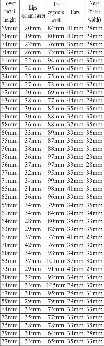
1. Lower facial height
2. Lips (commissure)
3. Bi-zygomatic width
4. Ears
5. Nose (nares width)
Cephalometric analysis
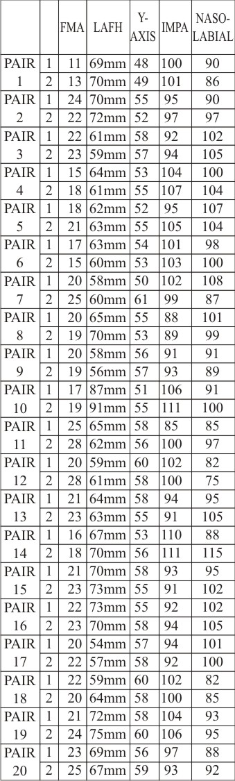
1. Mandibular plane angle (FMA)
2. LAFH
3. Y-AXIS
4. IMPA
5. Naso-labial angle
Preparation for standardized photograph :
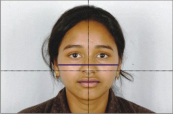
A photographic set up was done consisting of the following things in mind to make it strictly standardized in magnification and head position. For obtaining a true vertical line, a plumb line is created by suspending a weight of 200gms on a 0.016 inch string hung from ceiling. A true horizontal line is created by connecting two points on either side of the reflector board so that when the subject sits on the chair it touches the tip of the subject’s nose, using spirit level. The chair can be moved up and down in the vertical plane. A halogen light source (12v) is fixed on the ceiling with its focus on the reflector board. Two small halogen units are fixed on either side of the reflector board to provide additional illumination. The chair position is marked as well as the distance of the tripod stand from where the photographs of the subjects are taken. So that the individuals can be repositioned in the same place to check the precision of the method. The distance is further reconfirmed by marking the magnification number in the lens and moving manually.
Photograph (1) When all the above procedure is completed the subject is asked to sit on the chair and adjust the chair in the vertical plane so that the true horizontal at the tip of the nose dividing the face in to four halves. The subject is asked to maintain the natural head position. The frontal photographs are taken.
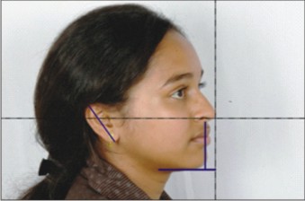
Photograph (2) Then the subject is asked to sit and adjust for the profile photograph with true horizontal parallel to ala-tragus line.
Photograph (3) The subject is then asked to determine the self-balanced position of the head by tilting the head backwards with increasing amplitude to a position of maximum flex for submentovertex view.
 | Photograph (1)
 |
 | Photograph (2)
|
 | Photograph (3)
|
Preparation for the cephalometric radiograph :
The subjects were oriented to natural head posture. Their mid-sagittal plane was placed at right angles to the path of x-ray. For obtaining the natural head posture, a modified approach as developed by Raju.S, Prasad K.G and Jayade.V.P. has been followed, in which the true vertical is captured on the patients face and the cephalogram was taken in a conventional manner. The procedure is as follows,
A mirror 4×2 feet was fixed on to the wall. To obtain a true vertical a plumb line is created by suspending a weight of 500gms on to A.J.Wilcock 0.016” Premium plus, Pulse straightened wire hung from the ceiling, 9 feet in front of the mirror. A kavo light source (12v) was fixed 7ft.6” to the right side of the wall., at a height of 5ft3”. The light source can move only in vertical direction to adjust the level of the light source depending on the subjects head. The light cast a shadow on the right side of the patients face, just distal to the lateral canthus of the eye. The light did not have horizontal movement so as to prevent shifting of the wire shadow in the horizontal direction which could alter the true vertical reference. The subjects foot mark were marked on the floor so that the individual could be repositioned in the same place to check the accuracy of the method. The subject was then asked to determine the self-balanced position of the head by tilting the head backward and forward with decreasing amplitude to find the most neutral position in between, as described by Solow and Tallgren. After determining the self-balanced neutral position, the subject was asked to look at his/her own eye in the mirror. At this juncture, two points were marked using the pen, along the wire shadow, one near the lateral canthus of eye and other at the corner of border of mandible. These areas were selected for placing the markers since no important landmarks are located here. Tiny circular metal markers (obtained by cutting the heads of safety pins) were fixed on each point using a transparent adhesive tape. These markers will give radio-opaque shadow on the lateral cephalogram. A line drawn to join two marker shadows thus gave a natural vertical line on the cephalometric tracing. Thus, the natural vertical axis on the face. After completing the above mentioned procedure, the subject was positioned in the cephalostat with the help of the ear rods and nosepiece while holding the teeth in centric occlusion position. Lateral cephalograms were then taken in a standard manner. This procedure was carried out in the Department of Oral Medicine and Radiology, V.S. dental college.
Standardized lateral cephalograms were taken with target film distance 5 feet, 80 KVP; 10mA and a exposure time of 0.63 – 0.8 sec. Extra oral cephalometric films of 8×10 inch size is designed to be used along with the intensifying screens, which is placed with the cassette between two screens under dark room condition. Green intensifying screens/films (Kodak ortho L) produces images of proper density with less exposure energy, with excellent soft tissue image and reduction of radiation dose received by the subjects. All the radiographs are developed under dark room condition.
Standard tracing of the soft tissue profiles as well as related to osseous and dental structures were made using 3H, 0.3mm leadpencil on the acetate matte tracing paper of thickness 0.003cm. All the cephalograms were traced by a single operator and were taken on the same radiographic unit.
Results
 | Cephalometric Measurements
|
 | Photographic Measurements
|
Statistical Analysis (T-test)
 | 1. Cephalometric Analysis
|
 | 1. Photographic Analysis
|
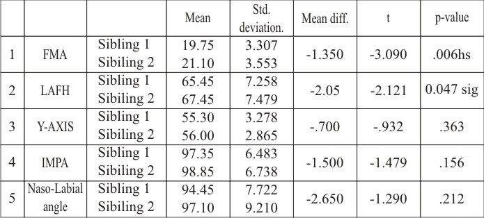
Based on statistical results for cephalometricanalysis :
It was found that Frankfort Mandibular Plane Angle(FMA) was highly variable which was followed by LAFH.
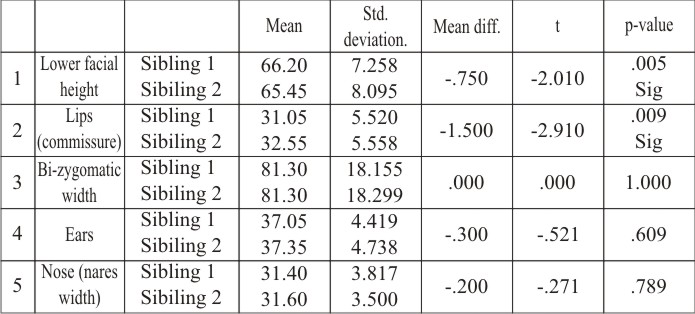
Based on statistical results for photographic analysis :
It was found that Lower Facial Height and Lips (Commissure) showed variability.
The remaining Cephalometric [Y-axis, IMPA, Naso-labial angle] and Photographic [Bi-zygomatic width, Ears, Nose (nares width)] parameters which were used in the study remained constant but their consistency varied slightly without showing any statistical significance.
Discussion
What are the facial features that give some amount of similarity among siblings/identical twins? And what are those factors that give them their individuality that is not shared among them?
An attempt has been made here to study the final facial form using both internal features (cephalometric) and external features (photographic) and strict standardization method. Nature seems to have adapted a dual model in giving the human face its final facial form, one of which is consistent & other is variable.
Although there have been few twin studies. Most of the twin studies done have been based on effects of hereditary and environmental factors on craniofacial morphology of the subject. Osborne R H, Horowitz S L and DeGeorge F V (1960), Dudas and Sassouni (1973); Nakata (1978); Ishikawa (1991).Ishikawa (1991)2, Jing Peng, Hui Deng, CaiFang Cao and Masaaki Ishikawa Ejo (2005)3. A recent cross-sectional twin study evaluated 10 to 13 year olds (Manfredi et al., 1997) using heritability for analysis and included different genders in the twin samples. Harris and Potter (1997),studied several intriguing observations relevant to the present research. These cross-sectional adolescent and adult twin studies reported the same findings related to facial depth and height (Horowitz et al., 1960; Hunter, 1965) which suggests that there might be a ‘universal influence’ of genetic and environmental factors in craniofacial dimensions for both children and adults.
In this investigation 20 pairs of similar looking siblings/twins within the age group 18-30 years were studied. Growth completion was important, otherwise some inconsistencies might arise due to difference in timing of onset, velocity and duration of adolescent growth spurts that some of the siblings had already entered.
Photographic analysis consisted of Frontal, profile and submentovertex views taken with Nikkon D70 camera and a 105mm lens by strict standardization in magnification and head position.
A plumb line was created by suspending a weight of 200gms on a 0.016 inch string hung from the ceiling to create a true vertical. A true horizontal line is created by connecting two points on either side of the reflector board so that it touches the tip of the subject’s nose using a spirit level, dividing the face into 4 quadrants.
For cephalometric analysis natural head position was established as described earlier, Photographs and Lateral cephalograms were strictly standardized. Routine relevant cephalometric land marks were assessed for the study.
The data collected was then subjected to statistical analysis. The T-test was carried out to determine the significance which later determined the variability.
Statistical analysis of cephalometric parameters revealed thatFrankfort Mandibular Plane Anglewas highly variable mong the siblings which was followed by LAFH.
Statistical analysis of Photographic parameters revealed that Lips(commissure) and Lower half of face showed variability.
The remaining Cephalometric as well as Photographic parameters which were used in the study remained constant but their consistency varied slightly without showing any statistical significance. Statistical analysis for cephalometric parameters revealed that, the Growth Pattern, the Inclination of the Lower Incisors, andNaso-labial angle remained consistent. Statistical analysis of photographic parameters revealed that the Bi-zygomatic width, Ears and Nose (nares width) remained constant.
Studies have shown us that although the external covering is made up of integument, adipose tissue, connective tissue, and muscle; does not always distribute itself in a uniform orderly manner on the skull structure. There is great variation in the amount and distribution of the soft tissue elements4. Therefore a cranio-facial assessment that is limited to the measurements of the skeletal structure would not appear to the standards of scientific accuracy, if a photographic evaluation were included.
Summary
Heredity has been investigated by racial, family-line and twin method. The influence of heredity can be assessed by studying the family members, observing the similarities and differences between mother-child, father-child, and sibling pairs. Siblings are special individuals who provide a wealth of information including intriguining and illuminating insights into the mechanism of human craniofacial growth and development. This study was carried out in Bangalore, Karnataka in order to determine the constant and variable features using Profilometrics, Cephalometrics and Standard Photographs of the craniofacial region in siblings. Five facial traits were studied cephalometrically and five facial traits were studied photographically. The study consisteda sample size of 20 pairs of similar looking siblings in the age group of 18-30 years. The cephalograms were taken in natural head position. All measurements were done with high level of accuracy and the photographs and lateral cephalograms with strict standardization. The facial traits that showed variability were Frankfort mandibular plane angle, LAFH, Lips (commissure), lower half of face. The facial traits showing similarity were Growth pattern, inclination of the lower incisors, Naso-labial angle, Bi-zygomatic width, ears, nose (nares width). Earlier studies have shown us that the external covering is made up of integument, adipose tissue connective tissue and muscle and it does not always distribute itself in a uniform orderly manner on the skull structure. There is great variation in the amount and distribution of the soft tissue elements.
Conclusion
An attempt was made to identify and isolate the constant features versus the variable features using Cephalometrics; Profilometrics and standardized Photographs using strict Standardization.
• The facial traits that showed variability were Frankfort mandibular plane angle, LAFH, lips (commissure), lower half of face.
• The facial traits showing similarity were Growth pattern, inclination of the lower incisors,the Naso-labial angle, Bi-zygomatic width, ears, nose (nares width).
References
1. S.Eugene Coben. The integration of Facial skeletal variants. Am J Orthod Dentofacial Orthop, 1955; 41(6):407-34
2. Horowitz S L, Osborne R H, DeGeorge F V. A cephalometric study of craniofacial variation in adult twins. Angle Orthod, 1960; 30(1): 1-5.
3. Jing Peng, Hui Deng, Cai Fang Cao and Masaaki Ishikawa. Craniofacial morphology in Chinese female twins: A semi-longitudinal cephalometric study. Eur J Orthod. 2005; 27:556-61.
4. Hwang HS, Kim WS, McNamara JA Jr, A comparative study of two methods of quantifying the soft tissue profile. Angle Orthod, 2000 jun; 70(3): 200-207
|