Introduction
The management of severely resorbed ridge has always posed a challenge to the dental profession. In particular, the resorption of the lower ridge is more common because the mandible atrophies at a greater rate than the maxilla.
The successful denture fabrication for such patients begins with an accurate impression coordinated with neuromuscular function so as to provide stability and physiological comfort to the patient. Complete dentures are primarily mechanical devices, but since they function in the oral cavity, they must be fashioned so that they are in harmony with normal neuromuscular function. All oral functions such as mast-ication, speech, swallowing and laughing involve the synergistic actions of tongue, lips, cheeks, and floor of the mouth which are very complex.[1] Failure to recognize the importance of tooth position and flange form and contour often results in dentures which are unstable and unsatisfactory.
After the loss of all remaining natural teeth, there exists within the oral cavity a void that may be called the potential denture space. This denture space is bounded by the tongue internally and by the muscles and tissues of the lips and cheeks externally. Within the denture space, there is an area that has been termed the neutral zone.[2] Fish first exposed the dental profession to the concept of neutral zone for complete denture treatment.[3] The neutral zone is the potential space between the lips and cheeks on one side and the tongue on the other, that area or the position where the forces between the tongue and cheeks or lips are equal.
Proper positioning of the artificial teeth within this neutral zone plays an important role in providing stability to prosthesis. Dental implants placed within the neutral zone stabilize the denture fabricated over atrophic mandibular ridge. However there may be certain medical, surgical or economical conditions when it is not possi-ble to place implants. In such complex cases, the placement of denture in neutral zone is the only option left for the stabilization ofcomplete denture.[4] This treatment modality can also be used in patients with partial glossectomy, motor nerve damage to the tongue which have led to either atypical movement or an unfavourable denture bearing area and surgical reconstruction of mandible.
Weighted mandibular dentures have been used for the management of severely resorbed mandibular residual alveolar ridges. Advocates claim that the denture aids in retention if it weighs approximately 30 dwt.[5] When the supporting edentulous alveolar mucosa is not appropriate for loa-ding with a cast metal base, an internally weighted mandibular denture can be used. This technique avoids direct contact of the metal base with the mucosa but provides the benefit of the additional weight to the denture.
This article describes a method for fabricating an internally weighted mandi-bular complete denture using neutral zone technique in management of severely reso-rbed mandibular ridge.
Case Report
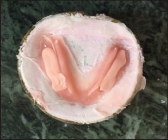
A 65-year-old-male patient reported to the Department of Prosthodontics, Himachal Dental College, Sundernagar, with a chief complaint of difficulty in mastication and speech.On clinical examination, the patient had no gross facial asymmetry or muscle tenderness. The Temporomandib-ular joint, muscles of mastication and facial expression were palpated and found to be asymptomatic. On intraoral examin-ation the maxillary and mandibular arches were completely edentulous. No gross abnormalities were detected in the overall soft tissue of the lips, cheeks, tongue and oral mucosa. The mandibular arch was severely resorbed (Atwood’s class VI) with shallow sulcus depth (Fig. 1).
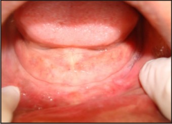 | Fig. 1 : Intra Oral View (Mandible)
 |
Treatment Procedure
The patient was explained about the treatment procedure. As the residual alveolar ridges were resorbed and the sulcus depth was shallow, a good preliminary impression with impression compound was difficult to achieve. To overcome this problem following procedure was planned.
1. In the first step, alginate impressions of maxillary and mandibular arches were made using patient’s old dentures as trays as shown in Fig.2. The impressions were poured in dental plaster. Over this cast a double thickness full spacer (to provide space for impression material) using modelling wax (Trulon) and a custom tray using autopolymerising resin (Rapid repair, DPI) was fabricated.
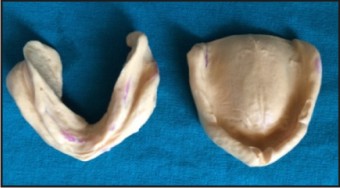 | Fig. 2 : Preliminary Impressions
|
2. The special tray was trimmed and checked in the patient’s mouth and then border moulding was done with green stick and final impressions were made in zincoxide eugenol impression paste (DentalProducts of India Ltd., Mumbai).
3. Record bases and wax occlusal rims were fabricated on the master casts. Jaw relations were recorded and mounted.
4. Following this the lower rim was removed and a second record base with a vertical occlusal stops and retentive loops to retain the material used to record the neutral zone was constructed(Fig. 3).
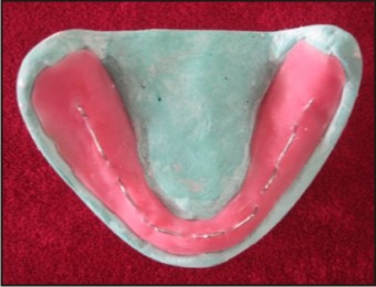 | Fig. 3 : Denture Base With Retentive Loops
|
5. This new record base was trimmed andchecked in the patient mouth and admix material (a mix of impression compound and low fusing compound in the ratio of 3:7) was placed over the retentive loops and the neutral zone was recorded (Fig. 4). During this procedure the patient was asked to make the movements like pucker the lips, swallowing and sucking to record the neutral zone.
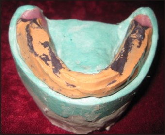 | Fig. 4 : Neutral Zone Recording
|
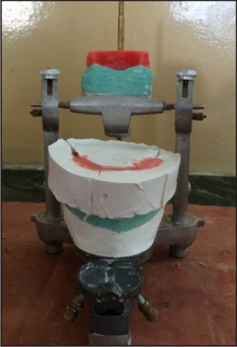
6. The baseplate carrying recording material was placed on the articula-ted mandibular cast and plaster index was made around the neutral zone record. Three orientation grooves were placed, that helped in repositioning the index on the master cast (Fig. 5).
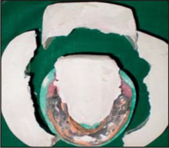 | Fig. 5 : Plaster Indices
|
7. The admix material was removed from the record bases and the indices rearranged. Then wax was made to flow into the space to make an occlusal rim to conform to the patient’s neutral zone (Fig. 6).
 | Fig. 6 : Admix Material Replaced By Wax
|
8. The teeth were arranged and the try-in was done in the patient’s mouth. (Fig. 7).
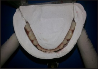 | Fig. 7 : Teeth Arrangement According To Plaster Indices
|
9. After the trial placement is assessed to be satisfactory, invest trial denture with dental stone (Kalabhai Karson Pvt. Ltd, India)for denture processing. Remove wax during the dewaxing stage.
10. After dewaxing, the flask was allowed to cool to room temperature. Separating medium (Cold mould seal, DPI) was applied with camel hair brush on exposed stone.
11. A heat cure acrylic resin (Dental Prod-ucts of India Ltd, India)dough was made by mixing the powder and liquid.When the mixture had reached the doughy stage, material was placed over the teeth in the flask and trial packing was done using cellophane sheet (DPI) (Fig. 8).
 | Fig. 8 : Trial Packing With Cellophane Sheet
|
12. Then the flask was opened and eight metal beads were placed evenly over the mandibular mold. Additional resin was added over the beads in the mold (Fig. 9 & 10).
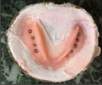 | Fig. 9 : Metal Beads Placed Over The Mold
|
 | Fig. 10 : Additional Resin Added
|
13. Then the flask was closed completely without the separating sheet and processing, finishing and polishing of dentures was done using conventional method (Fig. 11).
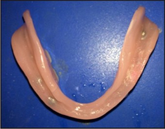 | Fig. 11 : Finished Mandibular Denture
|
14. Then the dentures were placed in the patient’s mouth and evaluated.
Discussion
For every patient the treatment requirements are different. So proper diagnosis and treatment planningform the first important step for the successful accomplishment of prosthodontic rehabi-litation. In patients with severely atrophic mandibular ridges, fabrication of stable mandibular denture is a great challenge. To overcome this problem, dentures are fabricated with their contours harmonizing neutral zone. A denture shaped by neutral zone technique will ensure that the mus-cular forces are working more effectively in harmony and gives advantage of stabilizing potential of oral and perioral musculature.[6] The neutral zone impression technique may be utilized for fabrication of any complete denture. Setting teeth and contouring polished surfaces of the lower complete denture within this zone makes the prosthesis less subjected to dislodging forces and adds more to stability.[7] Due to loss of teeth and supporting tissues, most lower dentures weight less than half the original. The reduction in weight in turn leads to improper muscle function and increased vertical distance between occlusal table and tissue support causing denture instability. These deficie-ncies can be overcome by reinforcing metal in lower denture base and adequate extension of the base.[8] The utilization of neutral zone technique along with internal metal reinfo-rcement provides a stable denture base foundation by neutralizing all internal and external forces directed against the denture with added weight.
Conclusion
The neutral zone is an alternative technique for the construction of lower complete dentures on highly atrophic ridges.This article describes a new fabrication technique for a weighted dent-ure base using neutral zone technique. A significant advantage of this technique is that the metal weight can be customized at the clinician’s request with minimal esthetic compromise. The predictable labo-ratory technique described may benefit the patient with a minimal residual ridge when implant therapy or preprosthetic surgery is not an option.
References
1. Beresin VE, Schiesser FJ. The neutral zone in complete dentures. J Prosthet Dent 1976; 36: 356-67.
2. Srivastava V, Gupta NK, Tandon A, Kaira LS, Chopra D. The neutral zone: Concept and technique.J Orafac Res 2012; 2: 42-7.
3. Wee A, Cwynar RB, Cheng AC. Utilization of the neutral zone technique for a maxillofacial patient. J Prosthodont 2000; 9: 2-7.
4. Porwal A, Jain P, Birader SP, Nelogi S, HC Naveen. Neutral zone approach for rehabilitation of severely atrophic ridge. Int J Dent Clin 2010; 2: 53-7.
5. Hurtado AJ. Internally weighted mandibular dentures. J Prosthet Dent 1988; 60: 122-23.
6. Agarwal S, Gangadhar P, Ahmad N, Bhardwaj A. A simplified approach for recording neutral zone. J Indian Prosthodont Soc 2010; 10: 102-04.
7. Jum’ah AA. Neutral zone in complete dentures: Systematic analysis of evidence and technique. Smile Dental Journal 2011; 6: 8-12.
8. Grunewald AH. Gold base lower dentures. J Prosthet Dent 1964; 14: 432-41.
|