Introduction
The failure of Hertwig’s epithelial root sheath to fuse onto the buccal or lingual root surface may be the main cause of the C-shaped root formation. The prevalence of C-shaped canal systems has been reported to range from 2.7% to 44.5% in mandibular second molars, depending on the population.[1] The main anatomic feature of C-shaped canals is the presence of a fin or web connecting the individual canals. The coronal orifice of these canals is usually located apically to the cement enamel junction level and may appear as a single, ribbon-shaped opening with a 180° arc linking all the main canals or a ribbon-shaped canal that includes the mesiobuccal and distal canals.[2] There are many variations in the anatomical configurations along the length of the canal system in these types of teeth. The C-shaped canal system might appear completely normal-looking at the level of the pulp chamber but the apical anatomy can be extremely complex.[3]
Cooke and Cox first described the clinical significance of C-shaped canals, which present major challenges with respect to their debridement and obturation. This is especially true when it is uncertain whether a C-shaped orifice found on the floor of the pulp chamber may continue to the apical third of the root. Irregular areas in a C-shaped canal that may house soft-tissue remnants or infected debris may escape thorough cleaning or filling and may be a source of bleeding and severe pain.[4]
Case Report
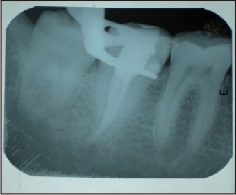
A 23-year-old woman presented with history of spontaneous pain on the right side of her face for past several days. The patient’s medical history was noncontributory . Clinically , the right mandibular second molar had a deep carious lesion. Intraoral periapical radiograph (Figure 1) revealed radiolucency involving pulp with periapical radiolucency. After extensive clinical and radiographic examination, diagnosis of acute apical periodontitis was made and the right mandibular second molar prepared for nonsurgical endodontic therapy.
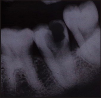 | Figure 1. Preoperative Iopa 47
 |
The patient received local anesthesia of 2% lidocaine with 1:100,000 epinephrine. A rubber dam was placed and a conventional endodontic access opening was made. Clinical evaluation of the internal anatomy revealed a continuous canal orifice extending from the distal aspect to mesiobuccal aspect. It was evident that in the present case, the canal system of the tooth assumed a C-shaped anatomy. The working length of the canal was estimated by means of an electronic apex locator (Root ZX; Morita, Tokyo, Japan), then confirmed by a radiograph. The canal was initially instrumented with #15 nickel titanium file (Dentsply Maillefer) under irrigation with 5% sodium hypochlorite. Coronal flaring was carried out using Gates-Glidden burs (nos. 3 and 2; Dentsply Maillefer).The canal was cleaned and prepared by hand nickel titanium files using a crown-down technique similar to that described by Saunders and Saunders. Intracanal medicament of calcium hydroxide was placed in the canal for a week.
One week later, the canal was obturated with conventional lateral compaction gutta-percha technique and AH-Plus sealer (Dentsply De-Trey, Konstanz, Germany) as used by most of the Indian dentists. A final radiograph was taken to establish the quality of the obturation (Fig.2). After completion of root canal treatment, the tooth was restored with a posterior composite filling (P60; 3M Dental Products, St. Paul, MN) (Fig.3).
 | Figure 2. Post Obturation 47
|
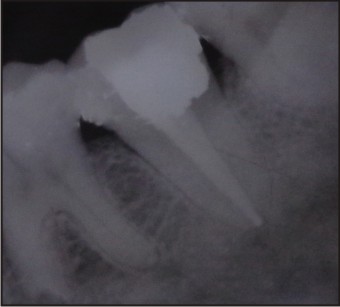 | Figure 3.Post Obturation Restoration 47
|
Case 2
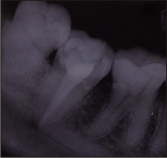
Twenty seven year old patient presented with the chief complaint of pain in lower left region of jaw for past several days. Clinical and radiographical examination revealed caries on the occlusal surface approaching pulp. The patient’s medical history was noncontributory. Clinically, the left mandibular second molar had a deep carious lesion. Intraoral periapical radiograph (Figure 4) revealed radiolucency involving pulp with periodontal widening. After extensive clinical and radiographic examination, diagnosis of acute apical periodontitis was made and the left mandibular second molar prepared for nonsurgical endodontic therapy.
 | Figure 4. Preoperative Iopa 37
|
The patient received local anesthesia of 2% lidocaine with 1:100,000 epinephrine. A rubber dam was placed and a conventional endodontic access opening was made. Clinical evaluation of the internal anatomy revealed two different canal orifices connected with a C configuration. The working length was estimated by means of an electronic apex locator (Root ZX; Morita, Tokyo, Japan), then confirmed by a radiograph. The canals were initially instrumented with #15 nickel titanium file (Dentsply Maillefer) under irrigation with 5% sodium hypochlorite. Coronal flaring was carried out using Gates-Glidden burs (nos. 3 and 2; Dentsply Maillefer).The canals were cleaned and prepared by hand nickel titanium files using a crown-down technique. Intracanal medicament of calcium hydroxide was placed in the canal for a week.
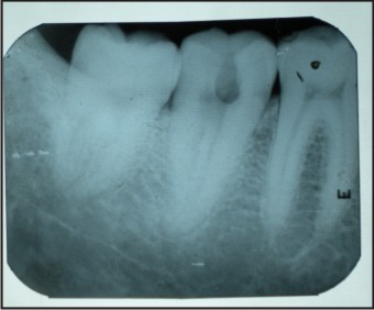
One week later, the canals were obturated with conventional lateral compaction gutta-percha technique and AH-Plus sealer (Dentsply De-Trey, Konstanz, Germany) as used by most of the Indian dentists. A final radiograph was taken to establish the quality of the obturation (Fig.5). After completion of root canal treatment, the tooth was restored with a posterior composite filling (P60; 3M Dental Products, St. Paul, MN)
 | Figure 5. Post Obturation 37
|
Discussion
The variability of the root canal system of multi rooted teeth represents a challenge to both endodontic diagnosis and treatment. The preoperative awareness of potential anatomic variations is essential for the success of the endodontic treatment. The C-shaped canal is not uncommon and this is confirmed by studies in which frequencies ranging from 2.7% to 8% [5] have been reported. The prevalence is higher in the middle Asia upto 10.6% in Saudi Arabians and 14% in Lebanese. In northeast Asia, the prevalence is 31.5% in Chinese and 32.7% in Koreans.[6] This variation may occur in mandibular first molars, maxillary molars, mandibular first premolars and even in maxillary lateral incisors, but it is most commonly found in mandibular second molars. In the view of above incidence and variability in canal configuration in our present case report, all the necessary steps to locate, debride and clean and shape the complex anatomy have been followed. Thermoplasticized gutta-percha technique is the recommended technique for obturation but as the most of dental practitioners use only lateral condensation technique so we used the same and found excellent results with gutta-percha and AH Plus sealer into the complex anatomy of the canal.
Conclusion
Although the prevalence is less, C-shaped canals can vary in number & shape along the length of the root, with the result that debridement, obturation & restoration in this group may be unusually difficult.Therefore, careful location & negotiation of canals & meticulous mechanical & chemical debridement of the pulp tissue should be carried out in order to successfully treat a C-shaped canal.
References
1. G . Seo, Y . Gu, A. Yi, J. Lee, S. Jeon, Y . Lee, W.Chang, K. Lee, W. Park, D. Kim and Y . Kum. A biometric study of C-shaped root canal systems in mandibular second molars using cone-beam computed tomography. Int Endod J 2012;45:807-14.
2. Michael Solomonov, Frank Paque, Bing Fan, Yuval Eilat, and Louis H. Berman. The Challenge of C-shaped Canal Systems: A Comparative Study of the Self-Adjusting File and ProTaper. J Endod 2012;38:209–14.
3. Xingzhe Yin, Gary Shun-pan Cheung, Chengfei Zhang,Yoshiko Murakami Masuda, Yuichi Kimura and Koukichi Matsumoto. Micro-computed Tomographic Comparison of Nickel-Titanium Rotary versus Traditional Instruments in C-Shaped Root Canal System. J Endod 2010;36:708–12.
4. Bing Fan, Yi Min, Guanfan Lu, Jun Yang, Gary S.P . Cheung and James L. Gutmann. Negotiation of C-Shaped Canal Systems in Mandibular Second Molars.J Endod 2009;35:1003–1008.
5. Weine FS, Pasiewicz RA, Rice RT : Canal configuration of the mandibular second molar using a clinically oriented in vitro method. J Endod 1988; 14 : 207–213.
6. Jafarzadeh Hamid, Wu Y.N. The C-shaped root canal configuration: A review. J Endod 2007;33: 517-23.
|