INTRODUCTION
Supernumerary teeth are described as the teeth formed in excess of the number found in a normal dentition and may or may not mimic the normal shape. Multiple supernumerary teeth are not a common occurrence and have been reported in the literature over the years as a well-recognized clinical phenomenon1. Multiple supernumerary teeth are commonly associated with vari-able syndromes. However the presence of multiple super-numerary teeth in the absence of any associated systemic condition/syndrome is unusual 2,3,4. Multiple supernumerary teeth are associated with leidocranial dysplasia and Gardner syndrome 5,6. In such cases, the mandibular premolar region is the common site
of occurrence 2. This case report presents a case of a non-syndrome male patient with multiple supplemental supernumerary teeth in two quadrants of his mouth
ETIOLOGY
The exact etiology of supernumerary teeth is still obscure although many theories have been proposed. Two popu-larly accepted theories are: The dichotomy theory of tooth germs states that the tooth bud splits into two equal or different sized parts, resulting in two teeth of equal size or one normal and one dimorphic tooth respectively. This hypothesis is supported by animal experiments in which split germs have been cultivated in vitro 7. Localized and independent hyperactivity of dental lamina is the other accepted theory, which suggests su-pernumerary teeth are formed as a
result of local, inde-pendent, conditioned hyperactivity of dental lamina 7.
Classification of supernumerary teeth may be on the basis of position 8 or form 9. Positional variations include:
1. Mesiodens - present in the incisor region.
2. Paramolars - present beside a molar.
3. Disto-molars - present distal to the last molar.
4. Parapremolars - present beside a premolar.
Based on the shape they can be of four types:
1. Conical: peg shaped teeth.
2. Tuberculate: made of more than one cusp or tubercle. They are barrel shaped, usually invaginated.
3. Supplemental: resemble normal teeth. May be an inci-sor, premolar or a molar.
4. Odontome: does not resemble any teeth but is only a mass of dental tissue
The supernumerary teeth can cause problems for the erup-tion and alignment of normal dentition. Associated prob-lems can range from failure of eruption, displacement, crowd-ing, adjacent teeth root resorption, formation of dentigerous cyst or they can be just asymptomatic.
is only a mass of dental tissue The supernumerary teeth can cause problems for the erup-tion and alignment of normal
dentition. Associated prob-lems can range from failure of eruption, displacement, crowd-ing, adjacent teeth root resorption, formation of dentigerous cyst or they can be just asymptomatic.
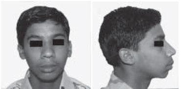 | Fig. 1 (a, b) Pretreatment Facial Photograph
 |
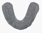 | Fig. 2 Lower model showing retained deciduous teeth
|
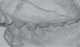 | Fig. 3 Tooth no 34 and 35 in crossbite
|
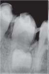
digital radiographic examination, three supernumerary teeth were found in the mandibular premolar region. There were two supernumerary on right side of mandible (Fig 4 a,b,c) and one on left side in the premolar region (Fig 5). All
supernumerary had rudimentary roots whereas crowns were well calcified. The crown of first supernumerary on right side was close to periapical area of 44 and second one was lingual to unerupted 45. This arrangement of addition
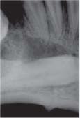
teeth was confirmed in occlusal view. There was a single supernumerary tooth on lingual aspect of 35. Radiographic evaluation also revealed
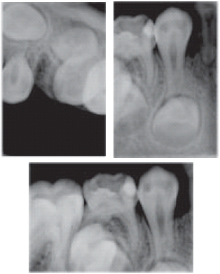 | Fig 4 (a,b,c) Intraoral periapical and occlusal digital radiographs showing two supernumeraries in 44 and 45 region
|
Fig 4 (a,b,c) Intraoral periapical and occlusal digital radiographs showing two supernumeraries in 44 and 45 region
 | Fig. 5 Supernumerary in 35 region
|
impacted right lower canine and associated cystic formation (Fig 6). A general physician was consulted who confirmed there was no associated syndrome. As the general and extra oral examination was non-contribu-tory, the diagnosis
of non-syndrome associated supernumer-ary teeth was made. The patient was advised extraction of supernumerary teeth prior to commencement of orthodontic treatment. The patient was referred to the Department of Oral Maxillofacial Surgery for surgical extraction of all supernumeraries and irretrievable 43. Cystic lesion associated with 43 was enucleated (Fig 7). The extracted supernumeraries resembled a lower premolar. The roots were underdeveloped and had capsule attached to them (Fig 8). The recovery was unremarkable. Since it was all first premolar extraction case according to the anchorage requirements, no extraction was done in fourth quadrant as 43 had been extracted. The patient is under active orthodontic treatment and follows up for the last 2 years and has not shown any signs
 | Fig. 6 Horizontally impacted 43 and Cystic formation
|
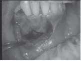 | Fig. 7 Surgical site after extraction of 43 and enucleation
|
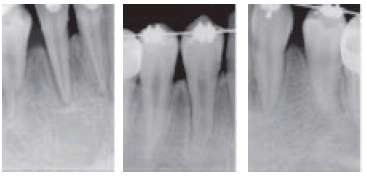
of recurrence and cystic lesion has recovered completely (Fig 9 a,b,c).
DISCUSSION
It is rare to find multiple supernumerary teeth with no associated diseases or syndromes. The few studies have found the prevalence of supernumerary teeth in permanent dentition to range from 0.15% to 3.8%.10 Scheiner11 reported
an occurrence of 11.1% for multiple supernumerary teeth, while Asaumim 14. found the prevalence for multiple supernumerary teeth to be 1%, and ArxN 12 found it to be 2%. However, where multiple supernumerary teeth’ is taken to
mean five or more supernumerary teeth, the prevalence has been reported as less than 1%.13 In the present case three supernumerary teeth were found.Several researchers have also proposed multiple supernumerary teeth are part of a post permanent dentition.10According to common opinion, multiple supernumerary teeth in the dental arch are mostly seen in the maxillary
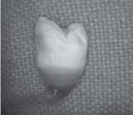 | Fig. 8 Extacted supernumerary
|
 | Fig. 9 (a, b, c)
|
anterior region followed by the maxillary molar region10 However, it has been reported multiple supernumerary teeth are very rarely seen, and the prevalence of supernumerary teeth in the premolar region has been reported as 0.2-10.9% in various studies.10 In this present case all three supernumerary teeth were seen in the premolar regions.It has been stated development of supernumerary teeth may cause various pathologies. Approximately 75% of supernumerary teeth are impacted and asymptomatic, and most of these teeth are diagnosed coincidentally during radiographic examination10. Early diagnosis is important in order to minimize the risk of complications resulting from supernumerary teeth. If they have caused delay or non-eruption of permanent teeth, displacement of permanent teeth, root resorption
of adjacent teeth due to the pressure and cystic formations, then extraction is recommended.1-14 .However, extraction of asymptomatic supernumerary teeth that do not affect the dentition may not always be necessary, but they should be followed through periodic examinations. Since there is a risk of tooth bud recurrence, follow-up on these patients is
recommended.
SUMMARY
Multiple supernumerary teeth are usually associated with syndromes but can occur, although rarely, without such an association. This case report presents a case of a non-syndrome male patient with multiple supplemental supernumerary teeth in two quadrants of his mouth. The importance of the use of a full mouth radiographs to evaluate a patient’s condition is emphasized whenever a supernumerary tooth is detected irrespective of whether the patient has any syndrome or not.
REFERENCES
1. Shafer WG, Hine MK, Levy BM. A textbook of Oral Pathology, 4th Ed. Philadelphia: WB Saunders Co.; 1993. p. 49.
2. Moore SR, Wilson DF, Kibble J. Sequential development of multiple supernumerary teeth in the mandibular premolar
region- a radiographic case report. Int J Paediatr Dent 2002;12:143-5.
3. King NM, Lee AM, Wan PK. Multiple supernumerary premolars: their occurrence in three patients. Aust Dent J
1993;38:11-6.
4. Hegde SV, Munshi AK. Late development of supernumerary teeth in premolar region: A case report. Quintessence Int
1996;27:479-81.
5. Shafer WG, Hine MK, Levy BM. (1993) A textbook of Oral Pathology, 4th edn. Philadelphia: WB Saunders Co.; p.49.
6. Yusof WZ. Non syndrome multiple supernumerary teeth literature review. J Can Dent Assoc 1990; 56: 147–149.
7. Liu JF. Characteristics of premaxillary supernumerary teeth: A survey of 112 cases. ASDC J Dent Child 1995; 62:262-26.
8. Thérèse MG, Barry HJ, Blake M. Supernumerary Teeth -An Overview of Classification, Diagnosis and Management, J
Can Dent Assoc 1999;65:612-6.
9. Mitchell L. Supernumerary teeth. Dent Update 1989;16:65-9.
10. Açikgöz A, Açikgöz G, Tunga U, Otan F. Characteristics and prevalence of non-syndrome multiple supernumerary teeth: a retrospective study. Dentomaxillofac Radiol 2006; 185-190
11. Scheiner MA, Sampson WJ. Supernumerary teeth: a review of the literature and four case reports. Aus Dent J 1997; 42:160-165.
12. Arx T. Anterior maxillary supernumerary teeth: a clinical and radiographic study. Aus Dent J 1992; 37: 189-195.
13. Rajab LD, Hamdan MAM. Supernumerary teeth: a review of the literature and a survey of 152 cases. Int Pediatr Dent 2002;12: 244-254.
14. Asaumi JI, Shibata Y, Yanagi Y, Hisatomi M, Matsuzaki H, Konouchi H, Kishi K. Radiographic examination of mesiodens and their associated complications. Dentomaxillofac Radiol 2004; 33: 125-127 |