Introduction
The soft palate establishes separation between oral and nasal cavities.[1] It moves in response to physiologic demand of speech, deglution and respiration. The soft palate, lateral and posterior pharyngeal walls forms the velopharyngeal (VP) closure so that all of them create a three dimensional muscular valve which is known as VP sphincter.[2] The VP closure pattern depends on the contraction degree of the sphincter components. Adequate VP closure is required during swallowing and production of all consonants except for the nasal ones.[3] Impairment of VP function can be due to insufficiency or incompetency.[3],[4],[5],[6],[7],[8] When some or all of the anatomic structure of the soft palate is absent, the term palatopharyngeal insufficiency applies.[9] Whereas when the soft palate is of adequate dimensions but lacks muscular and/or neurologic capacity, the term palatopharyngeal incompetence applies.[9] The term palatopharyngeal inadequacy includes incompetency and or in sufficiency but may also suggest a reduction or absence of pharyngeal wall function.[9]
The primary effects of the VP insufficiency are air-flow escape and hypernasality.[10],[11] VP insufficiency causes communication problems because of distortion in speech, resonance and articulation apart from swallowing disturbance.[11],[12] In this regard, patients usually have psychological problems together with physical difficulties.
Surgery in combination with speech therapy is a common approach to the treatment of VP dysfunction.[6],[11] There are several surgical procedures that can be performed to correct the physical malfunction. However, when surgical treatment is not considered as an option, prosthetic management of VP insufficiency is carried out by means of a pharyngeal obturator, whereas VP incompetence is traditionally managed by palatal lift prosthesis.[7],[8]
A pharyngeal obturator is a removable maxillary prosthesis which has a posterior extension to separate oropharynx and nasopharynx.[10] This obturator prosthesis restores the congenital or acquired defects of the soft palate and allows adequate closure of palatopharyngeal sphinter. The objectives of obturator are to provide the capability for the control of nasal emission and inappropriate nasal resonance during speech and to prevent the leakage of material into the nasal passage during deglutition.[9]
Case Report
A seventeen year old girl reported to our clinic with chief complaint of speech difficulty and communication problems. She had a history of cleft of hard and soft palate. In her medical history, she had undergone surgical correction of cleft palate two years back. (Fig. 1) She was speaking with soft intensity to decrease hypernasalance. Speech evaluation was performed by speech pathologists that assessed resonance, the occurrence of inappropriate nasal air emission, and articulation. (Fig. 2a, 2b).
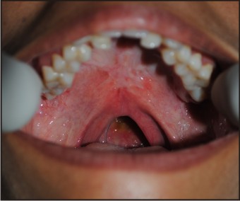 | Fig 1 : Velopharyngeal Defect
 |
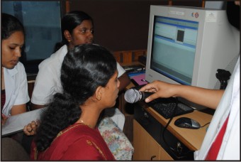 | Fig 2 (a) : Speech Analysis
|
 | Fig 2 (b) : Speech Analysis
|
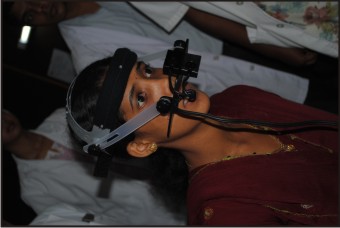
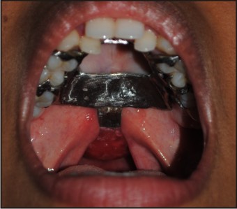
The patient refused to undergo second surgery, therefore a pharyngeal obturator with clasp retention was planned for the patient. The impression of pharyngeal bulb was made using putty elastomeric impression material (3M ESPE, St. Paul, USA) during the functional movements. The patient was instructed to flex the neck such that chin touches the chest. This movement will establish contacts of posterior aspect of obturator with the soft tissue covering the anterior tubercle of the atlas.[9] Lateral aspects of obturator are formed by rotation and flexion of neck. Patient is also asked to swallow. When border molding is complete patient’s speech should sound normal and there should be no escape of water between oropharynx and nasopharynx. Then the final impression is made with light body elastomeric impression material. (Fig 3).
 | Fig 3 : Impression of the defect
|
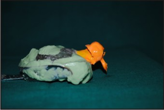
The framework with direct retainers on molar and premolar was fabricated. It consists of an extension on the pharyngeal part to support the acrylic bulb. The acrylic bulb was formed in wax, (Rolex, Ashoo Sons, Delhi, India) trial was done (Fig 4) and then it was fabricated with heat cure acrylic resin (Trevalon, dentsply, Gurgaon, India). The prosthesis was delivered to the patient with specific instruction for maintenance of the prosthesis. Speech pathologist assessed improvement in patient’s speech after wearing the prosthesis. (Fig 5).
 | Fig 4 : Wax Trial
|
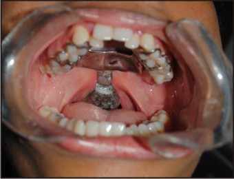 | Fig 5 : Final Prosthesis
|
Discussion
Prosthetic rehabilitation of the patients suffering from VP deficits with obturator prostheses varies according to the location and nature of the defect or deficiency.[4],[5],[7],[8] Pharyngeal obturator prosthesis may prevent the hypernasality and/or nasal emission associated with VP inadequacies.[5],[10] In order to obtain adequate VP closure during speech and swallowing a posterior extension is added to prosthesis. An acrylic resin extension must be formed functionally. This extension must be in static contact with the soft tissues and must not affect the stability of the prosthesis.[1],[10]
The success of the soft palate defect prosthesis depends on the functional adaptation of the impression material.[13] In this case elastomeric material was used to record the impression of the defect. High fusing compound, intra oral waxes can also be used for making impression.
Since the patient had complete maxillary dentition, direct and indirect retainers were used for retention. In case of partial or completely edentulous arches, the acrylic bulb can be a part of the partial or complete denture, however obtaining retention and stability can be difficult due to the weight of the prosthesis.[10] Dental implants have great importance for these patients.
The treatment of VP insufficiency requires multidisciplinary approach. Accordingly, a speech pathologist should participate in treatment of these cases to test articulation errors and inappropriate oro-nasal resonance balance.[7] Perceptual and instrumental measures of hypernasality and nasal escape along with a profile of the patient’s articulation provide the diagnostician information about the frequency and consistency of VP insufficiency. These measures, however, provide only limited information about the functioning of the VP mechanism. The use of multiview videofluroscopy (MVF) and/or nasendoscopy (NE) may contribute to the diagnostic confirmation of the assessment of velar mobility, pattern of velar elevation, size of residual VP gap and lateral pharyngeal wall displacement while the patient is producing a standardized sample of connected speech. It may also contribute the assessment of treated patients with VP insufficiency.[14] In present cases, no nasopharyngoscopic evaluations were made. However instrumental measures showed improvement in hypernasality of the patient.
Conclusion
In this report patient with soft palate defect was successfully treated by pharyngeal obturator. Success of such treatment largely depends on patient’s motivation and cooperation and ability to adapt with the prosthesis.
References
1. Zarb GA, Blonder CL. Prosthodontic Treatment for Edentulous Patient: Complete Dentures and Implant-Supported Prostheses. Maxillofacial prosthodontics for the edentulous patient.St. Louis:Mosby Inc; 2004.449–470.
2. Skolnick L, McCall GN, Barnes M. The sphincteric mechanism of velopharyngeal closure. Cleft Palate J. 1973;10:286–305.
3. Johns DF, Rohrich RJ, Awada M. Velopharyngeal incompetence: a guide for clinical evaluation. Plast Reconstr Surg J.2003;112:1890–1897.
4. Wolfaardt JF, Wilson FB, Rochet A, McPhee L. An appliance based approach to the management of palatopharyngeal incompetency: A clinical pilot project. J Prosthet Dent.1993;69:186–195.
5. Saunders TR, Oliver NA. A speech-aid prosthesis for anterior maxillary implant-supported prostheses. J Prosthet Dent.1993;70:546–547.
6. Ragab A. Cerclage sphincter pharyngoplasty: a new technique for velopharyngeal insufficiency. Int J Pediatr Otorhinolaryngol.2007;71:793–800.
7. Abreu A, Levy D, Rodriguez E, Rivera I. Oral rehabilitation of a patient with complete unilateral cleft lip and palate using an implant-retained speech-aid prosthesis: Clinical report. Cleft Palate Craniofac J.2007;44:673–677.
8. Shifman A, Finkelstein Y, Nachmani A, Ophir D. Speech-aid prostheses for neurogenic velopharyngeal incompetence. J Prosthet Dent. 2000;83:99–106.
9. Thomas D, Taylor. Clinical management of the soft palate defect. Clinical maxillofacial prosthetics. Illinios: Quintessence Publishing Co.Inc; 2000.127-137.
10. Beumer J, III, Curtis TA, Marunick MT. Maxillofacial Rehabilitation: Prosthodontic and Surgical Considerations; Speech, Velopharyngeal Function, and Restoration of Soft Palate Defects. St. Louis: Ishiyaku EuroAmerica, Inc; 1996. 285–324.
11. Yoshida H, Michi K, Yamashita Y, Ohno K. A comparison of surgical and prosthetic treatment for speech disorders attributable to surgically acquired soft palate defects. J Oral Maxillofacial Surg. 1993;51:361–365.
12. Werkmeister R, Szulczewski D, Walteros-Benz P, Joos UJ. Rehabilitation with dental implants of oral cancer patients. J Craniomaxillofac Surg. 1999;27:38–41.
13. Keyf F, Sahin N, Aslan Y. Alternative impression technique for a speech-aid prosthesis. Cleft Palate Craniofac J.2003;40:566–568
14. Lam DJ, Starr JR, Perkins JA, Lewis CW, Eblen LE, Dunlap J, Sie KC. A comparison of nasendoscopy and multiview videofluoroscopy in assessing velopharyngeal insufficiency. Otolaryngol Head Neck Surg.2006;134:394–402.
|