Introduction:
Ectodermal dysplasia (ED) is a hereditary disorder associated with dysplasia of tissues of ectodermal origin primarily nail, teeth, hairs and skin and occasionally dysplasia of mesoderm derived tissues. As de@257;ned by Freire-Maia the nosologic group of Ectodermal dysplasia is any syndrome that exhibits at least two of the following features, that is, abnormal hair (trichodysplasia), abnormal dentition, abnormal nails (onchodysplasia) and abnormal or missing sweat glands (dyshidrosis)[1]. More than 150 different variants of ED have been described [2].
EDs are a result of inheritance factors, rather than a single, direct genetic mechanism[3]. However, the mode of genetic transfer is debatable. Some are inherited as autosomal dominant or recessive trait disorders while others have a sex-linked mode of transmission. The majority of these abnormalities appear to be transferred by a dominant pattern with incomplete penetration and variable expression. The variable expression of the dominant trait explains the clinical diversity of the disease.
Hypohidrotic ectodermal dysplasia (HED) is one of the common types of ectodermal dysplasia. It is a specific syndrome manifested primarily by hypohidrosis, hypotricosis, and hypodontia[4]. In these patients the appearance of the teeth is extremely important because it can affect the patient’s self-esteem[1], which creates treatment challenges for the clinician. Since hypodontia leads to atrophy of the alveolar bone, prosthetic treatment is of great value to these patients, from the functional standpoint as well as for psychologic and psychosocial reasons.
Case Report:
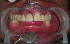
A 19-year-old male patient reported with the chief complaint of impaired esthetics, disturbed speech, and difficulty in chewing due to numerous missing teeth (Fig.1). Patient presented dryness of eyes, lips, thin eyebrows, sparse, thin and lusterless scalp hair, dry skin and repeated episodes of hyperthermia.
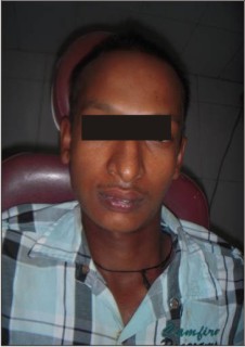 | Fig : 1
 |
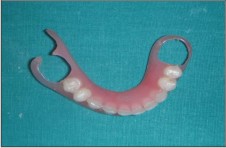
Intraoral examination revealed teeth no.12,14,15,22,24,25 to be clinically missing with 11 showing severe proclination (Fig. 2,3). In the lower arch 36, 46 and 47 were the only teeth present. The alveolar process in mandibular edentulous region was underdeveloped with minimal height and width (Fig. 4).
The patient’s initial psychologic evaluations revealed an introverted personality lacking in self-confidence.
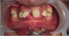 | Fig : 2
|
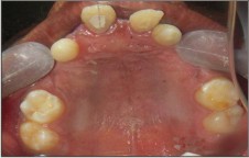 | Fig : 3
|
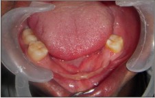 | Fig : 4
|
Several treatment options were presented to the patient which were as follows:
1) Fixed dental prosthesis (FDP) metal-ceramic/all-ceramic, tooth or implant supported [1],[5].
2) Overlay dentures[6] and teeth-supported overdentures[1],[7].
3) Removable dental prosthesis (RDP)[8], rigid and flexible.
After several considerations concerning advantages, disadvantages, prognosis and cost of the options, a combination of tooth supported metal-ceramic conventional FDP in upper arch and flexible RDP in lower arch was planned.
Prosthodontic Management:
After performing elective endodontics on 11,13,21 and 23, preliminary impressions were made with alginate impression material (Zelgan 2002, Dentsply) and poured in dental stone (Kalstone, Kalabhai). Full coverage crown peparations were done on 11,13,16,21,23,26. Final impressions were made using addition silicone putty-wash technique (Aquasil Soft Putty and Aquasil LV, Dentsply) and poured in die stone (Kalrock, Class IV, Kalabhai) and provisional FDP was fabricated and cemented. Try-in of metal framework FDP was done, and was followed by application of feldspathic-porcelain (VITA, VMK 95) and completed FDP 11 through 16 and 21 through 26 was cemented with glass-ionomer cement (Fig.5).
 | Fig : 5
|
Alginate impression of rehabilitated maxillary arch was made and poured.
Mandibular custom tray was prepared and impression was made with Aquasil Ultra Monophase (Dentsply) PVS impression material. On the master casts, acrylic bases with wax rims were made to establish maxillomandibular relations, and then they were mounted on an articulator. Try-in of waxed up mandibular PD was followed by fabrication of mandibular flexible (valplast) removable prosthesis. Final prosthesis (Fig. 6,7) was then adjusted and inserted in the patient’s mouth (Fig.8, 9).
 | Fig : 6
|
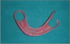 | Fig : 7
|
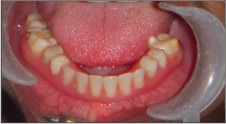 | Fig : 8
|
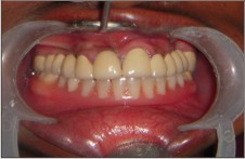 | Fig : 9
|
Prosthesis was placed in hot water for about 1 min prior to insertion in patient’s mouth. It was removed and allowed to cool just to the point when it was tolerated by the patient. This process made the PD as flexible as it would be at mouth temperature. The hot water treatment permitted a very smooth initial insertion and good adaptation with denture-bearing tissues. The clasps could be tightened or loosened by immersing clasp in hot water and bending the clasp inwards or outwards. After the final insertion of flexible PD the patient was instructed on the proper maintenance of the oral tissues and prostheses. The need for optimal oral hygiene and periodic check-up was emphasized to the patient.
Discussion:
An understanding of the ED patient’s psychosocial status is crucial to any prosthodontic treatment effort. The unesthetic appearance that accompanies ED syndrome often has a negative psychological effect on the patient. Poor self-image, peer pressure, and school/job related discrimination have been directly related to psychological scarring experienced by ED patients.
Oral rehabilitation of ED has historically evolved from partial and complete RDP supported by tissue and teeth to screw type osseointegrated dental implants[9].The most frequent prosthetic treatment for the dental management of ED is removable prosthodontics[1]. The need for partial or complete dentures is critical during the pre-school years and continues into adulthood. Since alveolar bone development is dependent on the presence of teeth, ED patient have little or no bone ridge upon which to construct dentures. Hence, the problems involved in attempting to restore function and appearance are greater than usual[10]. Although complete dentures are an acceptable form of treatment, overdentures that are supported by natural teeth will preserve the alveolar bone. Implant-supported restorations can improve physiological and psychosocial function to a greater degree than can complete dentures[1].
In the present case report, considering the clinical situation, maxillary FDP and mandibular flexible partial denture (PD) were determined to be the treatment of choice. Conventional FDP in the maxillary arch and Valplast @258;exible partial denture was planned for mandibular arch due to its favourable properties like esthetics, strength, biocompatibility, and comfort to the patient[11]. The finger like extension of the material into undercuts acts as a clasp and provides very good retention.
Higher body temperature can develop during illnesses, during strenuous physical activity, when the environmental temperature is elevated, or even when the patient is wearing heavy clothing. Treatments of these hyperthermic episodes are part preventive and part reactive in nature.
Conclusion:
This clinical report describes the characteristics and prosthodontic rehabilitation of a patient with ED. A combination of conventional fixed and removable prosthodontic therapy, as presented in this article, can be less complex and more affordable than some other therapies and resulted in a great improvement of the esthetics and function of the masticatory system.
References:
1. Pigno MA, Blackman RB, Cronin RJ, Cavazos E. Prosthodontic management of ectodermal dysplasia: a review of the literature. J Prosthet Dent 1996;76:541-545
2. Pinheiro M, Freire-Maia N. Ectodermal dysplasias: a clinical classi@257;cation and a casual review. Am J Med Genet 1994;53:153-162
3. Graber LW. Congenital absence of teeth: A review with emphasis on inheritance patterns. J Am Dent Assoc 1978;96:266-275.
4. Bartlett RC, Eversole LR, Adkins RS. Autosomal recessive hypohidrotic ectodermal dysplasia: Dental manifestations. Oral Surg Oral Med Oral Pathol 1972;33:736-742.
5. Heuberer S, Dvorak G, Zauza K, Watzek G. The use of onplants and implants in children with severe oligodontia: a retrospective evaluation. Clin Oral Impl Res 2012;23:827-31.
6. Pavarina AC, Machadi AL, Vergani CE, Giampolo ET. Overlay removable partial denture for a patient with ectodermal dysplasia: A clinical report. J Prosthet Dent 2001;86(6):574-7
7. Unger JW, Crabtree DG, Meyer M. Management of soft tissue problems in overdenture treatment of congenital and acquired defects: a case report. Quintessence Int 1990;21(5):353-56
8. Graser GN, Rogoff GS. Removable partial overdentures for special patients. Dent Clin North Am 1990;34:741-58.
9. Nallanchakrava S. Oral rehabilitation of a patient with ectodermal dysplasia with prosthodontics treatment. Indian J Dermat 2013;58(3):241.
10. Bolender CL, Law DB, Austin LB. Prosthodontic treatment of ectodermal dysplasia: A case report. J Prosthet Dent 1964;14:317–325
11. Bhargava A, Nagpal A, Kumar M, Bhargav R. “Flexible dentures demysti@257;ed.” Dental Technician 2010;2:18-21.
|