Introduction
Endodontic therapy enables several advantages including maintenance of a natural tooth, restoration of esthetics and function. But teeth treated by endodontic therapy are often mutilated by caries fracture or previous restoration. Historically, many methods have been attempted in the search for an ideal foundation or build-up design for endodontically treated teeth.
Post and core has been found to be the most appropriate method for reinforcing these teeth. Post is the portion embedded into the prepared root canal. Post can be either prefabricated or custom made. Custom made post can be fabricated either by direct or indirect technique. In indirect technique, impression taken should exactly replicate the parameters such as design, length, surface configuration and diameter. The major problem associated with making the impression of post space is incorporation of voids in the radicular portion and inability to produce accurate impression in all dimensionswhichaffects the cast post fabricated. With the indirect technique, less chair time is used when multiple cast dowel-cores are fabricated for the same patient. In addition, when radicular attachments are incorporated into partial denture designs, the indirect technique is invaluable to accurately align on the cast, the attachment system’s path of insertion with the prosthesis path of insertion.
Regardless of the selected technique, the impression must accurately reproduce the full length of the dowel space, as greater dowel length can contribute to more predictable retention of the core.
Materials And Method
To standardize the procedure standard instruments and equipments were used to make post-space. The extracted maxillary central incisors weresterilized for 48 hours in/ glutaraldehyde solution and stored in normal saline solution. The central incisor was prepared for complete porcelain fused to metal (PFM) crown. The facial and proximal reduction of 1.5 mm with a shoulder finish line and 0.5 mm lingual with a chamfer was given. An incisal reduction of 2 mm was done. A further incisal reduction of 4 mm was done on a flat plane for a core restoration. Access to root canal was made and pulp was extirpated. Biomechanical preparation was done upto K-file no. 80 using Schilder method and obturation was done with gutta percha usinglateral condensation technique.Post space was prepared with a No. 3 Paeso reamer (Dentsply Caulk, Milford, DE)leaving 4 mm of gutta percha at the apex and enlarged to No.5 Paeso reamer.Five such teeth were prepared.
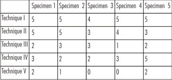
Custom trays (SR Ivoclar Custom Tray Material; Ivoclar, Liechtenstein) were made to carry heavy body impression material to make an impression with 2 mm wax relief (Figure I). Five different techniques were used to obtain the impression of post space by using light body addition polyvinyl silicone impression materialand fiveimpressions were made with each technique (Figure 2). Impression tray adhesive (VPS impression tray adhesive; Kerr Manufacturing Co, Romulus, MI) was applied to the inner surface of the tray 15 minutes before each dowel space impression procedure. Tests were conducted at a room temperature of 23°C and a relative humidity of approximately 70%.
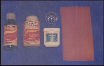 | Figure 1: Materials Used For Custom Tray Fabrication
 |
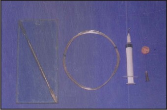 | Figure 2: Equipment Used For Making Impression
|
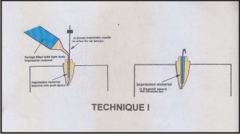
Technique I
In this technique a 24 gauge needle was used as a vent. It was inserted and held in the post-space with the help of tweezer and the light body addition silicone impression material was injected into the post space using disposable tip. Anesthetic needle was then removed and a half inch 24 gauze orthodontic wire bent slightly at tip was inserted into the light body impression material in the post-space. Then the custom tray filled with heavy body addition silicone impression material was used to pick up post impression and to record the prepared tooth.
The basic procedure for the post space impression was kept the same as in technique 1. Following alteration was done as mentioned in respective techniques. (Figure 3).
 | Figure 3: Using Both Needle And Wire.
|
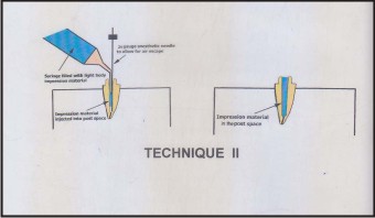
Technique II
In this technique, 24 No. anesthetic needle was used as a vent.But no orthodontic wire was used. (Figure 4).
 | Figure 4: Using Needle Only.
|
Technique III
In this technique, onlyorthodontic wire was inserted to recover the impression. (Figure 5).
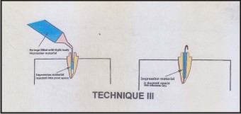 | Figure 5: Using Wire Only
|
Technique IV
In this technique, neither needle nor wire was used. (Figure 6).
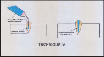 | Figure 6: Without Using Needle And Wire
|
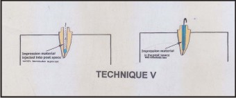
Technique V
In this technique, the light body addition silicone impression material was placed in the post space with the help of lentulo spiral with slow speed contra angle hand piece. Then orthodontic wire was inserted into the impression material. The impression was then made as in technique I and was recovered. (Figure 7).
A total of 125 samples obtained were evaluated for their accuracy using magnifying lenses of 3.5X. The clinical pictures of post space impression with and without voids are shown in Figure 8 and 9 respectively.
 | Figure 7: Using Lentulo Spiral
|
 | Figure 8: Showing Post Space Impression With Voids
|
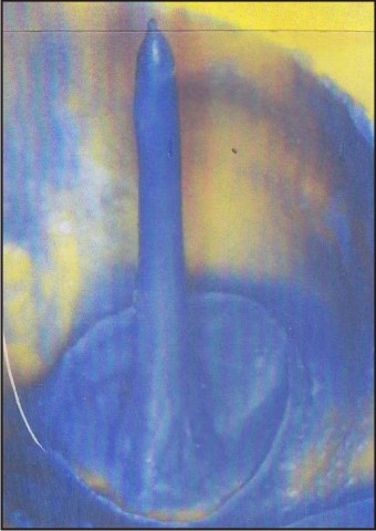 | Figure 9 : Showing Post Space Impression Without Voids
|
Observations And Results
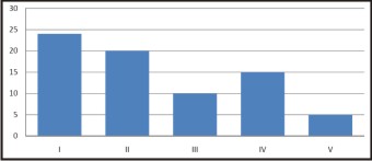
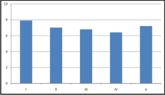
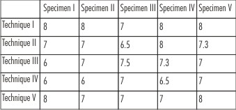
Impression technique directly influenced the proportion of impressions without voids. The maximum number of impressions without voids were in technique I where 24 out of 25 samples were void free. The number of impressions without voids made by each of the techniquewere in decreasing order of technique I > technique II > technique IV >technique III > technique V. (Table I, Figure 10).
 | Figure 10: Impressions Without Voids Using Different Techniques
|
 | Table I : Showing Number Of Samples Without Voids
|
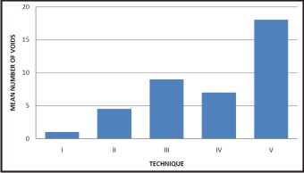
Further themean value of impression without voids using different techniques was found to be highest for technique I i.e. 4.80 and lowest for technique V i.e. 1.00. (Table II, Figure 11).
 | Figure 11: Mean Number Of Voids In Impression Using Different Techniques
|
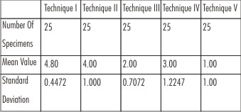 | Table II : Showing Mean (±SD) Of Impressions Without Voids Using Different Techniques
|
It was also found that maximum length covered by impression material was in technique I and minimum length was covered by technique IVas the root canals were prepared till the length of 8mm. (Table III, Figure 12).
 | Figure 12: Mean Number Of Complete Impressions Using Different Techniques
|
 | Table III : Showing completeness of impression in each of the technique (Total length of prepared post-space-8 mm)
|
Discussion
Endodontically treated teeth generally require post core system to develop a tooth preparation necessary to provide retention and adequate resistance form for the success of the definitive crown. Custom cast post-core is the method of choice to restore such teeth. There are two methods by which impression of post space core can be made - direct and indirect techniques. Out of these, indirect method is preferred as it takes lesser chair side time especially for multiple restorations.
In indirect technique, an impression of the prepared post space is made to form a working cast upon which the post-core pattern is then fabricated. Regardless of the technique employed, the impression for the post must record accurately the surface details along the entire length of the post space.While the impression is being made, due to insufficient escapement for air, voids commonly get incorporated in the radicular portion of the post impression leading to inaccurate fit of cast post.
This study was conducted to compare different post space impression techniques using polyvinyl siloxane impression material to determine which technique produced the most accurate impression of the post space.
Venting the dowel space with a needle while injecting impression material into the canal is an easy technique to predictably make void-free dowel space impressions. The use of local anesthetic needle allowed for air escape, leading to complete impressions of the apical portions of the dowel space. Voids on the lateral aspect of the intra radicular part of the impressions were found on the apical third of the dowel space with many impressions using Techniques II through V, and the voids largely resulted from air entrapment while the impression material was expressed into the canal.
When Technique I is used clinically, the local anesthetic needle should be secured with dental floss to prevent accidental swallowing or aspiration by the patient. Although use of vents with dowel space impression has not been previously reported, the use of vents to improve other aspects of fixed prosthodontics therapy has been described. For example, occlusal vents can be used to reduce hydraulic pressure during crown luting, thereby reducing vertical seating discrepancy and improving definitive restoration marginal adaptation.[10],[11] In this situation, the vent is an escape for excess luting agent during final restoration cementation. Likewise, the needle vent during a dowel space impression allows for air escape in the dowel space during impression material injection.
Summary And Conclusions
Based on the observation made, the following salient features can be deciphered:
1. In technique I, impression made using 24 gauge anesthetic needle which acted as a vent at the apex during injecting light body polyvinyl siloxane impression material followed by removal of the needle and placement of orthodontic wire to the depth of preparation resulted in maximum number of void free impressions.
2. In technique V, on the contrast when a lentulo spiral and orthodontic wire was used, the number of voids was maximum.
3. The void free impression with various techniques showed statistically significant difference.
4. In technique I, completeness of impression was maximum where as in technique IV the completeness of impression was minimum.
5. Completeness of impression with various techniques showed statistically significant difference.
6. When compared for the surface irregularity on the impression, the surface irregularity was maximum in apical third and minimum in cervical third in all the technique.
Refrences
1. Angmar-Mansson, Omnell K.A. and Rudd J. Root fractures due to corrosion: Metallurgical aspects. Odont. Revy. 1969; 20: 245.
2. Baraban D.J. The restoration of pulpless teeth. Dent. Clin. North Am. Nov. 1987, pp. 633-653.
3. Bergman, Peter Lundquist, Ulf Sjogren, Goran Sundquist. Restorative and endodontic results after treatment with cast posts and cores. J.Prosthet. Dent. 1989; 61: 10-5.
4. 7. Caputo A.A., Standlee J.P. Pins and posts - why, when and how. Dent Clin. North Am. 1976; 20: 299.
5. Ciesco et al. Comparison of elastomeric impression materials used in fixed prosthodontic. JP.D. 1981; 45: 85-94.
6. Cohen S., Burns R. Pathways of the pulp. 4th ed. St. Louis. CV Mosby Co., 1987; 640-84.
7. Hoag E. Patrick, Dwyer Thomas G. A comparative evaluation of three post and core techniques. J Prosthet. Dent. 1982; 47: 177-181.
8. Johnston J.F., Philips RW. and Dykema R.W. Modern Practice in Crown & Bridge Prosthodontics. 3rd ed.Philadelphia 1971, WB Saunders Co.
9. Lau V.M.S. The reinforcement of endodontically treated teeth. Dent Clin North Am 1976; 20: 313-328.
10. Perel M. and Muroff F.1. Clinical criteria for posts and cores. J Prosthet. Dent. 1972; 28: 405.
11. Phillips RW. Science of Dental Materials, Ninth Edition, WB Saunders Company, 1992, 141-149.
12. Rosensteil S.F., Land M.F., Fujimoto Junhei. Contemporary Fixed Prosthodontics. Third Edition, Mosby 2001, pp. 272-312.
13. Shillingburg HT., Hobo Sumiya, Whitsett L.D., Jacobi Richard, Brackett S.E. Fundamentals of fixed prosthodontics, 3rd Ed. quintessence Publishing Co., USA 1997, pp. 181-210.
14. Tylman S.D. Theory and Practice of Crown and Bridge Prosthodontics. Ed. 5, St. Louis, The CV Mosby Company, 1965, pg. 776-809.
15. Cho G.C., Donovan T.E., Chec WWL Tensile bond strength of polyvinyl siloxane impression bonded to a custom tray as a function of drying time: Part 1. J Prosthet Dent. 1955; 73: 415-23.
16. Ciesco et al. Comparison of elastomeric impression materials used in fixed prosthodontic. J Prosthet Dent. 1981; 45: 85-94.
|