Introduction:
Periodontology has grown exponentially incorporating various advances in technology available today, microsurgery being one of them.[1] Microsurgery refers to a surgical procedure performed under a microscope.[2] In 1979, Daniel defined microsurgery in broad terms as surgery performed under magnification by the microscope.
In 1980, microsurgery was described by Serafin as a methodology - a modification and refinement of existing surgical techniques using magnification to improve visualization, with applications to all specialties.[3] Historically microsurgery in general is not an independent discipline, but a technique that can be applied to different surgical disciplines. It is based on the fact that the human hand, by appropriate training, is capable of performing finer movements than the naked eye is able to control.[4]
As a treatment philosophy, microsurgery incorporates three important principles:-
1. Improvement of motor skills, thereby enhancing surgical ability. This is evident in the smooth hand movements accomplished with increased precision and reduced tremor.
2. An emphasis on passive wound closure with exact primary apposition of the wound edge. The aim is the elimination of the gaps and dead spaces at the wound edge to circumvent new tissue formation needed to fill surgical voids. A painful and inflammatory phase of healing is avoided.
3. The application of microsurgical instrumentation and suturing to reduce tissue trauma.
History:
In 1921, Carl Nylen, who is considered the father of microsurgery, first used a binocular microscope for ear surgery. First surgical operation with microscope was performed in Sweden to correct otosclerotic deafness (Nylen 1924). In 1950’s the first surgical microscope, OPMI[1], with a coaxial lighting system and option for stereoscopic view, was invented and commercialized by the Carl Zeiss company.[5] The micro vessel surgery revolutionized plastic and transplantation surgery was mainly developed by neurosurgeons (Jacobsen & Suarez1960, Donaghy & Yesargil 1967). Apotheker and Jako first introduced the microscope to dentistry in 1978.[3]
Clinical Philosophy:
Consistent application of the philosophy and techniques learned in basic microsurgery education is necessary for the operator to attain a level of experience and competence needed for various periodontal surgical procedures. Training with the microscope enhances the motor skills. The methods of precise, delicate tissue handling, wound closure, and suturing require concentration and practice. Attention must be paid to microanatomy, tissue manipulation, and surgical craftsmanship.[3]
Hand Control
Physiologic Tremor Physiologic tremor is the uncontrolled movement arising from both the intended and unintended actions of our bodies. Awareness of its effect is magnified by visual enhancement. During microsurgery, physiologic tremor manifests as a naturally occurring unwanted hand and finger movement. To minimize tremors, a microsurgeon must have a relaxed state of mind, good body comfort and posture, a well-supported hand, and a stable instrument-holding position. Finger movements controlled by the long flexor and extensor muscles that move our fingers are relatively crude. Thus, active finger extensions, or flexions, are likely to be relatively crude. However, when the wrist is stabilized by resting on a flat surface, angled in a dorsiflection position at approximately 20 degrees, more accurate, finely controlled finger movement can be accomplished because of the reduction in muscle tremor provided by this platform.[3] A microsurgeon’s chair is required to provide proper arm and hand support. The surgeon must be seated upright with the legs extending forward and with both feet flat on the floor so that the calf of each leg forms a right angle to the thigh. Support of the ulnar surface of the forearm and wrist is necessary to control or reduce tremor. The surgeon’s head should be held in a comfortable upright position (Fig 1). Proper ergonomics can help to prevent neck and back injuries resulting from poor chairside habits. During a surgical procedure, patient and chair position must be adjusted to the surgeon and the microscope. There are several factors that can influence a surgeon’s physiologic tremor, including anxiety, recent exercise, alcohol, smoking, caffeine, heavy meals, hypoglycemia, and medication usage.[1],[3] Hand Grips The most commonly used precision grip in microsurgery is the pen grip or internal precision grip, which gives greater stability than any other hand grip. In the three-digit grip, an instrument is held exactly as a pen would be held when writing. The thumb and index and middle fingers are used as a tripod (Fig 2). The forearm should be slightly supine, positioning the knuckles away from you, so that the ulnar border of your hand, wrist, and the elbow are all well supported, allowing the weight of the hand to be on the ulnar border. The middle finger should rest firmly and directly on either the working surface supporting the hand or indirectly on the ring finger. With the tripod formed by the fingers in the pen grip, the middle finger holds the instrument. The thumb and index finger are arranged on the instrument into contact with the underlying middle finger. When an instrument is held with the internal precision grip, the instrument can be opened and closed with very fine control. Any tremor resulting from the thumb or index finger is minimized by the contact with the supported, steady middle finger. Using the pen grip, the flexor and extensor muscles of the hand are relaxed, resisting fatigue, while the intrinsic muscles that rotate the hand are well postured, resulting in the most accurate motion of which the hand is capable of. The concept of microsurgery is based on three important elements which form the microsurgical triad that includes magnification, illumination and instruments. Without any one of these, microsurgery is not possible.[4],[6] Magnification Visual acuity is the ability to perceive two closely lying objects separately. It is influenced by various anatomic and physiologic factors, like density of cells packed on the retina, the electrophysiologic process of the image on the retina and illumination of the area. Visualization of fine details can be enhanced by increasing the image size of the object as well. Two obvious methods of increasing image size are either by getting closer to the objects or by magnification.[1]
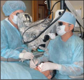 | Correct Posture And Arms Supported While Operating Microscope
 |
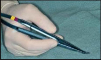 | Internal Precision Grip
|
Magnification systems:
A variety of simple and complex magnifications are available to dentists, ranging from simple loupes to prism telescopic loupes and surgical microscopes. Each magnification system has its specific advantages and limitations. Although magnification improves the accuracy of clinical and diagnostic skills, it requires an understanding of optical principles that govern all magnification systems. The assumption that “more magnification is better ‘’must always be weighed against the decrease in field of view and depth of focus that can occur as magnification increases which is a problem the most common with dental loupes than with operating microscopes. Dental loupes are the most common system of optical magnification used in Periodontics. Loupes are fundamentally dual monocular telescopes with side-by-side lenses convergent to focus on the operative field. The magnified image formed has stereoscopic properties by virtue of their convergence. A convergent lens optical system is called a Keplerian optical system.[4] Although dental loupes are widely used, they have disadvantages compared with the microscope. The clinician’s eyes must converge to view the operative field. This can result in eyestrain, fatigue, and even pathologic vision changes, especially after prolonged use. Three types of Keplerian loupes are typically used in Periodontics: simple or single-element loupes, compound loupes, and prism telescopic loupes.
Simple Loupes:
It consists of a pair of single meniscus lenses. Simple loupes are primitive magnifiers with limited capabilities. Each lens is limited to only two refracting surfaces. Their magnification can only increase by increasing lens diameter and thickness. Size and weight constraints make simple loupes impractical for magnification beyond 1.5X. Another disadvantage of simple loupes is that they are greatly affected by spherical and chromatic aberration. This distorts the image shape and color of objects being viewed.(Fig 3).
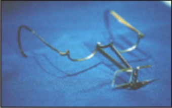 | Simple Loupes
|
Compound Loupes:
It consists of multiple lenses with intervening air spaces to gain additional refracting surfaces. This allows increased magnification with more favorable working distance and depth of field. Magnification of compound loupes can be increased by lengthening the distance between lenses, without excessive increase in size or weight. In addition to offering improved optical performance, compound lenses can be achromatic. This is an optical feature that clinicians should always choose when selecting magnifying loupes. Compound lenses can be Achromatic that consist of two glass lenses, joined together with clear resin. The specific density of each lens counteracts the chromatic aberration of its paired lens to produce a color-correct image. However multi element compound loupes become optically inefficient at magnifications above 3X.[5] (Fig 4).
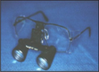 | Compound Loupes
|
Prism Telescopic Loupes:
The most optically advanced loupe optical magnification currently available is the prism telescopic loupe. Such loupes employ Schmidt or “rooftop” prisms to lengthen the light path through a series of switch back mirrors between the lenses. This arrangement folds the light so that the barrel of the loupes can be shortened. Prism loupes produce better magnification, wider depths of field, longer working distances, and larger fields of view than other types of loupes (Fig 5). The increased weight of prism telescope loupes with magnification above 4X makes headband mounting more comfortable and stable than eye glass frame mounting. Recent innovations in prism telescopic loupes include coaxial fiberoptic lighting incorporated in the lens elements to improve illumination.[3] HDTV Single camera 3D system This system involves a three dimensional High Definition Television attached to a stereoscopic microscope which enables 3D visualization and documentation. Viewing of the same stereoscopic vision by assistants and students proves to be advantageous to both clinicians and academicians. It also allows easy printing, organization of data base and adaption to tele-operations.1 Illumination Most of the manufacturers offer collateral lighting systems or suitable fixing options which are helpful, particularly for higher magnification in the range of 4× and more. Certain essential considerations need to be made in the selection of an accessory lighting source, these include total weight, quality, and the brightness of the light, ease of focusing and directing the light within the field of view of the magnifiers and ease of transport between surgeries.[1]
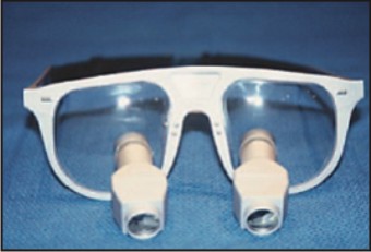 | Prismatic Loupes
|
Microsurgical instruments With addition to the use of magnification and reliance on atraumatic technique, microsurgery requires specially constructed instruments designed specifically to minimize trauma. An important characteristic of microsurgical instruments is their ability to create clean incisions that prepare wounds for healing by primary intention. Microsurgical incisions are established at a 90- degree angle to the surface using ophthalmic microsurgical scalpels.[1],[6] Microscopy permits easy identification of ragged wound edges for trimming and freshening. For primary wound closure, micro sutures in the range of 6-0 to 9-0 are needed to approximate the wound edges accurately (Fig 6). Microsurgical wound apposition minimizes gaps or voids at the wound edges. This encourages rapid healing with less postoperative pain.
 | Types Of Sutures
|
Basic Principles Of The Surgical Microscope
A microscope is nothing more than a monocular or binocular with a close-up lens. A binocular is simply mounted side-by-side for stereoscopic vision. In the binocular concept, the length of the telescope becomes condensed by the use of prisms. The components of microscope are the basic stereo microscope, the binocular head, and the objective lens. This microscope, however, contains two additional elements: a magnification changer and an illuminator which beams the light in through the objective lens. This type of illumination is desirable because the line of illumination is very close to the viewer’s line of vision. Therefore, the surgical field will be illuminated and free of shadows. Shadows would result if the line of illumination was at a large angle of incidence from the viewing axis.[4]
Advantages And Disadvantages Of The Surgical Microscope[4]
Less tissue trauma Less mobility Less patient anxiety Atraumatic tissue management. Accurate primary wound closure. Increased diagnostic skills. Minimally invasive. Improved cosmetic results. Increased surgical quality. Increased effectiveness of root debridementresults in greater predictability of a) Regeneration procedures, b) Cosmetic procedures. Improved documentation e.g. video, slide, digital.
Disadvantages
1. Educational requirements
A) Surgical technique
B) Understanding of optics
2. Long adjustment period for clinical proficiency.
3. Initial increased surgical time
4. igh patient cost
5. Limited surgical access.
MICROSURGERY IN PERIODONTICS
Periodontal flaps conventionally aid in access to root surface to perform both regenerative and respective surgical treatment. The introduction of surgical microscopes has led to considerably less invasive surgical incisions and flap reflections in periodontics.[1] Such procedures utilize microsurgical instruments to make incisions that are just long enough to afford access for operating space and exposure. Incisions should be clean and precise, with as little trauma as possible. Appropriately designed and sized microsurgical instruments produce minimal tissue trauma and promote healing. Tissue should be handled very gently and as little as possible. Retraction should be done carefully to avoid excess pressure, since tissue tension can alter the local physiologic state of the wound. Achieving a bloodless field should be a goal to avoid compromising the surgeon’s field of view.[3] Periodontal Plastic Microsurgery The current terminology, periodontal plastic surgery, is defined as surgical procedures performed to correct or eliminate anatomic, developmental, or traumatic deformities of the gingiva or alveolar mucosa. Periodontal plastic surgery is an integral aspect of periodontal education and practice. Knowledge of medical microsurgery offers a view as to what esthetic needs can be realistically achieved while treating periodontal problems. Improvement in esthetics is a major indication for periodontal plastic surgery.[3] Esthetic Surgical Procedures[3] When attempting to restore gingival esthetics, several periodontal plastic surgery procedures are helpful, including pedicle soft tissue grafts and free soft tissue grafts. The direction of transfer of the pedicle graft determines whether it is divided into rotational flaps (eg, laterally sliding flap, papilla flap, or double papilla flap) or advanced flaps without rotation or lateral movement (eg, coronally positioned flap). The pedicle soft tissue graft combined with the use of a membrane barrier, according to the principles of guided tissue regeneration, is also used as a treatment for root coverage. When using a guided tissue regeneration barrier, it is critical to maintain a space between the barrier membrane and the root surface for tissue regeneration. To correct small areas of recession without invasive major flaps, careful dissection and suturing can sometimes be used to place a graft. Microsurgically transferring donor tissue removed from one area of the mouth to a new microsurgically prepared recipient site allows for correction of gingival esthetic problems. Survival of the grafted tissue, whether the procedure is done macroscopically or microsurgically, is dependent on the recipient site having a blood supply to restore circulation to the transferred tissue. Periodontal microsurgery has proven to be an effective means of improving the predictability of gingival transplantation procedures used in treating recession with less operative trauma and discomfort. Accurate diagnosis with microsurgical techniques makes complete root coverage extremely predictable in Class I and Class II marginal tissue recessions. Partial root coverage results achieved in Class III & Class IV marginal recession with conventional surgery can also be greatly enhanced through the use of microsurgery.[1] Apart from the various root coverage procedures performed using the microsurgical armamentarium other mucogingival surgeries like papilla reconstructions and ridge augmentation around natural teeth and implants can also be carried out. Microsurgery has proved its potential in implant surgeries. It has established itself in implant site development and implant placement using both the flap and flapless techniques.
Sinus lift procedures using the microsurgical approach are also gaining recognition. The periodontal endoscope allows for subgingival visualization of the root surface at magnifications of 24x to 48x. This is accomplished through a 0.99 mm fiber optic bundle that is a combination of a 10,000-pixel capture bundle surrounded by multiple illumination fibers. This fiber is delivered to the gingival margin coupled into an instrument called an “explorer.” A single-use sterile sheath isolates the fiber so it can be used repeatedly (average use for the author has been 70 to 80 uses per fiber). The captured image is relayed to a screen so that the user can see “real time” video of the highly magnified environment (approximately 3 mm on screen at a time).[1]
Root Surface Conditioning Since root surface preparation addresses how the soft tissue attaches to the root of the tooth in root coverage surgery, it is of the utmost importance. In an attempt to get new periodontal ligament attachment of a graft to the tooth with new cementum and Sharpey’s fibers, several methods of root preparation have been suggested including mechanical root preparation, chemical root preparation, and biologic root preparation. The outcomes of some methods are based on histologic evidence and others on empirical observation, but all are important for successful root coverage.
Conclusions:
Periodontal microsurgery is in its infancy but will play a role in the future. The small scale of microsurgery presents special challenges in dexterity and perception. Its execution is technique sensitive and more demanding than are conventional periodontal procedures. As the benefits of the microscope are realized, it will be applied more universally. There are many indications in which periodontal microsurgery can be beneficial. It appears to be a natural evolution for the specialty of periodontics. Microsurgery offers new possibilities to improve periodontal care in a variety of ways. Its benefits include improved cosmetics, rapid healing, minimal discomfort, and enhanced patient acceptance. Periodontics of the future will see increasing use of magnification in all areas of practice, including implantology.
References:
1. Akbari G., Prabhuji MLV., Lavanya R.,(2012) Microsurgery: a clinical philosophy for surgical craftsmanship. e- journal of dentistry, 2(3), 233-236
2. Shanelec D,(2003) Periodontal microsurgery. J Esthet Restor Dent, 15,118-123.
3. Shanelec D , Tibbetts L., (2009) Principles and practice of periodontal microsurgery. Int J Microdent, 1, 13-24.
4. Satyanaraynan D., Vikram Reddy G., Raja Babu P. (2011) Periodontal microsurgery: a changing perspective. Indian J Dent Adv,3(4), 698-704.
5. Burkhardt R, Use of dental microscope in plastic periodontal therapy- evolution or revolution? Microscopes in dentistry.periodontology 22-25.
6. Belcher , (2001) A Perspective on Periodontal Microsurgery. Int J Periodontics Restorative Dent, 21, 191-196.
|