Introduction
Ectodermal dysplasia (ED) is defined by National foundation for ectodermal dysplasia as a genetic disorder in which there are congenital birth defects of two or more ectodermal structures These tissues primarily are the skin, hair, nails, eccrine glands, and teeth.[1] Charles Darwin first documented the earliest accessible account of ectodermal dysplasia in English as early as 1840s.To date more than 150 distinctive phenotypes have been described with all possible modes of inheritance. The most common group are hypohidrotic (anhidrotic) ED also known as Christ- Seimens –Touraine syndrome or hidrotic ED (Clouston syndrome).[2] Thurnam published the first report of a patient with ectodermal dysplasia in 1848,the term ectodermal dysplasia was not coined until 1929 by Weech. In hypohydrotic (anhidrotic) ectodermal dysplasia clinically there is anhidrosis, hypodontia and hypotrichosis .Other features include frontalbossing, thick lips, broad depressed nasal bridge of nose and deformed ears. Inheritance is usually X linked recessive trait. Ectodermal dysplasia results in aberrant development of ectodermal derivatives in early embryonic life. Hidrotic include the same without dental anomalies.[3] Dental manifestations include conical teeth , hypodontia or complete anodontia , and delayed eruption of permanent teeth.ED is caused by mutations in any of the three EDA pathway genes : ectodysplasin (EDA) ectodysplasin receptor (EDAR) , and EDAR associated death domain (EDARADD) which encode a ligand , a receptor , and an intracellular signal mediator of a single linear pathway , respectively.[2] Morbidity and mortality are related to absence or presence of eccrine and mucous glands In affected patients, the extensive lack of both deciduous and permanent teeth results in an atrophy and a reduced growth rate of the affected alveolar processes.[4]
Case Report
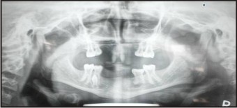
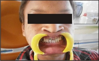
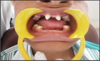
A 14 year old male patient reported to the department of Oral Pathology and Microbiology in Bhojia Dental College And Hospital, Baddi with the chief complaint of difficulty in mastication due to congenital missing teeth , lack of aesthetics , sparse hair on scalp and absence of sweating .Extra oral examination revealed thin eyebrow, fine linear wrinkles and sparse hair and thick everted lower lip (Fig 1). Past medical and treatment history was insignificant and no positive family history was found. He was the first of the two children in his family and was the only affected member of his family .Nails appeared normal (Fig 2). Intraorally the patient was partially edentulous .Tooth number 51 , 61 appeared conical and tooth number 52, 53,62,63,71,72,73,81,82,83 were absent (Fig 3). Upper and lower alveolar ridges appeared normal .His OPG was relevant to the above said findings (Fig 4). The combined dental and clinical findings pointed towards hypohidrotic ectodermal dysplasia. The treatment plan preferred was removable partial denture for both upper and lower arches and the upper midline diastema closure using elastic traction and restoration with composite resin (Fig 5).
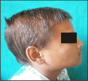 | Fig 1 - Sparse hair on scalp and thick everted lips
 |
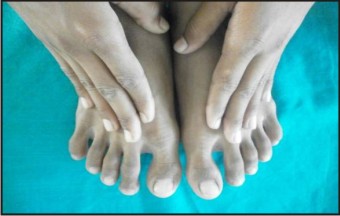 | Fig 2 – Nails appear normal.
|
 | Fig 3 – Tooth number 51, 61 appears conical and tooth number 52 , 53,62, 63 ,71 ,72,73,81,82,83 are absent.
|
 | Fig 4 – Partially edentulous on intra oral examination.
|
 | Fig 5 – Removable partial denture for both upper and lower arches and upper midline diastema closure using elastic traction.
|
Discussion
The ectodermal dysplasias comprise a large, heterogenous group of inherited disorders that are defined by primary defects in the development of two or more tissues derived from the embryonic ectoderm. The condition is thought to occur in approximately one in every 100,000 births. Genetic studies of more than 300 cases have revealed X linked mode of inheritance with its gene locus being Xq11-21.1, the gene is carried by the female but manifested in the male.[1] Current classification of ectodermal dysplasias is based on clinical features. Pure ectodermal dysplasias are manifested by defects in ectodermal structures alone , while ectodermal dysplasia syndromes are defined by the combination of ectodermal defects in association with other anomalies. Freire-Maia and Pinheiro proposed the first classification system of the ectodermal dysplasias in 1982,[5] with additional updates in 1994 and 2001.[6] The patients were stratified into subgroups based on presence or absence of the following :
1. Trichondysplasia (abnormal hair)
2. Abnormal dentition
3. Onchondysplasia (abnormal nails)
4. Dyshidrosis (abnormal or missing sweat glands)
Overall, the ectodermal dysplasias were classified into either group A disorders, which were manifested by defects in at least two of the four classic ectodermal structures as defined above ,with or without other defects, and group B disorders, which were manifested by a defect in one classic ectodermal structure (1-4 from above in combination with (5), a defect in one other ectodermal structure (i.e., ears, lips, dermatoglyphics). HED or anhidrotic ectodermal dysplasia is the most common syndrome among this large group of hereditary disorders. Hypohidrosis, hypotrichosis and hypodontia constitute the main symptoms of the syndrome.[7] In the hypohidrotic form, the skin is soft, thin, and dry. The sebaceous glands are also defective or absent .Palms and soles are hyperkeratotic, pseudorhagades are present around the eyes. In the oral cavity, the most striking feature is oligodontia. The teeth that are present have abnormal crown form. Teeth in the anterior region of maxilla and mandible are conical in shape.[8] In the above mentioned case this feature was prominent .Oligodontia with only presence of tooth number 51 and 61 was seen. There was a wide midline diastema. The characteristic facial features like frontal bossing, depressed nasal bridge, prominent supraorbital ridges, prominent and obliquely set ears were also present in this case. Also the midface is depressed, the lower third of the face appears small due to lack of alveolar bone development, and lips are protuberant.[9] The molecular pathogenesis of hypohidrotic ectodermaldysplasia (HED) is poorly understood. The gene responsible for X-linked HED, EDA, produces ectodysplasin-A, a protein that is important for normal development of ectodermal appendages including hair, teeth, and sweat glands. Evidence is accumulating that ectodysplasin-A is important in several pathways that involve ectodermal-mesodermal interactions during embryogenesis. Defects in the molecular structure of ectodysplasin-A may inhibit the action of enzymes necessary for normal development of the ectoderm and/or its interaction with the underlying mesoderm.[10] The dental literature describes the dental treatment and management for the patient with anodontia or severe oligodontia. There is not a definitive time to begin treatment, but Pigno . et al.[11] suggest that an initial prosthesis should be delivered before school age of the patient. As the child grows, the denture will have to be modified and replaced, because it is indicated in the longitudinal studies of anodontia that the growth of jaws is independent of existence of teeth.[12] When the patient is in the last stage, the removable prosthesis may be replaced by a fixed type restoration using osseointegrated implants. The implant-supported dentures reported are excellent in stability and retention but the placement of dental implants in young children generates several problems.[13]
Conclusion
Young patients with ED need to be evaluated by a dental professional to determine the oral ramifications of the condition. When indicated ,appropriate care needs to be rendered throughout the child’s growth cycle to maintain oral functions as well as to address the aesthetic needs of the patient.
References
1. Ranjan V. Ectodermal Dysplasia – A Case Report & Review of Literature. Journal of dentofacial sciences, 2013; 2 : 27-30
2. Kumar K.,Shetty D.,Dua M, Dua A,Dhanapal R. –An insight into the genesis of hypohidrotic ectodermal dysplasia I -A case report. Case reports in dentistry vol 2012 article ID 281074
3. J Jananee, M Satishkumar , Balaji S- Ectodermal dysplasia – a case report. Indian journal of multidisciplinary dentistry, 2012 ; 2 :465 – 7
4. Kohachiro Ohno, Ikuo Ohmori -Anodontia with hypohidrotic ectodermal dysplasia in a young female: a case report,Pediatric Dentistry 2000; 22:1
5. Pinhero M, Freire –Maia N.The ectodermal dysplasias. Arch . dermatol.1982 ;118 :125 – 6
6. Pinhero M, Freire – Maia N.The ectodermal dysplasias: a clinical classification and a casual review. Am j med genet 1994 ;53:153 – 62 (Freire – maia N, Lisboa – Costa T,Pagnan NA. Ectodermal dysplasias: how many?.Am j med genet . 2001;104 :84
7. G. Varghese and P. Sathyan, “Hypohidrotic ectodermal dysplasia—a case study,” Oral & Maxillofacial Pathology Journal,2011; 2 :124 - 6
8. P. J. M. Crawford, M. J. Aldred, and A. Clarke, “Clinical and radiographic dental findings in X linked hypohidrotic ectodermal dysplasia,” Journal of Medical Genetics, 1991;28:181–5 ( S. N. Ramraje, M. Wasnik, and Y. A. Momin, “Anhidrotic ectodermal dysplasia—a report of two cases,” BombayHospital Journal, vol. 5, article 12, 2009)
9. R. M. Shaw, “Prosthetic management of hypohydrotic ectodermal dysplasia with anodontia-Case report,” Australian Dental Journal. 1990 ; 35:113–6
10. T. Wright, D. K. Grange, and M. K. Richter, Hypohidrotic ectodermal Dysplasia, University of Washington, Seattle,Wash, USA, 1993.)
11. Pigno MA, Blackman RB, Cronin RJ, Cavazos E:Prosthodontic management of ectodermal dysplasia: a review of the literature. J Prosthet Dent. 1996; 76:541-5
12. Nomura S, Hasegawa S, Noda T, Ishioka K: Longitudinal study of jaw growth and prosthetic management in a patient with ectodermal dysplasia and anodontia. Int J Paediat Dent. 1993; 3:29-38
13. Guckes AD, Brahim JS, McCarthy GR, Rudy SF, CooperLF: Using endosseous dental implants for patients with ectodermal dysplasia. J Am Dent Assoc. 1991;122:59-62
|