|
|
|
| A Two Way Periodontal Approach For Preventing Relapse In Midline Diastema Cases |
Astha Agarwal 1 , Shankar T.Gokhale 2 , R.G.Shivamanjunath 3 , Hirak. S. Bhattacharya 4
1 Post Graduate Student, Department Of Periodontology - Institute Of Dental Sciences ,Bareilly
2 Professor , Department Of Periodontology - Institute Of Dental Sciences ,Bareilly
3 Head Of Department And Professor , Department Of Periodontology - Institute Of Dental Sciences ,Bareilly
4 Professor , Department Of Periodontology - Institute Of Dental Sciences ,Bareilly
|
| Address For Correspondence |
Dr. Astha Agarwal W/o Mr.Ashirwad agarwal
35-B/1/8, Rampur Garden
Bareilly 243006 (Uttar Pradesh)
Mobile no. 7895344444 |
| Abstract |
| Many orthodontic cases require periodontal intervention for successful treatment outcome.In the present case Frenectomy was performed for correction of maxillary midline diastema during the course of orthodontic treatment ,along with two techniques of circumferential supracrestal fibrotomy to prevent the relapse after completion of orthodontic procedure. Thus facilitating permanent closure of midline diastema along with desired esthetics. |
|
| Keywords |
| Circumferential supracrestal fibrotomy, frenectomy, midline diastema, papilla dividing procedure |
|
| Full Text |
Introduction
The increasing aesthetic concern ,with the purpose of achieving perfect smile has led to great interest in seeking dental treatment. The most common esthetic problem encountered is the spacing between the anterior teeth. This spacing can be due to many reasons like discrepancies between tooth size and dental arch length, abnormal large frenum/aberrant frenum, ectopic tooth eruption or congenitally missing tooth.[1] Thus the treatment modality for correction of midline diastema mainly depends upon the removal of the etiology behind the spacing.
Aberrant frenum can be treated by frenectomy or frenotomy procedures. The terms frenectomy and frenotomy signify operations that differ in degree of surgical approach. Frenectomy is a complete removal of the frenum, including its attachment to the underlying bone and may be required for the correction of midline diastema between central incisors.Whereas Frenotomy is the incision and relocation of the frenal attachment.[2]
To alleviate relapse after the complete closure of the maxillary midline diastema because of elastic supracrestal gingival fibres, circumferential supracrestal fibrotomy(CSF)[3] or papilla –dividing procedures[4] can be performed.
In this case report, frenectomy has been performed to remove aberrant frenum attachment and both CSF and or papilla –dividing procedure and have been performed to prevent the relapse after closure of midline diastema.
Case Report
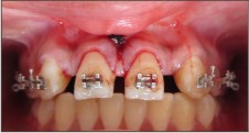
A 21 year old female patient was referred to the Department of Periodontics, Institute of Dental Sciences, Bareilly for the correction of aberrant frenum attachment. The patient was undergoing orthodontic treatment for correction of spacing between the maxillary anterior teeth. Clinical examination revealed a thick maxillary labial frenum as well as congenitally bilaterally missing maxillary lateral incisors, the main etiology for the spacing. The orthodontic treatment plan included the closure of midline diastema and to create appropriate space for the replacement of missing lateral incisors by fixed prosthesis (Fig.1). Patient was found to be systemically healthy. The abnormal frenum was detected by tension/blanch test. A written informed consent was undertaken from the patient to perform frenectomy, CSF as well as papilla dividing procedure in a single visit before the application of orthodontic forces.
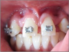 | Fig.1. Pre Operative Facial View Of Midline Diastema.
 |
The area was anaesthetized with a local infiltration by using 2% lignocaine with 1:80000 adrenaline. For frenectomy the classic[5] technique of frenum removal was applied .The frenum was engaged with a haemostat which was inserted into the depth of the vestibule and incisions were placed on the upper and the undersurface of the haemostat until the haemostat was free(Fig.2).The triangular resected portion of the frenum with the haemostat was removed(Fig.3). A blunt dissection was done on the bone to relieve the fibrous attachment and also scoring of the periosteum was done. The edges of the diamond shaped wound were approximated and then interrupted sutures were placed by using 3-0 black silk sutures(Fig.4).
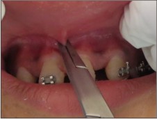 | Fig.2. Frenum With Hemostat In Place.
|
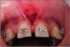 | Fig.3. Frenum Excised.
|
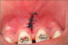 | Fig.4. Sutures Placed.
|
Then after suturing, CSF was performed by inserting the sharp 15 No. Bard Parker blade into the gingival sulcus down to the crest of alveolar bone (Fig.5). Cuts were made both interproximally, labially and palatally for both maxillary central incisors. An alternative method that is papilla dividing procedure was also performed in the space present between the central incisor and the canine that comprised of placement of incision in the centre of each gingival papilla,sparing the margin but separating the papilla from just below the margin to 1-2 mm below the height of the bone bucally and lingually(Fig.6).
 | Fig.5. Scalpel In Position Performing Conventional Csf.
|
 | Fig.6. Papilla Preserving Procedure Performed In The Interdental Area.
|
The area was covered by periodontal dressing. Antibiotics(Amoxicillin 500 mg,TDS for 5 days) and analgesic( Keterol Dt BD for 5 days) were prescribed after the surgical procedure and the post operative instructions were given to the patient. The dressing and the sutures were removed 15 days post operatively. Healing was found to be satisfactory and oral hygiene instructions were reinforced on every recall visit (Fig.7).
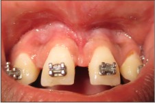 | Fig.7. Post Operative View After 15 Days.
|
Discussion
The desirable treatment outcome of diastema requires correct diagnosis and intervention based on the relevance of its specific etiology. The most common cause of midline diastema is abnormal frenal attachment that may require removal either before orthodontic tooth movement or after active orthodontic treatment. Since the chances of recurrence of midline diastema after the treatment are more, hence the permanent retention is required in most of the cases.
In this patient, following frenectomy procedure, the anticipated relapse would require a permanent retention by means of a fixed retainer or in combination with a short surgical procedure as reported in various studies. Inability to provide retainers due to congenitally bilaterally missing lateral incisors lead to a greater tendency for the teeth to return towards their initial position. Although the etiology of relapse is not fully understood, but relates to a number of factors, including periodontal and occlusal factors, soft tissue pressure and growth.[6] Specific periodontal surgical procedures are required to stabilize the results and enhance the aesthetics.
A major cause of rebound after orthodontic treatment is the network of elastic supracrestal gingival fibres. As teeth are moved to a new position, these fibres tend to stretch, and they remodel very slowly. If the pull of these elastic fibres could be eliminated , a major cause of relapse of previously treated irregular and rotated teeth can be prevented.[7] Thus CSF was performed to section the supracrestal gingival fibers.[3]
An alternative method that is “papilla split” or “papilla dividing procedure”is done in order to reduce the possibility that the height of the gingival attachment will be reduced after the surgery and it is particularly indicated for esthetically sensitive areas.[4]
Both the procedures are indicated until malaligned teeth have been corrected and thus performed towards the end of the finishing phase of the treatment. But in the present case spacing was present between the maxillary anteriors due to the missing lateral incisors and also the space has to be maintained for lateral incisors prosthesis by closing the midline diastema, thus both the procedures were performed along with the frenectomy at the same visit.
Conclusion
In the present case frenectomy was performed to facilitate for the closure of maxillary midline diastema. As well as Circumferential Supracrestal Fiberotomy procedure and papilla split procedure were done to prevent relapse after the completion of orthodontic treatment at the time of frenectomy only. The highlight of the present case was the excellent uneventful tissue healing inspite of combining all the 3 procedures in the same visit. So, subsequent surgical trauma can be avoided by combining above mentioned procedures in the same visit. However further case series with long term follow up is warranted.
References
1. Huang WJ, Creath CJ. The midline diastema: A review of its etiology and treatment. Pediatric Dent 1995;17:171-79.
2. Dibart S, Karima M. Labial frenectomy alone or in combination with a free gingival autograft. In: Serge Dibart, Mamdouth Karima (eds) Practical Periodontal Plastic Surgery. Germany: Blackwell Munksgaard: p53
3. Edwards JG.A long-term prospective evaluation of the circumferential supracrestal-fiberotomy in alleviating orthodontic relapse. Am J Orthod Dentofac Orthop 1988; 93:380-87
4. Edwards JG. Soft-tissue surgery to alleviate orthodontic relapse. Dent Clin North Am 1993;37:205-25.
5. Jhaveri H. The Aberrant Frenum. In: Dr. Hiral Jhaveri (ed), Dr. PD Miller the father of periodontal plastic surgery, 2006;29-34.
6. Melrose C, Millett DT. Toward a perspective on orthodontic retention? Am J Orthod Dentofacial Orthop 1998;13:507–14.
7. Campbell PM, Moore JW, Matthews JL. Orthodontically corrected midline diastemas. A histologic study and surgical procedure. Am J Orthodont 1975;67:139-58.
|
|
|
|
|
|
|