Introduction
Supernumerary teeth are those that are additional to the normal compliment. Supernumerary teeth, also called hyperdontia, may occur unilaterally or bilaterally, single or multiple and in one or both jaws. They may occur in any region of the dental arch with predilection for premaxilla [1],[2]. According to Bodin et.al[3] the commonest site for hyperdontia is premaxilla with prevalence of 1.57%. The etiology of hyperdontia is unclear.
Several theories have been suggested regarding their etiology , hyperactivity of dental lamina is being most supported by literature[1],[4],[5]. Among other theories are:
Atavism, a reversion to a more primitive type of dentition [6].
Dichotomy theory by Taylor[7], which states that tooth bud splits in to two equal or differently size parts, resulting in two teeth of equal size or one normal and one abnormal tooth.
Heredity: Many authors have suggested inheritance as a key factor in the development of supernumerary teeth, as these are more common in the family of the affected children than in general population [8],[9]. While there may be a genetic influence, this does not appear to follow a simple Mendelian pattern [10].
Prevalence
Supernumerary teeth have been reported in both the primary and the permanent dentitions. The reported prevalence of supernumerary teeth in the general Caucasian population for the permanent dentition ranges from 0.1 to 3.8% [1],[11]. The prevalence of supernumerary teeth is lower in the primary dentition and is said to be 0.3–0.8%[2],[7]. Hyperdontia in the primary dentition is often overlooked because supernumerary teeth are often of normal shape, erupt normally and appear to be in proper alignment; and can be mistaken for gemination or fusion anomalies[12]. Although cases of multiple supernumerary teeth have been reported [13],[14], they are rare, as are multiple supernumerary teeth in individuals with no other associated diseases or syndromes[15],[16]. The conditions commonly associated with an increased prevalence of supernumerary teeth include Cleft lip and palate, Cleidocranial dysostosis, Gardner’s syndrome, Fabry Anderson’s syndrome, Chondroectodermal dysplasia and Ehlers– Danlos syndrome.
Classification: Supernumerary teeth may be classified according to morphology and location[17],[18]. In the primary dentition, the morphology is usually normal or conical. The morphology of supernumerary teeth presenting in the permanent dentition is more variable, with the following four morphological types being described:
Conical: This small peg-shaped conical tooth is the most common supernumerary found in the permanent dentition. It develops with root formation ahead of, or at an equivalent stage to, that of permanent incisors and usually presents as a mesiodens between the maxillarycentral incisors, but rarely erupts labially.
Tuberculate: This type of supernumerary, that is larger in size than the conical tooth, possesses more than one cusp or tubercle.
Supplemental: The supplemental supernumerary refers to duplication of teeth in the normal series and is found at the end of a tooth series.
Odontoma: Despite not being universally accepted, most authors agree that odontoma represent a hamartomatous malformation. Bilateral supplemental maxillary lateral incisors have previously been described in the literature[19],[20]. but are regarded as an unusual finding. A case of non syndromic, bilateral supplemental type of maxillary lateral incisors is presented.
Case Report
An 8 year old boy reported to the department of Pedodontics, Himachal Dental College, Sundernagar with the chief complaint of irregularly placed upper front teeth. Patient had Angles class I molar relationship with marked upper arch crowding. On intraoral examination, he presented with mixed dentition in maxillary arch (11,12,12S,13,14,55,16,21,22,22S,63,24,25,26) along with presence of supernumerary supplemental bilateral lateral incisors having morphology similar to that of permanent maxillary lateral incisors ( Fig 1, 2 & 3). The lateral incisors were similar in size with marked anterior crowding. Supplemental right maxillary lateral incisor was in the normal alignment in the arch wheras supplemental left maxillary lateral incisor was rotated distobuccally and place buccally to normal alignment. Permanent maxillary right canine (13) was seen erupting buccally . There was no significant past medical history nor were there clinical signs of any recognized syndrome associated with multiple supernumeraries. The dentition present in the mandibular arch was mixed dentition (31, 32, 73, 75, 36, 41, 42, 44, 85, 46). An OPG radiograph was taken which had also revealed presence of supplement lateral incisor with incomplete root configuration (Fig 4). The crown and root morphology of both right and left lateral incisors and supplemental teeth were identical.
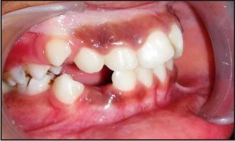 | Fig 1 : Intraoral Photographs Showing Supplemental Lateral Incisors
 |
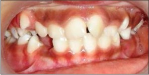 | Fig 2 : Intraoral Photographs Showing Supplemental Lateral Incisors
|
 | Fig 3 : Intraoral Photographs Showing Supplemental Lateral Incisors
|
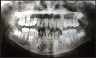 | Fig 4 : Opg Showing Supplemental Lateral Incisors
|
Management: Treatment depends on the type and position of supernumerary tooth and on its effect on adjacent teeth. Management of supernumerary tooth should be part of comprehensive treatment plan and should not be considered in isolation.
Usually it is difficult to distinguish the normal tooth from its supplemental twin. Supplemental supernumerary teeth should be observed till the child is old enough, if it is not interfering with the development and eruption of adjacent teeth. Removal of supernumerary teeth is recommended in cases where they are causing any pathological changes or crowding along with esthetical problem and difficulty in oral hygiene maintenance.
In the present case two supplemental lateral incisors were causing crowding along with esthetical problem in maxillary anterior region, so decision was taken to extract the supplemental teeth and wait for self alignment of the incisors and canine. In the present case as both the teeth were equally formed the teeth which are more displaced were extracted as reported by Hattab et al [21] (Fig 5, 6). After extraction, this patient was kept under follow up observation. After 6-months follow up, relieving of the crowding in maxillary anterior region and self alignment of buccally erupting canine in the arch was seen (Fig 7). If any minor orthodontic alignment would be required, decision will be taken after the eruption of full set of permanent dentition and patient is kept under follow up observation.
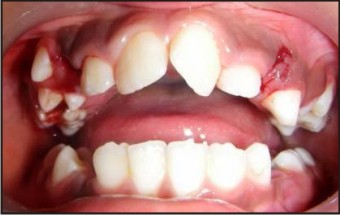 | Fig 5 : Intraoral Photographs Showing Extraction Of Supplemental Lateral Incisors
|
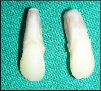 | Fig 6 : Intraoral Photographs Showing Extraction Of Supplemental Lateral Incisors
|
 | Fig 7 : Intraoral Photographs Showing Relieving Of Crowding With Self Alignment Of Buccally Erupting Canine (6 Months )
|
Discussion
Present case is unusual as it demonstrates multiple supernumerary teeth in the anterior maxilla in patient without any syndrome. Supplemental lateral incisors are rare, bilateral cases even rarer, only few cases having been reported in the literature to date. Yusof, in a literature review of multiple supernumerary teeth occurring in the absence of a syndrome, found the anterior maxilla to be an unusual site for this occurrence[22]. Extraction is not always treatment of choice for supernumerary teeth. Unerupted supernumerary teeth that are symptomless are sometimes best to left in place and kept under observation. Since these supplemental lateral incisors were causing crowding and esthetic problem, so extraction of supplemental teeth was done to prevent further worsing of malocclusion.
Why this paper is important to paediatric dentists?
Bilateral supplemental teeth are very rare.
Their differentiation from normal tooth is very difficult.
Treatment varies depending on the age and dentition of the child.
References
1. Primosch R. Anterior supernumerary teeth-assessment and surgical intervention in children. Pediatric Dentistry 1981; 3:204–215.
2. Nasif MM, Ruffalo RC, Zullo T. Impacted supernumerary teeth: a survey of 50 cases. Journal of the American Dental Association 1983; 106: 201–204.
3. Bodin I, Julin P, Thomsson M, Hyperodontia. I. Frequency and distribution of supernumerary teeth among 21 609 patients. Dentomaxillofacial Radiology 1978; 7 (1): 15–17.
4. Liu JF. Characteristics of premaxillary supernumerary teeth: a survey of 112 cases. ASDC Journal of Dentistry for Children 1995; 62: 262–265.
5. Levine N. The clinical management of supernumerary teeth. Journal of the Canadian Dental Association 1961; 28: 297– 303.
6. Smith JD. Hyperdontia: Report of a case. Journal of the American Dental Association 1969; 79:1191–1192.
7. Taylor GS. Characteristics of supernumerary teeth in the primary and permanent dentition. Dental Practitioner and Dental Record 1972; 22: 203–208
8. Sedano HO, Gorlin R. Familial occurrence of mesiodens. Oral Surgery, Oral Medicine, Oral Pathology1969; 27:360–362.
9. Marya CM, Kumar BR. Familial occurrence of mesiodentes with unusual findings: case reports. Quintessence International 1998; 29: 49–51.
10. Mitchell L. Supernumerary teeth. Dental Update 1989; 16:65– 69.
11. Mckibben DR, Brearly LJ. Radiographic determination of the prevalence of selected dental anomalies in children. ASDC Journal of Dentistry for Children 1971; 28: 390–398.
12. Humerfelt D, Hurlen B, Humerfelt S. Hyperdontia in children below four years of age: a radiographic study. ASDC Journal of Dentistry for Children 1985; 52: 121–124
13. Ruhlman DC, Neely AR. Multiple impacted and erupted supernumerary teeth. Oral Surgery, Oral Medicine, Oral Pathology 1964; 17: 199–202
14. So LLY. Unusual supernumerary teeth. Angle Orthodontics 1990; 60: 289 -292.
15. Scheiner MA, Sampson WJ. Supernumerary teeth: a review of the literature and four case reports. Australian Dental Journal 1997; 42: 160–165.
16. Yousof WZ. Non-syndromal multiple supernumerary teeth: literature review. Journal of the Canadian Dental Association 1990; 56: 147–149.
17. Brook AH. Dental anomalies of number, form, and size: their prevalence in British School children. Journal of the International Association of Dentistry for Children 1974; 5: 37–53.
18. Garvey MT, Barry HJ, Blake M. Supernumerary teeth – an overview of classification, diagnosis and management. Journal of the Canadian Dental Association 1999; 65: 612–616.
19. Robertson NRF. Supplemental incisors in the deciduous and permanent dentitions. Dental Practitioner 1962; 13 15C-151.
20. Townsend BR. Two cases of duplication of deciduous lateral incisors followed by permanent lateral incisors. British Dental Journal 1953; 95: 4748.
21. Hattab FN, Yassino M, Rawashdeh MA.Supernumerary teeth: Report of three cases and review of Literature. J Dent Child 1994:61:382-93
22. Yusof WZ. Non-syndrome multiple supernumerary teeth: literature review. Journal of the Canadian Dental Association 1990; 56: 147-149.
|