Introduction
Supernumerary teeth may be defined as any teeth or tooth substance in excess of the usual configuration of twenty deciduous and thirty two permanent teeth[1]. They might be single or multiple, fully or partially impacted, partially or totally erupted and unilateral or bilateral[2]. Multiple supernumerary teeth are usually associated with syndromes and the conditions commonly associated with an increased prevalence of these cases include cleft lip and palate, cleidocranial dysplasia and Gardner’s syndrome[3]. Supernumerary teeth can be classified in the following ways: chronologically, as predeciduos, similar to permanent teeth, post permanent or complimentary: morphologically similar to the regular teeth or completely different: and topographically as mesiodens, supernumerary premolar or supernumerary teeth in the molar region[4]. Supernumerary teeth in the molar region are paramolars or fourth molars (distomolars). Paramolars are rudimentary teeth situated lingually or buccally to the molar row. Fourth molars or distomolars are situated behind the third molar and are compressed mesiodistally. They are not completely developed and have a conical shape and may often be displaced palatally. They are more often smaller in maxilla than in the mandible, where they are equal to the normal molars in shape and size[4].
Supernumerary teeth are twice more common in males as compared to females [5]. Supernumerary molars are found more frequently in the maxilla than in mandible and their incidence is reported to be as high as 79% in the maxilla [4].
The exact etiology of supernumerary teeth is unknown but three basic theories for the pathogenesis of hyperdontia have been suggested by Price and Hoggins. The first concerns a horizontal proliferation of dental lamina and is possibly the cause of additional tooth families such as mesiodens and distomolars. The second theory concerns dichotomy of a tooth germ. Lastly an additional generation (predeciduous or post-permanent dentition) is an alternative mode of generation[6]. Supernumerary teeth can erupted, partially or fully impacted. If impacted they can be revealed only by roentgenograms. Radiographic means of diagnosis include orthopantomograms, occlusal view, lateral oblique and intraoral periapical radiographs.
Supernumerary teeth might cause dental abnormalities such as delayed eruption or impaction of permanent teeth, malposition of supernumerary teeth or displacement of adjacent teeth, root resorption and dental caries[7]. Supernumerary teeth may also cause cystic lesions, subacute pericornitis, gingival inflammation, periodontal abscesses, ameloblastomas, odontomas and fistulas[4]. Treatment of supernumerary tooth can take two forms (1) removal of the supernumerary tooth and (2) in selected cases maintenance of the tooth in the arch and frequent evaluation[3].
Case Report
24year old female was referred by her general dentist to our center for the removal of mandibular third molar as a part of treatment planning for the correction of her crowding in the permanent dentition in the upper and lower jaw.
On examination patient was found to be in good health, her medical and family histories were non contributory. Intraoral clinical examination showed no signs of caries or periodontal disease and revealed unerupted mandibular third molar. Panoramic radiograph (Figure 1) revealed presence of bilateral impacted mandibular third molar as well as fully formed and impacted 4th molar, both in a horizontal position, also seen was impacted 3rd and 4th molars in left maxilla. There was impacted third molar in the right maxilla.
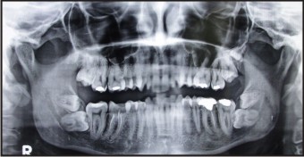 | Figure 1
 |
Surgical planning consisted of the removal of both third and fourth molars quadrant wise under local anesthesia in a clinical setup. The results of the preoperative lab tests (CBC, coagulation profile, urea, creatnine etc) were within normal limits and there were no local or systemic contraindications for surgery. Impacted fourth and third molars were successfully removed in two different sittings with no perioperative or postoperative complications. For the control of the post operative pain and infection the patient was medicated with oral antibiotics and analgesics. Sutures were removed one week after the surgery and the patient returned after one month for follow up and this course was uneventful.
Discussion
Supernumerary teeth are not uncommon and can occur almost anywhere in the mouth. Their prevalence in the permanent dentition has been estimated to range from 0.1% to 3.6% and are more frequently seen in the maxilla[8]. Grimanis[4] reported that supernumerary molars are found with a percentage of 79% in the maxilla. Cassetta claims the incidence of supernumerary molars among all the supernumerary teeth to be 75% in the maxilla[9]. The location of supernumerary can be presented in the decreasing order of frequency as follows: upper distomolar, upper paramolar and proportionally far behind are lower premolar, upper central incisor, lower fourth molar and lower central incisor[3]. Supernumerary teeth are two times more common in males as compared to females[5]. Yusuf[2] stated a 9:2 male-female ratio in the occurrence of supernumerary. In the present case, patient was 24 year old female with bilateral impacted 3rd and 4th mandibular molar along with unilateral left side maxillary impacted 3rd and fourth maxillary molars.
Supernumerary teeth can have normal morphology and are reffered to as supplementary teeth. On the otherhand, supernumerary teeth may be rudimentary in shape and smaller in size[10]. Four different forms of morphological types of supernumerary have been described ie conical, tuberculate, supplemental and odontome[3]. In the present case the 4th molar were of supplemental type as it closely resembled the 3rd molars in shape and size. Cases of three or four distomolar with normal morphology are extremely rare and have seldom reported in literature in USA, Israel and Italy[9].
Many hypothesis concerning the cause of supernumerary teeth have been suggested but the occurrence has not been fully clarified. Most authors point to phylogenetic factors, specifically the hyperactivity within the dental lamina as causing the appearance of additional tooth buds. It is also possible that supernumerary teeth may result from division of a tooth bud (dichotomy)[4]. It has also been suggested supernumerary teeth result from atavism or reversion. Aberration during embryological formation may cause supernumerary teeth formation and it is believed that supernumerary teeth arise from local independently conditioned hyperactivity of dental lamina and remnants of dental lamina[6].
In the case reported here, the presence of 4th molar was detected only on panoramic radiograph. Presence of impacted supernumerary teeth can be revealed only by a thorough panoramic radiographic examination. Mupparapu reported two cases of bilateral maxillary and mandibular fourth molar which were diagnosed on OPG, thus highlighting the limitations of intraoral radiographic examination and necessity for a more thorough examination of the jaw using panoramic and or other extra oral radiographic views[11].
Complications associated with presence of supernumerary teeth include enlarged follicular sacs, cystic degeneration, nasal eruptions, malpositioning of adjacent teeth, over retention of primary teeth, delayed eruption of permanent teeth, overcrowding, loss of space, impaction diastema, loss of tooth vitality and root resoption[12].
Treatment of supernumerary teeth depend on the type and position of supernumerary teeth and its effects or potential effects on the adjacent teeth. Treatment of supernumerary can be performed in two ways a) surgical extraction or b) maintenance of the asymptomatic tooth and periodic maintenance at least once a year[3].
The present case had a indication for surgical removal and treatment of the choice was surgical removal of bilateral impacted 3rd and 4th molar bilaterally in a quadrant wise sequential surgery in a clinical setup.
Conclusion
This case report documents a rare case in clinical practice. However general dentist must be able to correctly diagnose the presence of distomolar, recognize the possible complications arising from the presence of these supernumerary teeth and discuss with the patient possible treatment options and then execute the treatment plan.
References
1. Thomas oral pathology. St louis: CV Mosby, 1970: 112-22.
2. Yusuf WZ. Non-syndrome multiple supernumerary teeth : literature reveiw. J Can Dent assoc 1990 ;56 :417-9.
3. Garvey MT, Barry HJ and Blake M. Supernumerary teeth- an overview of classification, diagnosis and management. J Can Dent Assoc 1999;65:612-6.
4. Grimanis G A, Kyriakides A.T and Spyropoulous N.D. A survey on supernumerary molars. Quintessence Int 1991;22:989-995.
5. Goaz & White. Oral Radiology: principles and interpretetion. 3rd edition. St Louis :Mosby;1994.
6. Farman AG, Nortje CJ and Joubert J. Mandibular fourth molars. Ann Dent 1980;39:23-27.
7. Qaradaghi FI. Supernumeraray tooth: a report of a rare case of a fourth mandibular molar. Rev Clin Pesq Odontol, 2009;5:157-160.
8. Scheiner MA ans Sampson WJ. Supernumerary teeth: A review of the literature and four case reports. Aust Dent J 1997;42:180-5.
9. Cavalcanti AL, Alencer CRB and Neto LGC. Bilateral maxillary and mandibular fourth molars: a case report and literature review. J Investigative & Clinical Dentistry 2011;2:296-99.
10. Kokten G, Balcioglu H and Buyukertan. Supernumerary fourth and fifth molars: a report of two cases. J Contemp Dent Pract 2003;4:67-76.
11. Mupparapu M. Bilateral maxillary and mandibular fourth molars. Br Dent J 2002;193:363.
12. Salama FS. Supernumerary teeth: three case reports. Saudi Dent J 1994;6:173-8.
|