INTRODUCTION
A frenum is an anatomic structure formed by a fold of mucous membrane and connective tissue, sometimes muscle fibres. The superior labial frenum is triangular in shape and attaches the lip to the alveolar mucosa and/or gingiva. It extends over the alveolar process in infants and forms a raphe that reaches the palatal papilla. Through the growth of alveolar process as the teeth erupt, this attachment generally changes to assume the adult configuration.[1] Taylor has observed that a midline diastema is normal in about 98% children between 6 and 7 years of age but the incidence decreases to only 7 % in persons 12-18 yrs old.[2] But in some instances the infantile arrangement is retained. This high coronal attachment is generally associated with a hypertrophy of the frenum. Depending upon the extension of attachment of fibres, frena have been classified as-[3]
1. Mucosal – when the frenal fibres are attached up to mucogingival junction.
2. Gingival – when fibres are inserted within attached gingiva.
3. Papillary – when fibres are extending into interdental papilla; and
4. Papilla penetrating – when the frenal fibres cross the alveolar process and extend up to palatine papilla.
Clinically, papillary and papilla penetrating frena are considered as pathological and have been found to be associated with loss of papilla, recession, diastema, difficulty in brushing, alignment of teeth and psychological disturbances to individual.[4,5]
Abnormal or aberrant frena are detected visually, by applying tension over it to see the movement of papillary tip or blanch produced due to ischemia of the region.[6] Miller has recommended that the frenum should be characterised as pathogenic when it is unusually wide or there is no apparent zone of attached gingiva along the midline or the interdental papilla shifts when the frenum is extended.[7]
In such cases, it is necessary to perform a frenectomy for aesthetic, psychological and functional reasons. There are numerous surgical techniques for the removal of labial frenum. In the “classical frenectomy” by Archer [8] and Kruger [9] the frenum, interdental tissue and palatine papilla are completely excised leading to exposure of underlying alveolar bone and thus leading to scarring. Though this technique resulted into an unesthetic scar, but this approach was advocated to assure removal of muscle fibres, supposedly connecting the orbicularis oris with the palatine papilla. It was thought that if this was not done, the diastema would reopen.
Henry et al. studied thoroughly the histological constituents of frenum and found considerably dense collagenous tissue, loose connective tissue and elastic fibres but no muscle fibres.[1] So Edward[10], evaluating 308 patients who demonstrated either a diastema or an abnormal frenum or a combination of both, advocated a “conservative surgical procedure”. His method consisted of three procedures :
1. Apically repositioning of the frenum (with denudation of alveolar bone),
2. Destruction of the trans-septal fibres between the approximating central incisors,
3. Gingivoplasty of any excess labial and/or palatal tissue in the interdental area.
One of the salient aspects of Edward’s technique was the aesthetic maintenance of the interdental papilla. But the healed scar in the midline appeared unesthetic to the subjects.
Coleton[11] and Lawrence[12] have used free gingival graft combined with frenectomy. This procedure avoids the scar but a mismatched gingival colour in midline and need of a second surgical site to achieve donor tissue complicate the technique. Laser has been used by various clinicians which has its relative advantages and disadvantages.[13,14]
Miller has presented a surgical technique combining the frenectomy with a laterally positioned pedicle graft. Closure across the midline by laterally positioned gingiva and healing by primary intention resulted in aesthetically acceptable attached gingiva across the midline. No attempt was made to dissect the trans-septal fibres and hence, interdental papilla remained undisturbed. Aesthetically and functionally better results were obtained.[7]
So, in the following case-series this technique has been attempted and results are presented.
Materials and Methods
The present surgical technique was undertaken at Kothiwal Dental College and Research Centre, Moradabad. It was approved by Ethical Committee on Human and/or Animal Subjects’ Research, Kothiwal Dental College and Research Centre, Moradabad. The subjects underwent frenectomy for functional, aesthetic, periodontal or orthodontic reasons. A frenum was judged abnormal if it was unusually broad and there was no apparent attached gingiva in the midline and the interdental papilla could be stretched by the frenum.[7]
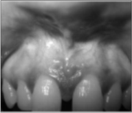
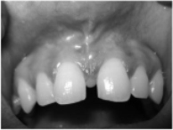
Case-1
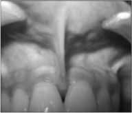
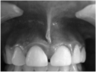
A 32-year male complained of receding gingival tissue in upper mid line region. On clinical examination, a papilla-penetrating upper mid frenum was found (Fig. 1a).
 | Fig 1a
 |
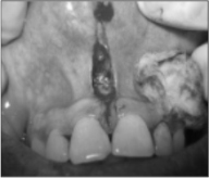
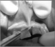
After local anaesthesia, a horizontal incision was taken to separate the frenum from the base of interdental papilla. This incision was extended apically up to the vestibular depth to completely separate the frenum from alveolar mucosa. Any remnant of frenal tissue in the mid line and on the under surface of lip was excised (Fig. 1b).
 | Fig 1b
|
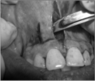
A vertical parallel incision was taken on the mesial side of lateral incisor, 2-3mm apical to marginal gingiva, up to vestibular depth. The gingiva and alveolar mucosa in between these two incisions were undermined by partial dissection to raise the flap (Fig. 1c).
 | Fig 1c
|
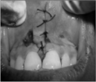
A horizontal incision was then given 1-2 mm apical to gingival sulcus in the attached gingiva, connecting the coronal ends of the two vertical incisions. Flap was raised, mobilised mesially and sutured to obtain primary closure across the midline (Fig. 1d).
 | Fig 1b
|
No attempt was made to dissect trans-septal fibres between approximating central incisors. Gingivoplasty of any excess labial and/or palatal tissue in the interdental area was done, preserving the integrity of the interdental papilla. The surgical area was dressed with COE PAK (GC America Inc., USA). Dressing and the sutures were removed 1 week later. A healing zone of attached gingiva was clearly visible with no loss of interdental papilla (Fig. 1e).
 | Fig 1e
|
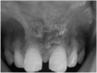
Case-2
In this case, high frenum was associated with the loss of interdental papilla and diastema (Fig. 2a).
 | Fig 2a
|
The same surgical steps were followed. 10 days post- operative observations show the formation and maturation of attached gingiva in the midline (Fig. 2b).
 | Fig 2b
|
Case-3
The patient had a complaint of a small nodular mass under upper lip. He had developed a habit of playing with it. On examination a high frenum with a nodule was found (Fig. 3a).
 | Fig 3a
|
Same surgical steps were followed. 10 days post-operative view (Fig. 3b)
 | Fig 3b
|
shows elimination of the nodule and healing with epithelialisation in midline apical to the interdental papilla.
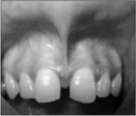
Case-4
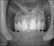
A 20-year male patient was referred from the Department of Orthodontics for frenectomy. On examination a papillary frenum associated with midline diastema was found (Fig. 4a ).
 | Fig 4a
|
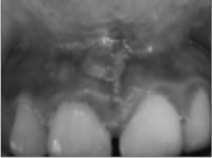
It was thereafter managed surgically following the above mentioned technique. Two weeks post-operative view (Fig. 4b)
 | Fig 4b
|
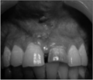
shows a healing zone of attached gingiva in midline. After complete healing no scar was observed. There was no loss of interdental gingiva. The zone of attached gingiva was increased and the colour was comparable to the adjacent tissue (Fig. 4c and 4d).
 | Fig 4c
|
 | Fig 4d
|
Results
The outcome of this surgical procedure shows this technique produced a pleasing aesthetic result. Scar formation in the midline could be avoided. On healing a wider zone of attached gingiva was obtained. It was colour matched with adjacent tissue. Healing was obtained by primary intention. No loss of interdental papilla was observed. No complication was noted during healing period. Patient’s compliance was also very good.
Discussion
In the era of periodontal plastic surgery, more conservative and precise techniques are being adopted to create more functional and aesthetic results. The management of aberrant frenum has travelled a long journey from Archer’s[8] and Kruger’s[9] “classical techniques” of total frenectomy to Edward’s[10] more conservative approach. Recent techniques added frenal relocation by Z-plasty[15], frenectomy with soft-tissue graft[11,12] and Laser[13,14] applications to avoid typical diamond-shaped scar and facilitate healing. Each method has its own advantages and disadvantages.
A frenum is evaluated in relation to vestibular depth, zone of attached gingiva, interdental papilla and diastema. If there is an adequate zone of attached gingiva, coronal to the frenum, it is of no clinical significance. A zone of attached gingiva is considered to prevent recession and it also gives an aesthetically pleasant appearance.
In this context, Miller’s technique combined with a laterally positioned pedicle graft[7] was attempted in this case series, due to its salient features. This technique offers two distinct advantages. First, on healing there is a continuous band of gingiva across the midline rather than unesthetic scar. The second advantage is that trans-septal fibres are not disrupted surgically, to avoid any trauma to interdental papilla. This prevents loss of interdental papilla. Though a continuous follow up is required for a long period to observe any loss or stability of interdental papilla, Miller, in his study observed that interdental papillae were maintained in all 27 cases surgically treated by this method.
The timing of frenectomy before or after closer of diastema has been controversial. Surgical resection of frenum prior to orthodontic treatment has been advocated since the frenum was considered to be the primary cause of diastema.[15] Edward suggested that during closure of diastema the different interdental fibres remained convoluted and compressed in a coiled up manner so they should be dissected during frenectomy.[10] On the other hand, others have felt difficulty in orthodontically moving the teeth through the scar tissue, if done afterwards.[16] At the same time, loss of interdental papilla due to destruction of trans-septal fibres is suspected. In the present case series no attempt was made to destroy the trans-septal fibres. Ten Cate et al.[17] have observed that there is simultaneous degradation and synthesis of collagen fibres in trans-septal area. So Miller feels that this physiological process may achieve remodelling of trans-septal fibres without surgical intervention. This will also prevent any loss of interdental papilla.[7]
In a study on 27 subjects with abnormal frenum who had undergone orthodontic closer of diastema, Miller did frenectomy combined with a laterally positioned pedicle graft. There was no loss of interdental papilla. No relapse of diastema was found in 24 cases and in three cases only minimal relapse (less than 1 mm) was noted. He has suggested that the newly formed broad attached gingiva contains collagenous fibres which may have a bracing effect and prevented reopening of diastema. He has further suggested that the ideal time for performing this surgery should be after orthodontic movement is complete and about 6 weeks before appliances are removed. This not only allows for healing and tissue maturation but also permits the surgeon to use orthodontic appliances as a means of retaining periodontal dressing.[7] This observations can be utilised during post-orthodontic retention period, but gain of attached gingiva in place of scar and no loss of interdental papilla are definite advantage of this technique.
CONCLUSION
The present study describes the surgical technique combining frenectomy with a lateral pedicle graft. This method has certain distinct advantages e.g.-
1. Healing takes place by primary intention.
2. A zone of attached gingiva, matching with adjacent tissue, forms in midline which is pleasing to the individual.
3. No unesthetic scar formation.
4. No recession of interdental papilla occurs because the transseptal fibres are not severed out.
5. The attached gingiva in midline may have a bracing effect which helps in prevention of orthodontic relapse.
REFERENCES
1. Henry SW, Levin MP, Tsaknis PJ. Histological features of superior labial frenum. J Periodontol 1976;47:25-28.
2. Taylor JE. Clinical observation relating to the normal and abnormal frenum labii superioris. Am J Orthod Oral Surg 1939;25:646.
3. Placek M, Miroslavs, Mrklas L. Significance of the labial frenal attachment in periodontal disease in man. Part 1; Classification and epidemiology of the labial frenum attachment. J Periodontol 1974;45:891-94.
4. Dewel B F,The labial frenum, midline diastema and palatine papilla: a clinical analysis. Dent Clin North Am 1966;175-84.
5. Maria E Diaz-Piaz, Manual O Lagravere, Rita Villera. Midline diastema and frenum morphology in the primary dentition. J Dent 2006;26:11-14.
6. Gottsegen R. Frenum position and vestibule depth in relation to gingival health. Oral Surg 1954;7:1069-72.
7. Miller P D Jr. The frenectomy combined with a laterally positioned pedicle graft; functional and aesthetic consideration. J Periodontol 1985;56:102-106.
8. Archer W H (ed). Oral surgery- a step by step atlas of operative techniques, 3rd edition. Philedelphia ,W B Saunders Co.,1961;192.
9. Kruger GO (ed). Oral surgery, 2nd edition. St. Louis, The C.V. Mosby Co.,1964;146.
10. Edwards J G. The diastema, the frenum, the frenectomy: a clinical study. Am J Ortho1977;71:489.
11. Colten S M. Mucogingival surgical procedures employed in re-establishing the integrity of the gingival unit(III):The frenectomy and the free mucosal graft. Quintessence Int 1977;8:53-61.
12. Lawrence G B, Fowler E B, Moore E A, Murray D J. The free gingival graft combined with the frenectomy; A Clinical review. Gen Dent 1999;47:514-18.
13. Coluzzi D J. Fundamentals of dental laser, science and instruments. Dent Clin North Am 2004;48:751-70.
14. Gontizo F, Navarro R S, Haypek P, Ciamponi A L, Haddad A S. The application of diode and Er;YAG lasers in labial frenectomy in infant patients. J Dent Child 2005;72:10-15.
15. Tait CH. Median frenum of upper lip and its influence on spacing of upper central incisor teeth. N Z Dent J 1929;25:116.
16. Finn SB. Clinical Pedodontics. Philadelphia, W. B. Saunders Co. 1973;416.
17. Ten Cate AR, Deporter DA, Freeman E. The role of fibroblasts in the remodelling of periodontal ligament during physiologic tooth movement. Am J Orthod 1976;69:155. |