Introduction
The discoloration of pulpless teeth could be a result of an etiological factor (trauma) or the endodontic procedure itself. A hemorrhage in the pulp chamber may result from either a blow, death of the pulp or a failure in controlling the bleeding during endodontic therapy. The penetration of blood into the dentinal tubules, followed by haemolysis of the red cells, which results in the release of haemoglobin and its breakdown products, produces a yellowish brown discoloration. This discoloration occurs when the iron pigments get degraded to iron sulfide. Such a discoloration may appear some months after the endodontic treatment is completed and it is similar to hemorrhagic discoloration[1],[2]. The failure of the operator to remove blood or other organic material completely from the pulp chamber during the treatment appears to be the most important and common reason for this post endodontic discoloration[1]. Inadequate access to the cavity preparation results in the presence of shelves of dentin which make it difficult to remove the debris from the pulp horns and the lingual area of the pulp chamber. Therefore, adequate access for the complete debridement of the pulp chamber is essential. Medication and sealing pastes must be removed from the coronal pulp space subsequent to the completion of the root canal therapy. Many of these agents contain silver, which, if left in the crown, will cause discoloration[2]. One material which has been used extensively in the past, for the restoration of lingual access cavities, subsequent to the root canal treatment, is silver amalgam. This material causes a grayish discoloration of the tooth, probably as a result of the penetration of the sulfidized by products of the corrosion process into the dentin tubules. The use of resin materials without the acid-etch technique, usually results in a marginal percolation, with the eventual internal staining of the crown. Extensively discolored, non vital teeth are highly receptive to the bleaching techniques. But the clinical situation must be carefully assessed before the bleaching treatment is considered. The quality and the type of the root canal filling that has been employed are of primary importance in this regards. Proper apical sealing is necessary to prevent percolation of the bleaching agents into the periodical tissues. A pretreatment is indicated in the cases where the root canal filling is inadequate or where it is improperly condensed. Also, the crown should be relatively intact, since a crown with large carious lesions or restorations can be better treated by means of a cast post and core, together with full coverage[3],[4],[5]. The dental practitioner is provided with a variety of post endodontic treatment options which range from invasive methods like full veneer crowns to least invasive procedures like bleaching[4]. Though non-vital bleaching has been widely mentioned in the literature as an option in the post endodontic management, an extensive review of the literature surprisingly showed us that there were very scanty case reports and follow up reports on non-vital bleaching. The main reason for this fear of cervical resorption following non-vital bleaching is that adequate precautions are not taken during the procedure. This article is aimed towards filling that void by presenting a case of non vital bleaching and its follow up using Portland cement as an intracoronal barrier which proved as an effective barrier material and a cheaper alternative to MTA.
Case Report
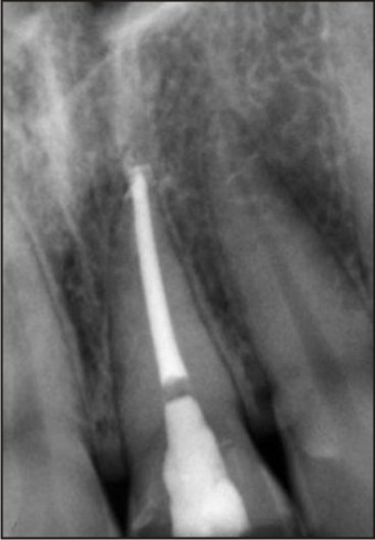
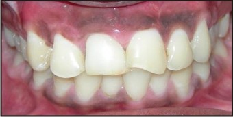
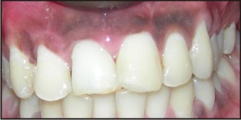
A 24-year old male patient reported to the Department of Conservative Dentistry and Endodontics of the SGT University with complaints of the discolored and unaesthetic appearance of his upper tooth. Clinical and radiographic examinations were carried out [Fig-1 and 2]. A diagnosis of non vital maxillary right central incisor was made, based on the vitality testing which was performed by using an electric pulp tester. [Fig-1]: Pre-operative image of patient’s tooth. [Fig-2]: P re-operative radiograph of patient’s tooth. The endodontic treatment was carried out under a rubber dam. The access cavity was temporarily sealed with Cavit (3M ESPE, St. Paul, MN, USA). The patient was recalled after one week for the bleaching procedure. In the subsequent visit, the tooth was cleaned with pumice and the shade was noted (VITA shade guide). A rubber dam was applied to ensure the complete isolation of the tooth. The coronal third of the gutta-percha was removed 3mm apical to the cementoenamel junction with hot pluggers. Remnants of the gutta percha and sealer were removed from access cavity with a cotton pellet soaked in 90% alcohol. The pulp chamber was rinsed thoroughly with distilled water. A cervical base of sterilized Portland cement was placed 1mm apical to cementoenamel junction level. The Portland cement was packed into place with plugger and was allowed to set against moist cotton for a period of 140 minutes. The access cavity was sealed with gutta percha temporary . Following a lapse of 140 minutes the gutta percha temporary was removed .The chamber was etched with 37 % phosphoric acid (Total Etch, Ivoclar Vivadent, Liechtenstein) for 30 seconds and it was washed and dried. A mixture of sodium perborate (in the tetrahydrate form) (Degussa, Hanau, Germany) and 30 % hydrogen peroxide (in the ratio, 1 g of powder: 0.5 ml of liquid) was made and it was placed in the pulp chamber and condensed with a wet cotton pellet[7]. A piece of dry cotton was placed over this mixture and the access cavity was sealed with modified zinc oxide eugenol cement (IRM, Dentsply). The patient was recalled after 2 weeks for a review. After 2 weeks, the tooth showed a definitive improvement in the shade, except at the incisal third tooth]. So, the internal bleaching procedure was repeated and the patient was recalled after 1 week. The sodium perborate – water mixture was removed from the pulp chamber and the access cavity was sealed with composite resin (Filtek Z350, 3MESPE, MN, USA)). Radiographs were taken to serve as a comparison for the subsequent follow up visits. The patient was asked to report after 12 months and 24 months for reviews. The bleached shade was maintained even 24 months after the bleaching and the radiographs showed no evidence of the cervical resorption. Fig 3: Radiographic Image after root canal treatment and placement of barrier. Fig 4: Post-operative image after 2 weeks follow-up
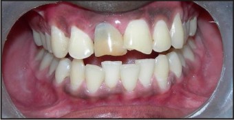 | Fig 1 : Pre-operative image of patient's tooth
 |
 | Fig 2 : Pre-operative radiograph of patient's tooth
|
 | Fig 3 : Radiographic Image after root canal treatment and placement of barrier
|
 | Fig 4 : Post-operative image after 2 weeks follow-up
|
Discussion
The literature has reported numerous reviews on the bleaching of vital and non vital teeth; yet, there are extremely few published case reports on successful non-vital bleaching. One factor which stops the dental practitioner from performing this procedure in the clinical practice is the fear of invasive cervical resorption, which has been reported to occur in several cases following internal bleaching[8],[9],[10],[11]. The “walking bleach” technique that was introduced in 1961 involved the placement of a mixture of sodium perborate and water into the pulp chamber that was sealed off between the patient’s visits to the clinician[12]. The method was later modified and water was replaced by 30–35% hydrogen peroxide, to improve the whitening effect[13],[14]. Some reports have suggested the use of a mixture of sodium perborate and water because of its decreased potential to cause cervical resorption[10],[13],[15]. Non vital bleaching has not found much favour amongst the clinicians because of the fear of resorption following the procedure, which has a poor prognosis. But our case report has shown that adhering to the proper barrier placement methods can definitely prevent the development of the resorption. It has been demonstrated that root canal fillings do not effectively prevent diffusion of bleaching agents from the pulp chamber to apical foramen. Therefore it has recommended that that a protective barrier be placed over the root canal obturation to prevent penetration of bleaching agents along the filled root canal to apical region and inside the dentinal tubules, from the canal walls to the outer surface. 20The guttapercha following obturation was removed 3mm apical to cementoenamel junction so as to enable placement of 2mm thick protective base barrier as the efficacy of the base in preventing the hydrogen peroxide leakage has been demonstrated to be superior when the thickness of the layer exceeded 1mm.24, 25, 26. Portland cement a material available in masonry supply houses has recently been compared with mineral trioxide aggregate and findings suggest that they are similar materials. In a chemical evaluation of the elements present in MTA and Portland cement it was observed that the later contains the same chemical elements as MTA except MTA contains bismuth. Thus it was considered a possible substitute for MTA in present study given low cost and similar properties. Another reason for its use was to counteract the decrease in ph caused by bleaching with hydrogen peroxide.6 Portland cement is slightly radiopaque but does not meet the minimum requirements for radiopacity set out in ISO 6876/2001, which is a major disadvantage of Portland cement if it is to be used clinically. MTA consists of 75 wt% Portland cement, 20 wt% bismuthoxide, and 5 wt% calcium sulfate as a setting modifier. Bismuth oxide is a necessary radiopacifier to enable a radiographic assessment of dental materials. In the present study, experimental cement containing Portland cement and bismuth oxide at a ratio of 4:1 was prepared. Contamination of the cement may occur during packaging, shipping and storage so Portland cement also needs to be sterilized before it can be used in vivo. Due to the effects of water in the set of the cement we have used“dry heat” as the method of choice for sterilization. Cement was subjected to to a dry heat cycle: 170 degrees Centigrade (340 degrees F) for Ihour rendering it sterilized. This is the first clinical case report to describe the use of Portland cement as a barrier material in vivo prior to the non-vital bleaching. Several barrier materials and supplementary barriers have been proposed in the literature. They range from materials like Cavit to Modified Zinc Oxide eugenol (IRM), glass ionomer cement, calcium hydroxide and resin modified glass ionomer cement[11],[14],[15]. But taking into consideration the properties of Portland cement, like its high alkaline pH, insolubility and reduced moisture permeability, it appears to be a favorable material which can serve as an intracoronal isolating barrier and as a cheaper alternative to MTA prior to the bleaching[15],[16]. After placing the Portland cement, the patient was asked to report after 140 minutes for the bleaching procedure, as Portland cement took 140 minutes to set according to manufacture instructions. Non vital bleaching has several advantages over other post endodontic treatment options like full veneer crowns. Difficulties in shade matching and achieving the life like appearance and the emergence profile of the natural teeth are the possible drawbacks of the full coverage restorations. In contrast, non vital bleaching is a non invasive procedure and it is also less time consuming and economical. Also, the patient’s natural tooth structure is preserved.
Conclusion
This case report demonstrates the successful management of a discolored, endodontically treated tooth by non-vital bleaching. Non vital bleaching can be used as a very effective and safe post -endodontic treatment for discolored anterior teeth, provided the procedural protocol and precautions are strictly adhered to.
References
1. Ingle J, Bakland: Endodontics, ed 5. Philadelphia, Lea and Febiger, 1976.
2. Cohen SC, Bums RC. Pathways of the Pulp. St. Louis, C.V. Mosby Co,1980.
3. Fisher NL, Radford JR. Internal bleaching of discolored teeth. Dent Update, 1990; 110–14.
4. Wray A, Welbury R. Treatment of the intrinsic discoloration in the permanent anterior teeth in children and adolescents. Int J Paediatr Dent 2001; 11: 309–31.
5. Nutting EB, Poe GS. Chemical bleaching of discolored, endodontically treated teeth. Dent Clin North Am 1967: 655–62.
6. Steiner DR, West JD. A method to determine the location and shape of an intracoronal bleach barrier. J Endod 1994; 20: 304 – 06.
7. Weiger R, Kuhn A, Lost C. Radicular penetration of hydrogen peroxide during intra-coronal bleaching with various forms of sodium perborate. Int Endod J 1994 ;27:313-17.
8. Heithersay GS, Dahlstrom SW, Marin PD. Incidence of invasive cervical resorption in bleached, root-filled teeth. Aust Dent J 1994 ;39:82-87.
9. Lee GP, Lee MY,Lum SO, Poh RS, Lim KC. Extra-radicular diffusion of hydrogen peroxide and the pH changes which were associated with the intracoronal bleaching of discolored teeth by using different bleaching agents. Int Endod J 2004 ;37:500-06.
10. Rotstein I, Lewinstein I, Zuwabi O, Stabholz A, Friedman M. Role of the cementoenamel junction on the radicular penetration of 30% hydrogen peroxide during intracoronal bleaching in vitro. Endod Dent Traumatol 1996;12:146-50.
11. Costas FL, Wong M. Intra-coronal isolating barriers: effect of location on the root leakage and the effectiveness of the bleaching agents. J Endod 1991 ;17:365-68.
12. Spasser HF. A simple bleaching technique by using sodium perborate. NY State Dent J 1961; 27:332–34.
13. Nutting EB, Poe GS. A new combination for bleaching teeth. J So Calif Dent Assoc 1963; 31:289–91.
14. Lambrianidis T, Kapalas A, Mazinis M. Effect of calcium hydroxide as a supplementary barrier in the radicular penetration of hydrogen peroxide during intra-coronal bleaching in vitro. Int Endod J 2002; 35:985-90.
15. Tselnik M, Baumgartner J, Marshall J. Bacterial leakage with mineral trioxide aggregate or a resin-modified glass ionomer which was used as a coronal barrier. J Endod 2004 ;30:782-84.
16. Barrieshi – Nusair KM, Hammad HM. Intra-coronal sealing comparison of mineral trioxide aggregate and glass ionomer. Quintessence Int 2005; 36: 539-45.
|