Introduction
Orthodontic extrusion/ forced eruption is a well documented clinical method for altering the relation between a non restorable tooth and its attachment apparatus, elevating sound tooth material from within the alveolar socket[1],[2],[3]. It has some advantages over surgical crown lengthening, which is less conservative considering the sacrifice of supporting bone and the negative change in the length of the clinical crowns of the tooth and its neighbors[4], [5].
Case Report - 1
A 20 year boy reported to the Department of Conservative and Endodontics, 6 months after trauma to anterior teeth due to road side accident leading to its fracture.
Clinical Examination
Crown fractures were seen with 21, 22,31,32,41. Ellis class III fracture of 21, 22. Incomplete treatment of lower anterior teeth was found. Access opening along with crown preparation seen in relation to 31, 32, 41(Fig. 1a). Ellis class II fracture in relation to 42. Vitality test of 11 was performed and reported to show delayed response. There was no mobility of any of the teeth. IOPA in relation to 21, 22 (Fig. 1b) showed incomplete obturation and 41,31,32 showed no obturation. On the basis of clinical and radiographic findings following treatment plan was made and patients consent was taken,
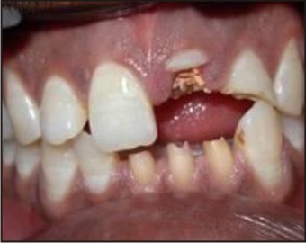 | Figure 1a : Pre Operative - Intraoral Photograph;
 |
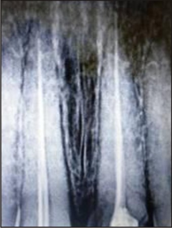 | Figure 1b : IOPA Of 21,22
|
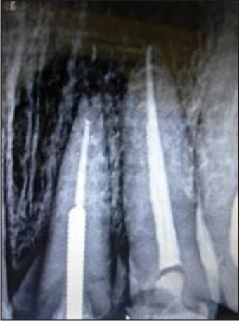
Endodontic therapy for 11,31,32,41. Re RCT for 21,22. Modified Post and core for Orthodontic extrusion of 21 (Fig. 1c). Composite build up for 42. All ceramic crown in relation to 21,22,31,32,41 (Fig. 1 d, e).
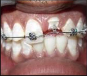 | Figure 1c : Modified Post Used During Orthodontic Extrusion
|
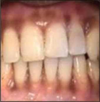
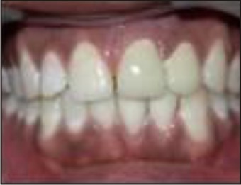 | Figure 1d : Post Operative Intraoral Photograph
|
 | Figure 1e : Post Operative Radiograph
|
Case Report - 2
An 18 year boy reported to the Department of Conservative and Endodontics, 2 years after trauma to maxillary tooth -22 due to road side accident leading to its fracture and intrusion.
Clinical Examination
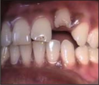
Ellis class II fracture at the middle third in relation to 22 (Fig. 2a). The gingival around the intruded tooth was normal and well contoured. There was no mobility of any of the teeth. Vitality test was done for 22 and it showed no response. IOPA Radiograph showed calcified root canal in relation to 22 (Fig. 2b). On the basis of clinical and radiographic findings following treatment plan was made, Orthodontic extrusion of 22 (Fig. 2c) and then Composite build up was done. The treatment plan and time required for the treatment was explained to the patients consent was taken. Post operative photograph showed an esthetic smile (Fig. 2d).
 | Figure 2a : Pre Operative - Intraoral Photograph
|
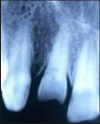 | Figure 2b : Pre Operative - Iopa Of 22
|
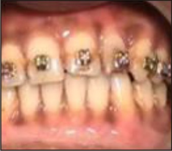 | Figure 2c : Orthodontic Extrusion
|
 | Figure 2d : Post Operative - Intraoral Photograph.
|
Conclusion
We report these cases of Interdisciplinary approach between orthodontist and restorative dentist. The careful integration of multiple specialists suggests the possibility of optimal results with high predictability[6],[7],[8].
References
1. Jain V, Gupta R, Duggal R, Prakash H. Restoration of traumatized anterior teeth by interdisciplinary approach: report of three cases. J Indian Soc Pedo Preven Dentistry 2005 Oct-Dec;23(4):193-7.
2. Kocadereli I, Ta#1;man F, Güner SB.Combined endodontic-orthodontic and prosthodontic treatment of fractured teeth- Case report. Aust Dent J. 1998 Feb;43(1):28-31
3. Koyuturk AE, Malkoc S. Orthodontic extrusion of subgingivally fractured incisor before restoration. A case report: 3 years follow-up. Dental Traumatology 2005; 21:174-78.
4. Frank Spear. A patient with a central incisor fractured apically in relation to the gingival margin. J Am Dent Assoc; 140(3),395-399.
5. Addy LD, Thomas MB. Orthodontic extrusion: an interdisciplinary approach to patient management. Dental Update 2009;36(4): 212-4.
6. Delivanis P, Delivanis H, Kuftinec MM. Endodontic-orthodontic management of fractured anterior teeth. J Am Dent Assoc. 1978 Sep; 97(3):483-5.
7. Ivey DW, Calhoun RL, Kemp WB, Dorfman HS, Wheless JE. Forced eruption Orthodontic extrusion: its use in restorative dentistry. J Prosthet Dent. 1980 Apr; 43(4):401-7.
8. Smidt A, Lachish-Tandlich M, Venezia E. Orthodontic extrusion of an extensively broken down anterior tooth: a clinical report. Quintessence Int. 2005 Feb; 36(2):89-95.
|