Introduction
Dentistry has changed tremendously over the past decade to the benefit of both the clinician and the patient. New materials and technologies have improved the efficiency of restorative dentistry for clinicians. One technology that has become increasingly popular in clinical dentistry is that of the laser. Initially introduced as an alternative to the traditional halogen curing light, the laser now has become the instrument of choice in many applications for both periodontal and restorative care. Although the laser offers many advantages over other modalities of treatment, probably the greatest impact it has made is in its ability to be used for both hard and soft tissues often without the need of anaesthesia[9]. The field of lasers in general practice essentially began with the introduction of the Nd: YAG laser in 1990[3].With dentistry in the high tech era of 1990s, we are fortunate to have, at our disposal many technologic innovations to enhance treatment, including intraoral camera, computer imaging ,CAD-CAM and air abrasive units[12] Today more experience and knowledge in applying lasers in conservative dentistry and Endodontics is available [10] Lasers now a days are widely used in conservative dentistry and endodontics for Cavity preparation, Tooth Etching, Disinfection of the root canal, Sealing of the root canal, Removal of the Gutta-Percha in Re-Treatment, Bleaching ,Crown lengthening and for treating Dentin hypersensitivity.[10]Lasers are employed in the restorative dentistry for the removal of incipent caries,for curing composite resins, enamel etching.[3]. When lasers are properly used within the ethical envelope of dentistry, they offer present day dentists a superb treatment modality for various common clinical conditions.[12] The promise that laser offers to both dentists and the patients of simple, painless, dental treatments has stimulated continuing research into their use for the removal of dental hard tissue.[13]. The aim of this review is to describe the application of lasers in dental hard tissue procedures.
Review
The first experiment with lasers in dentistry was reported in a study about the effects of a pulsed ruby laser on human caries [7]. The results of that study showed that the effects varied from small 2-mm deep holes to complete disappearance of the carious tissue, with some whitening of the surrounding rim of enamel, indicating extensive destruction of carious areas along with crater formation and melting of dentine. Further work in the 1970's focused on the effects of neodymium (Nd) and carbon dioxide (CO2) lasers on dental hard tissues. Early researches found that CO2 lasers produced cracking and disruption of enamel rods, incineration of dentinal tubule contents, excessive loss of tooth structure, carbonisation and fissuring and increased mineralization caused by the removal of organic contents[6]. It was also reported that the use of the CO2 laser was unfavourable because of the loss of the odontoblastic layer[16].
Therefore, it was concluded that, unless heat-related structural changes and damage to dentinal tissues could be reduced, laser technology could not replace the conventional dental drill. Further advances in laser technology however, have identified acceptable biologic interactions. For example, the Er: YAG laser was tested for its ability to ablate (or vapourise) dental hard tissues [6]. Enamel and dentine cavities were successfully prepared using the Er: YAG laser. Since then, this laser has been used for caries removal and cavity preparation, soft tissue minor surgery and scaling [1]
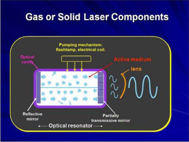
How Laser Works[8][12]
Lasers Consist of the Following Components
1. Optical cavity:
Lasing medium
Parallel mirrors
2. Pump energy source
3. Cooling system
Optical Cavity
An Optical cavity is at the center of the device.
 | Gas Or Solid Laser Components
 |
The core of the cavity is comprised of chemical elements, molecules, or compounds and is called the Active medium.
Lasers are generically named for the material of the Active medium, which can be a container of gas, a crystal or a solid state semiconductor( e.g, a crystal)
Lasing Medium
There are two Active medium gaseous lasers used in dentistry:
Argon and Co2
The remainder that are a available are solid state semiconductor wafers made with multiple layers of metals such as gallium, aluminum, indium, and arsenic or solid rods of garnet crystal grown with various combinations of yttrium, aluminum, scandium and gallium and then doped with the elements of chromium, neodymium, or erbium. ;
Mirrors: On either side of the lasing medium there are placed 2 parallel mirrors. In this configuration, photons bounce off the mirrors and re-enters the medium to stimulate the release of more photons. The mirrors collimate the light that is photons exactly perpendicular to the mirrors re-enter the active medium, while those off axis leave the lasing process. If one mirror is totally reflective and other mirror partially transmissive, the light that escapes through first mirror becomes the laser beam.
Pump energy source: The Pump Source is the Energy provided by the Flash lamp or electrical coil.High energy radiation is pumped into active medium by medium of the pump source. (flash lamp or electrical coil)
Cooling System:
Because the process is not 100 % efficient and the remaining energy is converted into heat, it is necessary to provide some form of cooling. Cooling is provided by water. Thus, stimulated emission within an optical cavity generates a collimated, coherent, and monochromatic beam of light
Clinical Applications Of Lasersin Endodontics[2][3]
Pulpal Diagnosis
Laser Doppler flowmetry, which was developed to assess blood flow in microvascular systems , also can be used for diagnosis of blood flow in the dental pulp . This technique uses helium-neon and diode lasers at a low power of 1 or 2 mW. The laser beam is directed through the crown of the tooth to the blood vessels within the pulp. Moving red blood cells causes the frequency of the laser beam to be Doppler shifted and some of the light to be backscattered out of the tooth .The reflected light is detected by a photocell on the tooth surface
The main advantage of this technique, in comparison with electric pulp testing or other vitality tests, is that it does not rely on the occurrence of a painful sensation to determine the vitality of a tooth. Laser Doppler flowmetry has some limitations. It may be difficult to obtain laser reflection from certain teeth. Generally, the anterior teeth, in which the enamel and dentin are thin, do not present a problem. Molars, with their thicker enamel and dentin and the variability in the position of the pulp within the tooth, may cause variations in pulpal blood flow.
Cleaning and Shaping of Root Canal System
Successful endodontic therapy, which mainly depends on the elimination of microorganisms from the root canal system, is accomplished by means of biomechanical instrumentation of the root canal system. In various laser systems used in dentistry, the emitted energy can be delivered into the root canal system by a thin optical fiber (Nd:YAG), erbium, chromium:yttrium-scandium-gallium-garnet [Er,Cr:YSGG], argon, and diode) or by a hollow tube (CO2 and Er:YAG). Thus, the potential bactericidal effect of laser irradiation can be used effectively for additional cleansing of the root canal system following biomechanical instrumentation .This effect was studied extensively using lasers such as CO2 ,Nd:YAG , excimer, diode , and Er:YAG
Endodontic Surgery
Surgical endodontic therapy is the treatment of choice when teeth have responded poorly to conventional treatment or when they cannot be treated appropriately by nonsurgical means. The goal of endodontic surgery is to eliminate the disease and to prevent it from recurring. The surgical option should be considered only when a better result cannot be achieved by nonsurgical treatment.Nd:YAG lasers have shown a reduction in the penetration of dye or bacteria through resected roots..
Root Canal Irrigation in Combination with laser
Some laser devices produce cavitation effects in root canals in a manner similar to that of the ultrasonic irrigation. At present, the effect is weaker than that of ultrasonic irrigation. This laser technique is likely to be improved in the future. Straight and slightly curved root canals as well as wide root canals are indications for this treatment.The pulsed Nd:YAG laser, Er:YAG laser, and Er,Cr:YSGG laser are recommended.The laser irradiation is not carried out by the laser alone; a solution such as 5.25% sodium hypochloride or 14% ethylenediaminetetra-acetic acid (EDTA) also must be used. A power of 2 to 5 W usually is used for approximately 2 minutes.
Sterlization or disinfection of Infected Root Canals
The laser is an effective tool for killing microorganisms because of the laser energy and wavelength characteristics. Infected root canals are an indication for this laser treatment, but application to extremely curved and narrow infected root canals appears difficult .Pulsed Nd:YAG , argon, semiconductor diode, C02, Er:YAG, have been considered for use in this treatment.
Root Canal Obturation using Gutta percha and Laser:
Gutta-percha is thought to be melted by laser heat energy.Anic and Matsumoto, attempted to investigate whether it is possible to perform the root canal filling using sectioned gutta-percha segments and a pulsed Nd:YAG laser. This was shown to be possible by the vertical condensation method, but the technique required too much time. At present, this technique is not practical. Although a method combining an argon laser and light-curable resin is in the literature, proper application of this method requires further research.
Treatment by Laser in Case of Indirect Pulp Capping
As lasers were introduced to dentistry, nobody thought that laser could perform the treatment of indirect pulp capping. The discovery of closure of dentinal tubules by laser energy and the sedative effects on pulpitis has led to the development of several new treatments that are soon to be put into practice. Deep cavities, hypersensitive cavities, and cavities that require sedative treatment are some of the indications for this treatment.
Treatment by laser in Case of Direct Pulp Capping
Because laser treatment has advantages with respect to control of hemorrhage and sterilization, laser use for direct pulp capping has attracted dentists' attention. Various studies have examined this treatment, and some researchers have recommended the laser as a treatment method for direct pulp capping.. Laser irradiation should be performed at I or 2W irrigating alternatively with 5.25% sodium hypochlorite and 3% hydrogen peroxide for more than 5 minutes.
Conclusion
Scientific research and papers describing the dental applications of lasers have appeared in dental literature for more than 30 years. However due to vast and contradictory nature of this subject very few comprehensive compilations are available on their use in dentistry.
Dentistry like other health care profession is in midst of major transitions. In the fast changing arena of dentistry the laser can be very useful tool for dental practitioners.The high quality state of art has been employed in various procedures such as in diagnostics ,the laser is used for examining the caries, Pulpal blood flow using laser Doppler flowmetry.[11] From the vast literature on lasers it is understood that it is not only important to realize the various potential uses but also the necessity to select proper wavelength, understand laser tissue interactions and not over enthusiastically jump into laser dentistry before science properly supports it.
This review is a very humble effort to provide an overview of applications of lasers in endodontics as technology advances and our understanding of laser light expands its necessary to revise and update from time to time.
Researchers and clinicians must continue to explore before the investigational uses of lasers are brought from the researchers lab to the clinicians doorstep.
References
1. AOKI A et al: Lasers in Nonsurgical Periodontal Therapy,Periodontology 2000,Vol 36,2004,59-97
2. COLUZZI.J.D & CONVISSOR. R.A: Atlas of laser applications in dentistry, Quintessence International,2007
3. CONVISSOR. R.A & COLUZZI.D J: The biological rationale for the use of lasers in dentistry; Dental clinics of North America,48,4,771-794,2004
4. COURROLL.L.C: Spectroscopy characterization of sound and carious ablated dental tissue after Er:YAG laser interaction ; J in applied research in CD, 1,1 , 29-34. 2004
5. FRENTZEN.M & KOORT.H.J: Lasers in dentistry: new possibilities with advancing laser technology?,IDJ,40,1990,323-332
6. GIMBEL et al: Ablation of Dentin by Various Er:YSGG laser ,Academy of Laser Dentistry,2000.
7. GOLDMAN, et al, . Impact of the laser on dental caries. Nature, 203: 417-417,1964.
8. GUTTENBERG.S.A: Laser Physics and tissue interaction; Oral & Maxillofacial clinics of North America,16,143-147,2004
9. HORNBROOK DAVID : Lasers in dentistry: Good through, June 30,2008,1-4.
10. MERCHANT.N: Lasers in conservative dentistry and endodontics ; IDRR 25-26, 2007
11. MISERENDINO. L.J: History & Development of laser dentistry; Lasers in dentistry
12. PICK.M.R: The use of laser for treatment of gingival diseases : “Oral & Maxillofacial clinics of North America”;9,1,1-20,1997
13. RODE.A V et al.: Precision Ablation of Dental Enamel using a Sub-Picosecond pulsed laser; ADJ, 48,4, 233-239,2003
14. STABHOLTZ.A et al: Lasers in endodontics; Dental clinics of North America,48,4,2004,809-832
15. WALSH.L.J: The current status of laser application in dentistry, Australian Dental journal , 143- 154,2003
16. WIGDOR et al : The Effect of lasers on Dental Hard Tissues,JADA,124,65-70,1993.
|