Introduction:
Self Injurious Behavior (SIB) is a deliberate alteration or damage to oneself without a suicidal intent.[1] SIB is the deliberate alteration to one’s own body part without suicidal intent. Self Harm, Deliberate Self Harm, Self Injury, and Self Poisoning are other terms used to describe the same condition. Self mutilation patients violently inflict lesions to their own bodies with no intent to commit suicide.[2]
Self inflicted injuries are very common and range from simple to severe forms of mutilation.Various foreign objects are reported to be lodged like pencil leads, darning needles, metal screws, beads and stapler pins.[3] Self inflicting injuries can lead to accidental grave problems. Several aero-digestive accidents can result and accidental inhalation of foreign bodies can lead to accidental death during childhood.[4] Foreign bodies in pediatric airway is potentially life-threatening. Coughing, choking, and wheezing are some of the presenting symptoms seen for 95% of the patients.[5] The manifestations again depends on type and severity of etiology.
Case Report:
A 5 year old boy presented with the chief complaint of pain in the upper front tooth. Pain was present and severe past 2 days. Past history revealed dull aching constant pain past 1 year in the same tooth and occasional pus discharge from adjacent gum region. General examination revealed an otherwise healthy child. Child’s behavior assessment showed a definitely negative behavior which could be related to severe pain at the time of presentation to clinic.
Intra-oral examination showed dentition corresponding to 6 years, tooth number 51 showed intrinsic black discoloration, grade II mobility, tenderness to percussion and presence of a yellow metallic object in the lingual aspect of tooth number was noticed. The metallic object was tightly wedged preventing easy removal and attempts to touch the tooth caused severe pain leading to behavior problems in the child. Tooth 21 had erupted but 11 was missing due to retained 51 (Figure 1-labial view & Figure 2- Palatal view). Soft tissue findings included presence of gingival-vestibular abscess surrounding tooth number 51.
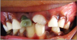 | Figure 1 - Labial View
 |
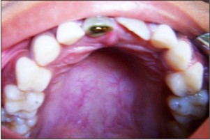 | Figure 2 - Palatal Intra-oral View
|
When the child was enquired regarding the placement of metallic object, he was initially reluctant to inform regarding the incident due to shame. Once the parents were separated; the child revealed that he regularly indulged in placement of metallic objects like pins into the tooth to remove impacted food particles. Unfortunately the last act resulted in tight wedging of the metal; self removal of which was unsuccessful. The child did not notify parents regarding the mischief as he feared punishment by parents.
Intraoral–IOPA view was advised (Figure 3-IntraOral Periapical Radiograph of maxillary anterior segment). IOPA view revealed presence of sharp radio-opaque mass wedged within the pulpal outlines of the tooth 51. Presence of peri-apical radiolucency surrounding tooth 51, high amount of bone loss apically and laterally to the tooth indicative of peri-apical pathology. Tooth 11 was impacted. A diagnosis of foreign body impaction and periapical abscess with respect to retained tooth 51 was given.
Treatment:Vaccination history was enquired and tetanus booster dose was administered to the patient. Based on prognosis of the tooth and dental age of the patient, extraction of the tooth was carried out followed by drainage of the abscess. Since the patient indulged in the habit for a very long time, care was take to carefully inspect the local site for presence of any other foreign bodies Careful debridement of socket was carried out to clear any other foreign body remnants (Figure 4 & 5- Labial and Palatal view of extracted teeth). Patient was followed at 1 week interval. At follow-up period there was absence of symptoms and there was presence of good healing socket. Child was counseled regarding the ill effects of placing foreign bodies in the mouth.
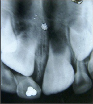 | Figure 3 - Intraoral Periapical Radiograph Of Maxillary Anterior Segment
|
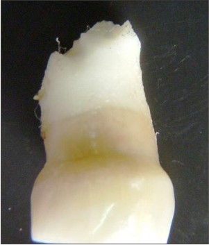 | Figure 4 - Labial And Palatal View Of Extracted Teeth
|
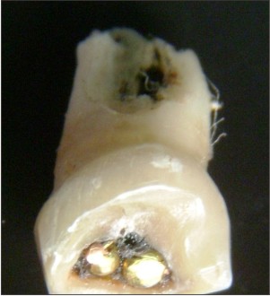 | Figure 5 - Labial And Palatal View Of Extracted Teeth
|
Discussion:
In clinical situations presence of foreign body in oral cavity of a child is usually associated with accidental etiology; none think about the role of self inflicted injury. Foreign bodies get deposited accidentally due to trauma or sometimes due to self mutilation.[2]Foreign bodies can cause acute problems like atelactasis, bleeding into tracheo-bronchial tree or act as potential source of chronic infection.[4] Although accidental deposition of foreign body occur at all ages, self-mutilation type is more common in childhood because children have the habit of placing various foreign bodies in the mouth.
Presence of self mutilation or self-injurious behavior might go un-noticed if history is not enquired properly, in such conditions clinicians usually tend to regard presence of foreign body as accidental injury rather than self-mutilation injuries.[6],[7] Rarely parents are aware of child’s self injurious habit; Parental narration does not ascertain the condition. Initial misdiagnosis can lead to delay in treatment, complications or worsening of the situations.[8] Early diagnosis and treatment can decrease morbidity and length of hospital stay in these children.[8]
Radiographic examination is very useful especially when the foreign body is metallic or radio-opaque. Various radiographic techniques can come handy in diagnosis & treatment. Importantly, a negative radiographic finding does not rule out the absence of foreign body in aero-digestive tract foreign body as many of these foreign bodies are radiolucent.[9] clinician should not totally rely on the use of radiography to detect foreign bodies.
As a whole, absence of positive history, inconsistent clinical features and radiolucencies of objects cause difficulty in diagnosis.[10] Since the aetilogy of both differ, the treatment also differs- In accidental foreign body impaction treatment is only limited to symptomatic treatment but in the SIB, a psychological counseling of patient might be required to prevent repetition of the habit.[11]
References:
1. In: Riding, Swann, Swann. Ed. The Handbook of forensic learning disabilities. 1st ed. Lancaster England. Radcliffe Medical PR; 2005: 97-120
2. ErdurB, TurkcuerI, HerkenH. An unusual form of self-mutilation: tongue amputation with local anesthesia. Am J Emerg Med.2006 Sep;24(5):625-8.
3. AduriR, ReddyRE, KiranK. Foreign objects in teeth: retrieval and management. J Indian Soc Pedod Prev Dent.2009 Jul-Sep;27(3):179-83.
4. Aytaç A, Yurdakul Y, Ikizler C, Olga R, Saylam A. Inhalation of foreign bodies in children. Report of 500 cases.J Thorac Cardiovasc Surg.1977 Jul;74(1):145-51.
5. Black RE, Johnson DG, Matlak ME. Bronchoscopic removal of aspirated foreign bodies in children. J Pediatr Surg.1994 May;29(5):682-4.
6. Papsin BC, Friedberg J. Aerodigestive-tract foreign bodies in children: pitfalls in management.J Otolaryngol.1994 Apr;23(2):102-8.
7. Josell SD, Abrams RG. Traumatic injuries to the dentition and its supporting structures. Pediatr Clin North Am.1982 Jun;29(3):717-41.
8. Bhatia PL. Problems in the management of aspirated foreign bodies.West Afr J Med.1991 Apr-Jun;10(2):158-67.
9. Holla et al. Unusual objects in root canals of deciduous teeth: report of 2 cases. Contemp Clin Dent 2010;1(4):246-48.
10. Gujjar KR, Algali G, Omar SM, Amith HV, Anegundi RT. Foreign bodies in primary molars: a report of 2 cases.J Dent Child (Chic).2012 Jan-Apr;79(1):40-3.
11. Blanton PL, Hurt WC, Largent MD. Oral factitious injuries. J Periodontol.1977 Jan;48(1):33-7.
|