INTRODUCTION
Attempts to re-implant teeth have been carried out from time almost immemorial. Ambroisepare Para1 who practiced in France in 1579 described replanting, extracting and putting back into the socket again.
In 1785, Dr. Josiahflagg1 of Boston informed the public, that he could replant the teeth both dead and alive.
Since then many dentists resorted to these techniques with not much success.
Today, however, with the modern prophylactic use of antibiotics, improved techniques of asepsis and advanced knowledge of tissue response, better results should be obtained.
CASE REPORT
A 21 year old girl reported the department with the complaint that she had an ailing upper tooth, which was source of spordiac swelling, pain and tooth was shaky and discolored.
INTRA-ORAL EXAMINATION
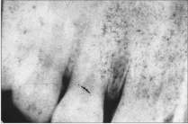
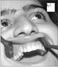
On making the intra oral examination, the patient had all the permanent teeth erupted, in excellent health. There were no caries, no gingival disease, excepting that upper right milk canine was retained, shaky with gingival abscess around it. (Fig 1)
 | Fig 1
 |
Extraction of the tooth was advised to relieve the recurrent abscess in the mouth of the patient. The patient however, enquired about the post extraction options for filling the space.
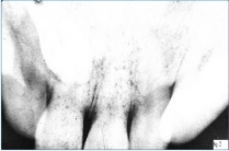
In 1999 implants were not being practiced in Jammu, therefore a removable partial denture or a crown or bridge were explained and suggested to the patient. The patient declined both the alternatives and enquired the reasons of not having her permanent canine. An intra oral radiograph was taken, which revealed upper right canine to be embedded deep in the palate. (Fig 2)
 | Fig 2
|
The patient insisted that somehow this tooth should be utilized in place of milk canine. All aspects regarding a difficult surgery and re-implantation of permanent canine were explained to the patient and her parent’s accompanying her. The patient and parent’s agreed to it, a written consent was taken for surgery and photography of the patient.
As the patient was healthy, without any physical ailment, she was put on antibiotic for performing the surgery next day. On the following day milk canine was extracted and flap was raised to locate the upper permanent canine. Bone cutting was done from buccal side but the tooth could not be located, therefore palatal assess was taken. After raising palatal flap, a bulge in the palatal plate could be seen. Bone cutting was done; the tooth was lying deep along the roots of upper right central and lateral incisors and was more in horizontal placement to the long axis of central and lateral incisors, quite contrary to how it looked in the radiograph. With a lot of manipulation tooth was extracted and kept in a tray containing
normal saline in it. This tooth was quite a long and could not be accommodated in socket of milk canine; therefore 3-4 mm apex was cut and root canal filling was done after the removal of neurovascular tissue. Apex was shaped to avoid mechanical irritation.
The socket was also modified to accommodate the tooth which was re-implanted and splinted with acrylic. Patient was kept on antibiotic and anti-inflammatory drugs for one week. After 8 weeks the splint was removed. Tooth had significant stability and patient was satisfied.
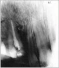
A radiograph was repeated after three months (fig 3)
 | Fig 3
|
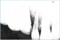
bone formation was taking place, there was no soft tissue problem excepting little of gingival shrinkage. After one year another x ray was taken. (Fig 4)
 | Fig 4
|
Crestal bone formation was quite significant, there was no mobility of the tooth, a porcelain cap was given and patient felt quite happy and satisfied. Fig 5
 | Fig 5
|
(without crown). The patient was recalled after 11 years Dec 2009 for a follow up and the canine is seen to be perfectly well placed with excellent aesthetics.
DISCUSSION
Utilization of embedded teeth is quite an old technique. In the absence of appropriate option available and the patient not accepting partial denture or crown and bridge to avoid drilling of the healthy teeth, the embedded tooth can be used for the satisfaction of the patient. If possible tooth should be moved to place by orthodontic treatment, otherwise extraction and re-implantation is also a good option with quite a good success as experienced in the above case. It is however imperative that one must be quite experienced to extract such embedded teeth placed remotely in the jaw without damaging the adjacent teeth
and there is an utter need for the complete asepsis and adequate handling of extracted tooth for success of such operation.
REFERENCE
1. Oral surgery, Kurt H Thoma 4th Edition.
2. Peterson's Principles of Oral and Maxillofacial Surgery, 2nd Edition.
3. Spiechowicz E, Gawor E et al. Reimplantation, bone augmentation, and implantation procedures for impacted maxillary canines: a clinical report. J Prosthet Dent. 2004 Mar;91(3):223-7.
4. Penarrocha M, Garcia-Mira B et al. Extraction of impacted maxillary canines with simultaneous implant placement. J Oral Maxillo fac Surg. 2007 Nov;65(11):2336-9. |