Introduction
Recurrent dental caries has been associated with the deterioration of dental restorative materials. Breakdown in marginal areas between cavity preparation and restorative materials can provide potential for reinfection. Cariogenic micro-organisms can easily penetrate into the underlying dentine through these defects. Reducing or preferably preventing such marginal breakdowns is an important element in reducing the incidence of recurrent caries.[1]
Dentin bonding agents were developed for the purpose of direct bonding of the hydrophobic composite resin materials to the hydrophilic dentin. It can be accomplished by means of etch and rinse or self etch adhesive system.[2]
Settembrini et al reported that the strong acids of the total etch technique removed bacteria on the cavity walls. However with self etching primers, the demineralised smear layer which may contain bacteria is not removed and is incorporated into the hybrid layer. This implies that residual bacteria are not washed away and thus cannot be eliminated. The dentin primer of these systems is the component that comes into contact and reacts with the dentin substrate at first stage of restoration in adhesive systems. Consequently when these systems are used antibacterial activity is considered to be an important factor.[3]
To enhance antibacterial activity, first the incorporation of fluorides into the dentin bonding agents may be one way to inhibit bacterial growth. Furthermore monomer by themselves by a low pH or through the addition of special antibacterial groups, may produce antibacterial effect. The latter approach was followed by Imazao et al who developed a monomer 12-Methacryloxydodecylpyridinumbromide[MDPB] that consistently demonstrated antibacterial effects.[4]
Single step self-etch adhesives can be classified as mild [pH > 2],intermediate [pH=1.5] and strong [pH < 1] depending on their pH.5 Not only the antimicrobial agents but also other substances commonly found in the adhesive systems formulas such as adhesion promoting monomers that are acidic in different degrees might be able to exert some activity against bacterial growth.[6]
Streptococcus mutans has been chosen as a representative oral bacterium as it has been counted among the early colonizing oral bacteria and has been identified as one of the major causative agents for the development of dental caries.[7]
Limited study has been done to evaluate whether acetone/ethanol based single bottle self-etch adhesive agents inhibit Streptococcus mutans, when compare to fluoride containing Prime and Bond NT a total etch adhesive agent.
So the purpose of this study is to know the efficiency of single bottle self-etch adhesive agents namely Xeno V and G-Bond respectively depending on their pH in inhibiting Streptococcus mutans compared to Prime and Bond NT a total etch adhesive agent incorporating fluoride as an antibacterial agent.
Materials & Methods
The methodology of this study was divided into two parts adherence testing and agar disk diffusion tests.
Adherence Test
Freshly extracted human molars were hand scaled ,cleaned with an aqueous slurry of pumice and strored in 0.85% NaCl at room temperature.60 enamel specimens were obtained from the proximal surface of crown portion measuring 6x 5mm using a diamond disc. Enamel specimens were coated with nail polish leaving a window of 4mm. Enamel specimens put into test tube containing 0.85% NaCl to prevent dehydration and sterilized in an autoclave at 1210C for 15 mins. Enamel specimens carried into laminated air flow chamber and divided into 4 groups. One control and three experimental groups.
In the prime and bond NT [PB NT] group specimens were etched with 37% phosphoric acid for 15 sec washed with sterile distilled water for 15 sec and dried with sterile absorbent paper. One coat of prime and bond NT was applied using microbrush tips and light cured for 10 sec using light curing unit.
For Xeno V and G bond[GB] one coat of adhesive agent applied with microbrush tip and light cured for 10 sec using light curing unit.
Each enamel specimens were placed in individual test tube containing 1.5ml of brain heart infusion broth and 0.1ml of Streptococcus mutans strain. Incubated at 370 C for 24 hrs at 5% CO2. After incubation enamel specimens were transferred into test tube containing 3ml of sterile saline solution and adhered bacterial cells dispersed. Intial suspension is diluted 1:10,1:100 & 1:1000 times in saline solution. 0.1ml of solution from each concentration is duplicated on brain heart infusion agar and incubated for 48hrs and number of colony forming units determined.
Agar disk diffusion method
Sterile filter paper disks of 8mm in diameter and 1mm in thickness [N=15] were impregnated with 10µl of each adhesive systems and placed on an agar plate inoculated with 0.1ml of S mutans.15 disks were soaked in sterile distilled water as negative control and 15 disks were soaked in 0.2% chlorhexidine as a positive control. The size of inhibition zone produced around the disks was measured after 48hrs of incubation using the formula size of inhibiton zone in mm = [diameter of halo –diameter of specimen] x ½
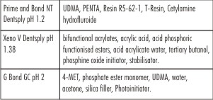 | Composition Of Materials Used
 |
Adherence test and agar disk diffusion method were statistically analyzed with ANOVA and scheff’s post hoc test.
Results
Agar disc diffusion method
The mean differed statistically significant between the positive control and all the other study groups.
The CTR group [Control] had the highest mean of about 7.033 followed by PBNT and Xeno V which had mean value of 5.33 and 4.1333 respectively. GB group had the lowest mean value 2.333 (Table 1)
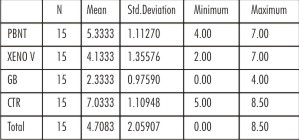 | Table 1 - Agar Disc Diffusion Method Descriptive Method
|
When compared between the groups the observed F value was found to be highly significant.[ P<0.000] (Table 2)
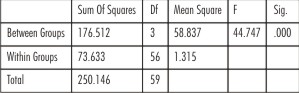 | Table 2 - Anova
|
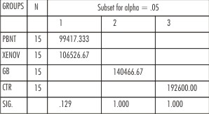
Further Schffe’s post hoc test indicates that group GB had least value, followed by Xeno V and PBNT. CTR group had the highest value. However, no significant difference was observed between Xeno V and PBNT group. (Table 3)
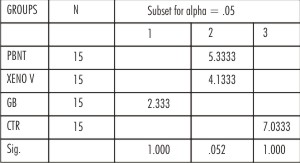 | Table 3 - SCHEFFEaPOST HOC TEST
|
Adherence Test
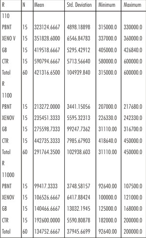
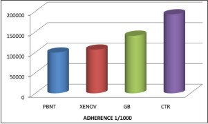
The statistical analysis of the adherence data showed that CTR [Control] group had the highest mean value followed by GB and Xeno V. PBNT had lowest mean value in all the three ratio’s of 1;10, 1;100 and 1;1000. (Table 4)
 | Table 4 - Adherence Test Descriptive Method
|
ANOVA indicates significant difference between groups in all the ratio 1;10, 1;100 and 1;1000.[P< 0.000] (Table 5)
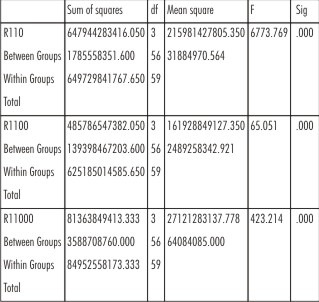 | Table 5 - Anova
|
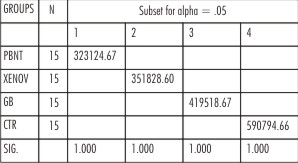
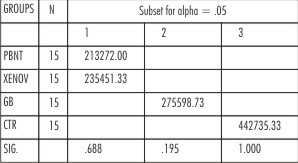
Further scheffe’s post hoc test indicates that group PBNT ,Xeno V had least value followed by GB.CTR group had the highest value. However no significant difference was observed between the groups. (Table 6,7,8) [P>0.05]
 | Table 6 - R110 Scheffe
|
 | Table 7 - R1100 Scheffe
|
 | Table 8 - R11000 Scheffe
|
Discussion
Concerns have been expressed about residual bacteria in cavity preparations. Not only do residual bacteria reside in the dentinal tubules and can therefore be a source of infection to the pulp but bacteria are also present at the area of the contraction gap between tooth and composite resin. Brannstrom [1987] characterizes cavity infection by bacteria as being due to multiple sources [1].invasion from the tooth surfaces via marginal gaps between tooth and restorative material [2].bacteria present in the smear layer [3]. bacteria present in the dentinal tubules [4]. bacteria at the gap at the dentin enamel junction [5]. microbes recontaminating the surface after cleaning. After cavity preparation assessments of residual caries and bacteria has in most cases been a subjective assessment. A number of different investigators have described procedure for the detection of carious dentin using dyes that assist visual detection[8]. But it has been suggested that the use of disclosing dye doesn’t completely eliminate the chances of bacteria remaining in a cavity and the accuracy of this material in reflecting the true bacterial condition has been questioned. Based on this evidence the need for a dentin adhesive with a potential antibacterial effect is of interest because it is in direct contact with the residual bacteria infected dentin in a prepared cavity.[9]
![Fig 1 : Co1lony forming units formed [CFU/ml], 1. Control](article-image-2864-FIG_1_CO1LONY_FORMING_UNITS_FORMED_[CFU_AND_ML]_AN.jpg) | Fig 1 : Co1lony forming units formed [CFU/ml], 1. Control
|
![Fig 1 : Co1lony forming units formed [CFU/ml], 2. Prime And Bond Nt](article-image-2865-FIG_1_CO1LONY_FORMING_UNITS_FORMED_[CFU_AND_ML]_AN.jpg) | Fig 1 : Co1lony forming units formed [CFU/ml], 2. Prime And Bond Nt
|
![Fig 1 : Co1lony forming units formed [CFU/ml], 3. Xeno V](article-image-2866-FIG_1_CO1LONY_FORMING_UNITS_FORMED_[CFU_AND_ML]_AN.jpg) | Fig 1 : Co1lony forming units formed [CFU/ml], 3. Xeno V
|
[Image 13]
Settembrini et al found that many of the commercially available acid etchants demonstrated antimicrobial activity against several bacteria commonly found in the oral cavity. So the lack of antibacterial activity of one bottle adhesives or the addition of antimicrobial agents to one bottle dentin adhesives might not be necessary in light of the antimicrobial activity of phosphoric acid etchants.
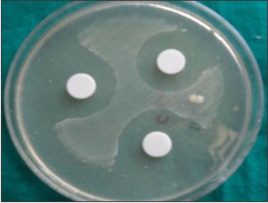 | Fig 2 : Inhibition zone formed, 1 .0.2% chlorexidine
|
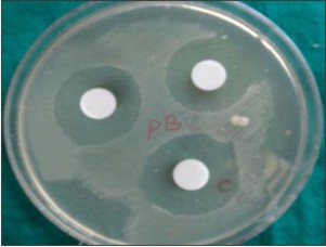 | Fig 2 : Inhibition zone formed, 2. Prime and bond NT
|
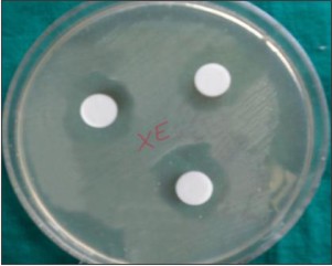 | Fig 2 : Inhibition zone formed, 3. Xeno V
|
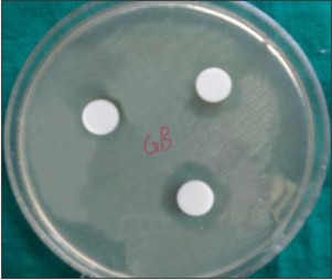 | Fig 2 : Inhibition zone formed, 4. G Bond
|
Self-etching adhesives have been introduced in an attempt to simplify the clinical application procedure and reduce the technique sensitivity and risk of the primed surface being contaminated. In self etching adhesive systems the pH of self etching primer solution is sufficiently low to demineralise the smear layer and underlying dentin surfaces, so etching and priming of the cavity can be accomplished simultaneously. Therefore the separate acid etching step is omitted. Because of the non rinsing procedure residual bacteria may remain at the interface between the tooth and the restorative material.
The dentin primer is the compound that comes into contact and reacts with the dentin substrate at the first stage of restoration in an adhesive system. If tooth conditioners such as primer posses antibacterial activity these bacteria could be eliminated there by preventing the secondary caries. So the antibacterial activity of these adhesive systems primers which are directly applied to the dentin plays an important role in the restoration’s longetivity.[10]
In the present study a comparison was made to check the antimicrobial efficacy of total etch adhesive agent Prime and Bond NT with self etch adhesive agents namely Xeno V and G Bond on streptococcus mutans.
Total etch with a 37% phosphoric acid etchant was used as that the etchant usually >30% phosphoric acid would exert a significant antibacterial effect on the microflora of the infected dentin.[2] Even studies have shown that phosphoric acid etchant materials demonstrated antimicrobial activity against several bacteria commonly found in the oral cavity.[8]
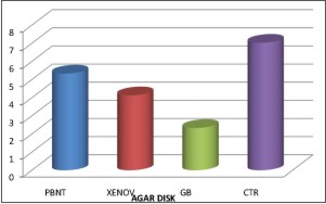 | Graph Table 1 : Inhibition Zone (Mm)
|
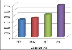 | Graph Table 2 : Number Of Bacterial Recovery (Cfu/Ml)
|
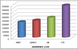 | Graph Table 3 : Number Of Bacterial Recovery (Cfu/Ml)
|
 | Graph Table 4 : Number Of Bacterial Recovery (Cfu/Ml)
|
Previous studies have reported that the antibacterial effect of some adhesive system is due to their low pH.3 .Based on this low pH of the primer namely Xeno V [pH 1.38] and G Bond [pH 2] was selected in the present study to check the antimicrobial efficacy.
Chlorhexidine is an antiseptic with a wide spectrum of action and it’s use has been generalized over the past two decades for the chemical control of bacterial plaque and disinfection of therapeutic cavities.[9] Silva et al reported a significant decrease in the number of bacteria in the dentinal tubules after application 0.2% chlorhexidine for 5 mins. It is also effective in reducing the levels of streptococcus mutans found on exposed carious root surfaces. For this reason it is adapted as a positive control for studies on bacterial growth or antibacterial activity.[11]
In terms of adherence test the CTR [Control] group showed more adherence of micro-organisms followed by G Bond and Xeno V. The PBNT group showed less adherence, because it is a total etch adhesive agent and about 83% of microorganisms are eliminated by acid etching. [2] Moreover it’s pH is about 1.2 and incorporation of antibacterial agent fluoride into the primer also inhibit the growth of microorganisms. But in Xeno V and G Bond their pH is about 1.38 and 2 respectively, the acidic nature of the primer [acrylic acid, 4-Methacryloxy ethyltrimellitic acid] itself acts as an antibacterial agent. But when compared with the adhesive agent incorporating fluoride as an antibacterial agent, the acidic nature of these bonding agents antibacterial property in inhibiting streptococcus mutans is less. But statistically the difference between the groups is insignificant. This is because even though total etch technique is capable of complete removal of microorganisms –contaminated smear layer with phosphoric acid, it is a multistep procedure and there is a chance of cross contamination. So self etching adhesives belong to the new generation of bonding agents that modify and incorporate the bacteria containing smear layer into their mechanism[11] was used to compare with total etch adhesive agent.
In addition to adherence testing agar disk diffusion testing was included. The agar diffusion method is generally used to investigate the antibacterial activity of materials from which an antibacterial component leaches out and the activity is determined based on the size of the inhibition zones.[6]
In this method 0.2% Chlorexidine showed highest inhibition zone which is used as positive control followed by PBNT and Xeno V.GB showed the least inhibition zone. The statistical result showed that PBNT and Xeno V is significant when compare to other groups. The reason for PBNT showing good inhibition zone is due to the fluoride content and it’s low pH [1.2] and Xeno V is due to its low pH 1.38. But the drawback of this method is, it did not differentiate between bactericidal and bacteriostatic activity and finally the method is at best semi quantitative.[2]
Thus within the parameter of this invitro study adhesive agent incorporating fluoride or the low pH of the monomer itself ,even 37% of phosphoric acid etchant in etch and rinse technique can reduce the occurrence of secondary caries .
Conclusion
Within the limitations of this invitro study the following conclusions can be drawn
Total etch adhesive agent Prime and Bond NT and self etch adhesive agent Xeno V showed better antibacterial activity against streptococcus mutans.
When compared between them total etch adhesive agent Prime and Bond NT showed highest antibacterial efficacy followed by self etch adhesive agent Xeno V with pH 1.38 and G Bond with pH 2 which showed less antibacterial efficacy.
References
1. C J Palanik, JC Setcost Antimicrobial Abilities Of Various Dentine Bonding Agents And Restorative Materials Journal Of Dentistry 1996; 24; 4;289-295.
2. M Vaidyanathan,ECSheehy Antimicrobial Properties Of Dentine Bonding Agents Determined Using Invitro And Ex-Vivo Methods Journal Of Dentistry 2009;37;514-521.
3. L Sebnem Turkuna,Mustafa Ates Antibacterial Activity Of Two Adhesives Systems Using Various Microbiological Methods Journal Of Adhesive Dentistry ;2005; 7;315-320.
4. G Schmalz,Z.Ergucu, Effect Of Dentin On The Antimicrobial Activity Of Dentin Bonding Agents Journal Of Endodontics 2004;30;5;339-344.
5. H Tsuchiyal,K Tsubota, Influence of adhesive application time on enamel bond strength of single step self etch adhesive systems operative dentistry 2010;35;77-83.
6. Cm Esteves,C Ota Tsuzuki, Antibacterial Activity Of Various Self-Etching Adhesives Systems Against Oral Streptococci Operative Dentistry;2010;35;4;448-453.
7. Thais Cachute Paradella,Cristiane Yumi Koga Ito Invitro Antibacterial Activity Of Adhesive Systems On Streptococcus Mutans Journal Of Adhesive Dentistry 2009;11;95-99.
8. L.Settembrini ,R Boylan A comparison of Antimicrobial Activity of Etchants used for a total etch technique Journal of Operative Dentistry 1997;22;84-88.
9. Atila Stephan Atac,C.Cehelis,Antibacterial activity of fifth generation dentin bonding systems Journal of Endodontics 2001;27;12;730-733.
10. Meserret Baseren ,Ruya Yazici, Antibacterial activity of different generation dentin bonding systemsJournal of Quintessence International 2005;36;339-344.
11. Ibrahim M Hamouda,Adel AL-Khodary Degree Of Conversion And Antimicrobial Activity Of Etch And Rinse Versus Self Etching Adhesives Journal Of Adhesive Dentistry 2010;12;33-38.
|