Introduction:
One of the difficult aspects of complete denture prosthodontics is the selection of appropriately sized maxillary anterior teeth, especially the central incisors. When no pre extraction records are available, selecting the proper anterior teeth size for edentulous patients can be difficult[1].
The mesiodistal width of central incisor can be determined by certain anthropometric measurements of face. Scandrett et al[2] have also studied the relationship between width of maxillary anterior teeth and the central incisor to that of intercommisural width, bizygomatic width, sagital cranial diameter and philtrum width. These investigators hypothesized that two or more of these predictor variables would provide a better prediction of maxillary tooth width than any of them individually.
Another anthropometric measurement of face is the distance between inner canthus of the eyes. The inner canthal distance (ICD) is defined as the distance between the medial angles of the palpebral fissure[3]. Laestadius et al[4] reported that in 78% of the population evaluated, the ICD was attained by the time the subject was 1 year old. Subsequently, the growth rate in that area was slow in contrast to the outer orbital dimensions. The previous anthropometric parametric measurements have not been evaluated with geometric progression[5] to determine proportionality between them and maxillary central incisor width.
Proportion is the study of the harmony of structures in space.[6],[7] When the proportion or ratio of a smaller to greater part is the same as the ratio of the greater part to the whole, it is said to be in geometric progression.[5],[7],[8] Because of their immense importance in geometry and architecture and their manifestations in nature, these ratios are called “Golden proportions”.
A proportion between two adjacent parts which is repeated across, enhances the unity within the diverse parts of a composition, and Lombardi[9] states that when this repeated ratio is equal to the Golden Mean, the composition is said to be esthetic.
Levin[10] has described the use of this proportion in dental esthetics. Any finite line can be arbitrarily divided into two segments by a point and a ratio of the smaller segment to the larger, and the ratio of the larger to the entire segment can be calculated. Only when the line is divided mathematically into segments that exhibit the proportion 0.618:1 does the golden proportion exist. The Golden Proportion is described as follows: The “proportion of the smaller to the greater is the same as the proportion of the greater to the whole.”
If, in a sequence of terms, the ratio between 2 consecutive terms is consistent, it is said to be in geometric progression (GP).
The ratio is called a common ratio.1 For example:
3,6,12,24, ;common ratio 2
8,4,2,1,1/2, ;common ratio 1/2
2,-10,+50, ;common ratio -5
In general, if a is the first term and r is the common ratio, then the GP may be written as follows:
a + ar+ ar2 +ar3 + ar n-1 to ‘n’ terms
if x , y and z are 3 terms in a GP , then the common ratio is equal to the following:
y/x = z/y or y2 = xz
Y, the square root of the product of 2 numbers x and y,is called the geometric mean. For example, the geometric mean of 4 and 9 is 4x9 = 6.
A straight line may be divided into two unequal parts (AB & BC) in any ratio. In a specific case when AB = 0.618 and BC=1, BC is the geometric mean of AB and AC.
0.618 1
________________________________
A . B. C.
AB/BC = smaller / larger = 0.618 / 1=0.618 and BC / AC = larger / whole = 1 / 1.618 = 0.618
OR
AB/BC=BC/AC
OR
BC2=AB X AC 1
Hence, AB, BC and AC are three consecutive terms of a geometric series in which the first term AB = a = 0.618, the second term = BC=ar=1, so that the common ratio r =1/0.618. Hence the third term AC = ar2 = ar X r = 1 X 1.618 = 1.618, as shown below:
0.618 1 1.618
________________________________
A . B. C. D.
The fourth term will be ar3 = 2.618. Thus we can construct the whole series as a+ar+ar2+ar3+ar4+ar5+ to n terms.
Or
0.618+ 1+1.618 +2.618 + 4.236 + 6.854+ to n terms.
Similarly, if we take 1 as the first term and the common ratio as 1.618 or its reciprocal 1/0.618, then we get the following geometric series:
a+ ar +ar2+ar3+ar4+ar5+ to n terms.
Or
1+ 1x 1.618 + 1x1.6182 +1x1.6183 + 1x1.6184 + 1x1.6185+ to n terms.
Or
1+1.618+ 2.618+4.236+6.854 + 11.090 + to n terms.
Hence, if the common ratios are 0.618 or 1.618, geometric progression results. For any decreasing function, we multiply by 0.618, and for any increasing function, we multiply by 1.618 to get the next result. As mentioned above, these ratios are called the golden proportion.
Some parts of the face have been reported to manifest golden proportions. The width of maxillary central incisor is in golden proportion to the width of lateral incisor, and the width of the lateral incisor is in golden proportion to the width of canine[8]. The purpose of this study was to determine the relationship between inner canthal distance and the mesiodistal width of the maxillary central incisors in terms of geometric progression.
Material And Methods:
The study was conducted on 250 subjects of Sri Guru Das Institute of dental/ medical college, Amritsar (Punjab). The subjects comprised 121 males and 129 females in the age group of 18-25 years. Young adults were selected because the ICD is established by 1 year of age, after which the rate of growth in the area is slow in contrast to the outer orbital dimensions[4]. Subjects with caries, restorations, severe attrition, congenital facial defects, orthodontic or crown restorations were excluded.
Natural Tooth Measurements:
1. The mesiodistal width of each of the maxillary central incisor (CIW) was measured at the contact points intraorally with the spring loaded divider, pointed members held parallel to the incisal edges and vertical to the facial surface of tooth interdentally.[Fig.1&2] The combined mesiodistal width of maxillary central incisors, thus obtained was divided by 2 to obtain the mesiodistal width of a single tooth. This procedure was similar to that reported by Garn et al,[11] who found no significant difference in the mesiodistal diameter of the right and left maxillary central incisors and thus justified the common practice of combining these tooth measurements
2. After the measurement procedure, the pointed members of the divider were placed on a piece of white paper, which was placed over a cork board, so that the pointed members perforated the paper when gentle pressure was applied. The perforations were joined by a straight line, which was measured to an accuracy of 0.1mm by a digital caliper. Each tooth was measured five times and the average of these five values were recorded. Impressions of the maxillary and mandibular arches were made in alginate. [Zelgan, Dentsply India Pvt. Ltd.] Instructions by the manufacturer regarding powder water ratio [22gms of powder and 57ml of water approximately] and mixing of alginate material and dental stone were precisely followed. The impression was poured within 5 minutes and cast was made. The mesiodistal width of maxillary central incisor was measured on the cast as done intraorally.
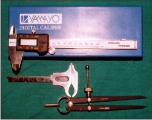 | Fig 1. : Armamentarium used
 |
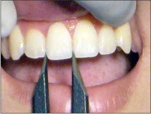 | Fig 2. : Measurement of mesiodistal width of central incisor at the contact points
|
Measurement Of Innercanthal Distance
1. Subjects were seated in a dental chair with their heads supported in an upright position so that they looked forward at the horizon.
2. The Boley gauge was placed against the forehead and lowered towards eyes. The arms of the caliper were adjusted so that they were in a gentle contact with the median angle of the palpable fissures of the eyes.[Fig.3]
3. The distance between the two anatomical landmarks was recorded as the inner canthal distance to an accuracy of 0.1 mm.
4. The inner canthal distance was measured five times for each subject and the values were averaged. The common ratio of geometric progression are 0.618 and 1.618.Any decreasing function is multiplied by 0.618 and increasing function by 1.618 to get the next result. As the inner canthal distance was greater than the combined widths of the maxillary central incisors, it was multiplied by 0.618. The resultant product was divided by 2 to obtain the width of a single central incisor. The formula can be expressed as follows:
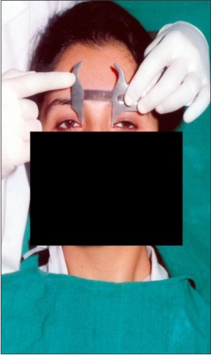 | Fig 3. : Measurement of innercanthal distance with Boley’s Gauge
|
FCIW=ICD/2x0.618, where FCIW is the calculated width of a maxillary central incisor. The calculated width was compared with the natural tooth measurement for each subject. The data thus obtained was put to a statistical analysis.
Results
Descriptive statistics for mean innercanthal distance and mean mesiodistal width of maxillary central incisor values using ICD and G.P for male and female subjects are presented in Table 1 & Graph 1,2,3.
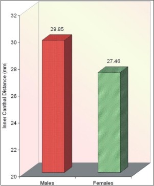 | Graphs 1. : Mean Inner Canthal distance in males and females
|
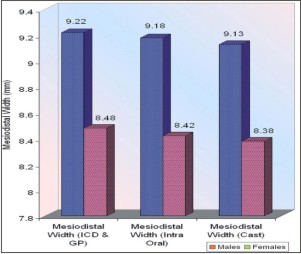 | Graphs 2. : Mean mesiodistal width of central incisor calculated by inner canthal distance and geometric progression, intraoral and cast (in males and females)
|
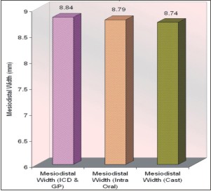 | Graphs 3. : Mean mesiodistal width of central incisor (combined) calculated by inner canthal distance and geometric progression, (intraoral and cast)
|
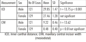 | Table- 1 : Mean Inner Canthal Distance And Mean Mesiodistal Width Of Maxillary Central Incisor Using Innercanthal Distance (ICD) And Geometric Progression (GP). (In Mm)
|
Means for both measurements were significantly higher for males than females. The correlation between the measured and calculated central incisor width values for all subjects was 0.96(0.964 for males and 0.963 for females). A paired t test revealed no significant difference between the mean width of natural tooth and its calculated size.
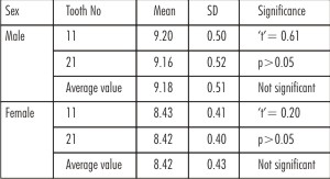
There was no satisfactory significant difference in the mesiodistal width of right and left central incisor measured intraorally and on the cast for both males and females(p>0.05) (Table 2 and 3)
Similarly, in all subjects, there was no satisfactory significant difference in the combined mesiodistal width of right and left central incisor measured intraorally and on the cast.[Table 4 and 5]
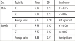
 | Table- 2 : Mean Mesiodistal Width Of Central Incisor In Males And Females (Intraoral).(In Mm)
|
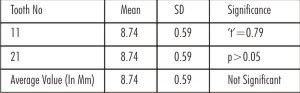
 | Table- 3 : Mean Mesiodistal Width Of Central Incisor In Males And Females (Cast).(In Mm)
|
 | Table- 4 : Combined Mesiodistal Width Of Tooth No.11 And 21 (Intraoral)
|
 | Table- 5 : Combined Mesiodistal Width Of Tooth No 11& 21(Cast).(In mm)
|
Discussion
As the subjects were from the same population, the results of this study are more applicable to the Indian population. In the present study, all the tooth dimension were significantly higher in males than in females. Variation based on gender has also been reported by Garn et al[11] (males 8.78mm, females 8.50 mm);Sanin and Savara[12](males 8.70,females 8.54 mm).The mesiodistal width of each of maxillary central incisor was measured at the contact points. The contact areas of central incisor was chosen, as the width is maximum at these contact areas; which was in accordance with the statement made by Wheeler[13]. The combined mesiodistal width of maxillary central incisors, thus obtained was divided by 2 to obtain the mesiodistal width of a single tooth. A similar study reported an insignificant difference in the mean value of mesiodistal central incisor crown diameter for 297 left and 297 right incisors (8.86 to 8.83 mm for men and 8.59 to 8.58 for women), justifying the common practice of combining left and right tooth measurements in comparative population studies[14].
The results of the present study showed that the measured mean value of central incisor width to be 8.79 ±0.59 mm, whereas the calculated width by means of ICD and GP was 8.84 ± 0.56 mm. The difference in the measured and calculated central incisor width was not statistically significant (p>0.05), which does concur with the findings of previous study,[1] in which the actual and calculated width of the maxillary central incisor were found to be highly correlated.
The mean ICD recorded in the present study (28.62mm) was less than the value reported by Laestadius et al (30.00 mm), who- in contrast to the present study found no significant difference between mean value for male and females.
The results of the present study suggest that ICD and Geometric prorression may be a reliable predictor of the width of the maxillary central incisors. Interpretation and extrapolation of the results may be tempered, however, by an acknowledgment of the study’s limitations. Only one ethnic group (Indian) was evaluated, and the subjects were selected within a narrow age range. It is possible that ethnic- related difference in ICD may exist. Further research is necessary to validate the outcome of this investigation.
Conclusion
With in the limitations of this study, the following conclusions were drawn:
1. The mesiodistal width of maxillary central incisor measured intra orally and on the cast were highly correlated with the calculated width of central incisor by means of inner canthal distance and geometric progression in both males and females.
1. Sex related difference in the mesiodistal width of the central incisor was reported. The mean central incisor width was significantly higher in males and females
2. Inner canthal distance when multiplied by a decreasing function value of this geometric progression term 0.618 and divided by 2, was a reliable predictor of maxillary central incisor width.
References
1. Abdullah M A: Inner canthal distance and geometric progression as a predictor of central incisor width. J.Prosthet.Dent.2002; 88(1):16-20.
2. Scandrett F R.,Kerber P E.,Umrigar Z A. A clinical evaluation of techniques to determine the combined width of the maxillary anterior teeth and the maxillary central incisor. J.Prosthet.Dent.1982;48:15-22.
3. Farkas L G. Anthropometry of the head and face in medicine. New York; Elsevier Sciences: 1981.P.11-2.
4. Laestadius N D., Aase JM., Smith DW. Normal inner canthal and outer orbital dimensions. J Pediatr.1969;74:465-8.
5. Huntley H E. The divine proportion: a study in mathematical beauty. New York; Dover Publications: 1970. P.22-9.
6. Lombardi R E. The principles of visual perception and their clinical application to denture esthetics. J.Prosthet.Dent.1973; 29:358-82.
7. Young H A. Denture esthetics. J.Prosthet.Dent.1956; 6:748-55.
8. Levin E I. Dental esthetics and the golden proportion. J. Prosthet. Dent. 1978; 40:244-52.
9. Behanam M., Padmanabhan T V., Subramanian,R. The golden proportion in esthetic treatment planning. Jr.of Indian prosthodontic society.2004;4(4):5-8.
10. Minoo M., Alireza K., Masoud V., Naser V. Evaluation of “Golden Proportion” in individuals with an esthetic smile. J. Esthet Restor Dent. 2004;16:185-93.
11. Garn SM., Lewis AB., Swindler DR., Kerewsky RS. Genetic control of sexual dimorphism in tooth size. J Dent Res. 1967;46: 963-72.
12. Sanin C., Savara BS. Permanent mesiodistal crown size. Am J Orthod.1971; 59:488-500.
13. Major M A., Stanley J N. The permanent maxillary central incisors. St. Louis 8th Ed. Wheeler’s Dental anatomy, Physiology and Occlusion. Elsevier,2003:149-70.
14. Varjao F M.,Nogueira S S. Inter-commisural width in 4 racial groups for the selection of maxillary anterior teeth in complete dentures. Int.J. Prosthodont. 2005; 18(6):513-515.
|