Introduction
The root apex is of interest to endodontics because the stages of root development and the type of tissue present within the roots of teeth are significant to the practice of endodontics. The development of the root begins after the enamel and the dentin formation has reached the future cemento-enamel junction. At the time of tooth eruption root development is only 62-80% i.e., 2/3rd of the root isformed[1]. If due to trauma or caries exposure occurs, the pulp undergoes necrosis, dentin formation ceases and root growth is arrested. The resultant immature root will have an open apex which is also called as Blunder Buss Canal.[2]
The word ‘blunderbuss’ basically refers to an 18th century weapon with a short and wide barrel. It derives its origin from the Dutch word ‘DONDERBUS’ which means ‘thunder gun’.
The walls of the canal are divergent and flaring, more especially in the buccolingual direction
The apex is funnel shaped and typically wider than the coronal aspect of the canal.[1]
Stages Of Root Development
According to the width of the apical foramen and the length of the root, Cvek has classified 5 stages of root development.
Stage 1 Teeth with wide divergent apical opening and a root length estimated to less than half of the final root length.
Stage 2 Teeth with wide divergent apical opening and a root length estimated to half of the final root length.
Stage 3 Teeth with wide divergent apical opening and a root length estimated to two thirds of the final root length.
Stage 4 Teeth with wide open apical foramen and nearly completed root length.
Stage 5 Teeth with closed apical foramen and completed root development.[3]
Causes of Open Apices
The open apex typically occurs when the pulp undergoes necrosis as a result of caries or trauma, before root growth and development are complete
(i.e. during stages 1-4)
An open apex can also occasionally form in a mature apex (stage 5) as a result of extensive apical resorption due to orthodontic treatment, periapical pathosis or trauma and over-instrumentation duringconventional root canal therapy.
Problems faced with open apex
Due to large apical diameter and smaller coronal canal diameter debridement is difficult.
Lack of apical stop makes working length determination and obturation difficult.
The thin root canal walls are always prone to fracture[3].
Various conventional treatment modalities for treatment of mature teeth with open apices are:
1. The use of Calcium hydroxide is well documented and most widespread treatment modality practiced to stimulate hard tissue deposition at the apex[3].
Drawbacks of this conventional therapy are:
The length of treatment time with calcium hydroxide is sometimes upto 18 months which is too long for patient motivation. Calcium hydroxide may not be effective on large fibrous periapical lesions. The fragility and porosity of the calcified barrier.
2. Periapical surgery with a root end filling.
Several materials that have been used over the years as root end filling materials are: Amalgam, Gold Foil Pellets, Polycarboxylate Cements, Titanium Screws, Bone Cement, Cavit, Composite Resin, Zinc Oxide Eugenol Cement, Reinforced ZOE Cements I.E., Super-EBA, Intermediate Restorative Materials (IRM), Ketac Silver, Glass Ionomer Cement.[4]
However none has been found to be satisfactory to be able to fullfill the desired criteria.
The newer materials being advocated as the materials of choice for root end repair are:
Mineral trioxide aggregate (MTA)
Dentinal shavings
Resorbable ceramic.
Freeze-dried cortical bone and freezed dried dentin=472;
Bone morphogenic protein
Case Report
A 27 year old male patient, reported to the Department of Conservative Dentistry and Endodontics, Seema Dental College and Hospital ,Rishikesh, with a chief compliant discoloredupper front tooth since 10-12 years and recurrent episodes of sinus formation and pus discharge with respect to the same tooth since last 8-10 months which subsides after medication.
Clinical examination revealed a discolored maxillary right central incisor with presence of intra oral sinus as shown in Figure 1. On palpation/percussionno tenderness was detected.
The concerned tooth did not respond to both electric and heat test.
Radiographic Evaluation
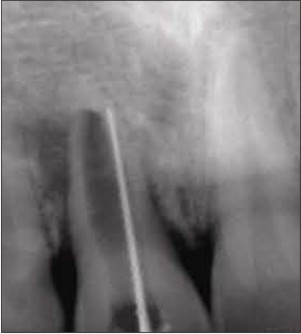
Periapical radiolusency involving distal surface of the root of maxillary right central incisor Figure 2.
Thickening of periodontal ligament and total loss of lamina dura
Internal root resorption along with blunt open apex i.r.t. maxillary right central incisor
Treatment Protocol
It was decided that the treatment would be carried out in two stages .First stage being conventional endodontic instrumentation followed by the three dimensional obturation with thermoplasticized gutta percha to manage the internal root resorption and the Second stage
being surgical intervention with the manipulation of the open apex followed by placement of suitable retrograde restorative material.
In First Visit
Access Opening was done and the chemomechanical preparation was carried out using conventional hand instruments. Passive irrigation was done using side-vented needle with 3% Sodium Hypochlorite and normal saline to prevent pushing of irrigant apically.
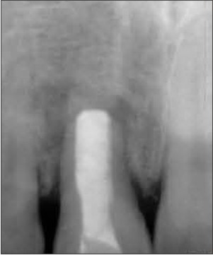
Working length determination was done by radiographic method .Canal was dried using paper points.
To obtain canal disinfection slurry of calcium hydroxide was temporized in the canal and left for one week.
Working length determination
After one week it was observed that the intra oral sinus had markedly reduced in size which could be considered as an indication of initiation of healing.
In Second Visit
Pulp space was prepared and irrigated with normal saline. Calcium Hydroxide sealer Sealapex ( kerr) in conjuction with thermoplasticized gutta percha Calamus ( Dentsply ) was used to obdurate the pulp space.
In Third Visit
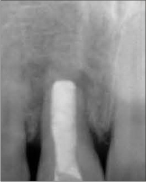
48 hours after obturation surgical intervention was done. After raising the trapezoidal flap, periapical currettage along with resection of apical third of the concerned tooth. Retrograde Mineral Trioxide Aggregate plug of 3-4mm was introduced into canal using MAP system (P.D. Systems) .The coronal access cavity was then restored with composite restoration.
Placement of 3-4 mm of MTA Plug
Results
1. Radiographs show decrease in radiolucency after 1 month and 3 months recall and review.
2. Clinically, complete retraction of intraoral sinus which is quite suggestive of healing.
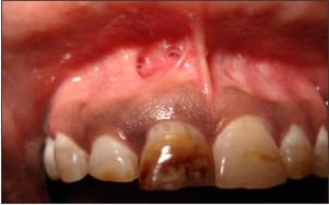 | Figure 1
 |
 | Figure 2
|
 | Working Length Determination
|
 | Placement Of 3-4 Mm Of Mta Plug
|
 | After 1 Month
|
Discussion
Teeth with incomplete rhizogenesis, requiring endodontic management, leave the dentists in a dilemma. In addition to presenting with an open apex, we are frequently faced with a large, long, irregular canal which lends itself to a compromised three-dimensional obturation. The condition is further compromised in those cases where a large, wide-open canal exists, predisposing it to root fracture, when conventional lateral obturation techniques are used.
Achievement of a perfect seal at the apex using an osteoinductive and biocompatible filling material is the critical factor affecting success in endodontics. The endodontic treatment of non vital immature anterior teeth after trauma remains complicated because of necrotic pulp tissue, large open apices, divergent root walls, thin dentinal walls and frequent periapical lesion.
The main aim of root end filling material is to fill the apical canal space and obtain hermetic seal between periodontium and the root canal system. As an endodontist, we should be careful to adopt the best available evidence for supporting clinical treatment plans.
Mineral trioxide aggregate (MTA) was developed in the 1990s initially for use as a root-end filling material due to its ability to set in the presence of moisture.[6] Whilst its chemistry was based on that of ordinary Portland cement, significant differences preclude use of the latter as a clinical substitute.[7] MTA has been shown capable of inducing mineralised tissue formation at a variety of oral and dental tissue sites and subsequently its potential applications within dentistry have expanded[8]
Bates et al found MTA superior to the other traditional root-end filling materials. MTA expands during setting which may be the reason for its excellent sealing ability[9].
Torabinejad et al. 1995,Xavier et al. 2005 suggested that mineral trioxide aggregate is most biocompatible and bacteriostatic material with good sealing property, which stimulates cell growth, adhesion and proliferation.[10]
MTA composed of:
Fine hydrophilic particles of:
Tricalcium silicate,
Tricalcium aluminates,
Ttricalcium oxide,
Silicate oxide and
Other mineral oxides (responsible for chemical and physical properties), which sets in the presence of moisture.[7],[9]
Calcium hydroxide is the main compound released by MTA in water. The calcium hydroxide
is biocompatible with tissue. The initial pH of MTA is 10.2 with an increase to 12.5, 3 hours after mixing.
MTA offers a biologically active substrate for bone cells and stimulates interleukin production because of its alkaline pH and calcium ion release.
The most characteristic tissue reaction of MTA is the presence of connective tissue after the first postoperative week and with a longer duration MTA which allows the overgrowth of cementum and formation of bone which facilitates the regeneration of periodontal ligament.[11]
Conclusion
During the last 20 years there have been many changes in the rationale governing the Treatment of teeth with open apex[5]
It is essential to have thorough understanding of the compatibility of the material, its physiological response, and the histological changes that takes place during and after the use of selected materials.
References
1. Beena Phillip Mathew, Mithra N. Hedge. Management of non vital immature teeth-case reports and review. Endodontology 18-21
2. Avinash R. Salgar, Manoj G . Chandak.N U Manwar. Blunderbuss canal :a challenge for endodontist. Endodontology 77-81
3. Mechanism of tooth eruption – T.B. Kandos. B.D.J. Vol 181, No. 3 Aug. 10 : 1996 (91-95).
4. Biocompatibility of retrograde root filling materials: a review. Australian Endodontic Journal 2008; 34: 30–35.
5. Periradicular surgery, curratage, root-end resection, root end fillings. Pg no.230-261, Gutmans surgical endodontics by Gutmann J.L.
6. Torabinejad M, Watson TF, Pitt Ford TR. Sealing ability of a mineral trioxide aggregate when used as a root end filling material. Journal of Endodontics 1993;19:591–5.
7. Dammaschke T, Gerth HU, Zuchner H, Scha¨ fer E. Chemical and physical surface and bulk material characterization ofwhite ProRoot MTA and two Portland cements. Dental Materials 2005;21:731–8.
8. Camilleri J, Pitt Ford TR. Mineral trioxide aggregate: a review of the constituents and biological properties of the material.International Endodontic Journal 2006; 39:747–54.
9. Mineral tioxide aggregate – review .The Journal of Clinical Pediatric Dentistry . 2009; 34(1);1-7
10. Kogan P, He J, Glickman GN, Watanabe I. Effects of various additives on setting properties of MTA. J Endod. 2006;32:569-572.
11. Cellular response to mineral trioxide aggregate. Journal of endodontics 1998;24(8):543-547.
|