Introduction
Odontomas are considered to be developmental anomalies resulting from the growth of completely differentiated epithelial and mesenchymal cells that give rise to ameloblasts and odontoblasts. These tumors are basically formed of enamel and dentin but they can also have variables amounts of cement and pulp tissue.[1] During the development of the tumor, enamel and dentin can be deposited in such a way that the resulting structures show an anatomic similarity to normal teeth, in which case the lesion is classified as a compound odontoma. However, when the dental tissues form a simple irregular mass occurring in a disorderly pattern, it is described as a complex odontoma.[2] Compound odontomas appear more frequently than complex odontomas.[3],[4] These odontogenic tumors can be found anywhere in the dental arches. The majority of odontomas which are located in the anterior region of the maxilla are compound, while the great majority of odontomas located in the posterior areas, especially in the mandible, are complex odontomas.[1],[5],[6] The etiology of the odontoma is unknown.[7] However, it has been suggested that trauma and infection at the place of the lesion can offer ideal conditions for its appearance.[7],[8] In general they are asymptomatic, have slow growth,[1] and seldom exceed the size of a tooth, but when large can cause expansion of the cortical bone.[1],[2]
Odontomas may be diagnosed at any age but they are usually detected during the first two decades of life.[1],[3] One study analyzed 396 cases and showed that diagnosis usually happens between ages 11 and 15 years.[4] Another study comprising 149 cases concluded that the lesions are detected most often during the second decade of life.[6] Many times odontomas are found associated with unerupted teeth. The canines, followed by upper central incisors and third molars, are the most frequent teeth impacted by odontomas.[4] In a very few instances odontomas are related to missing teeth.[8] Generally these malformations are intraosseous, but occasionally they may erupt into the oral cavity. Radiographic aspects of odontoma are characteristic. The complex odontoma appears as an irregular mass of calcified material surrounded by a thin radiolucent area with smooth periphery, and the compound type shows calcified structures resembling teeth in the center of a well-defined radiolucent lesion. A periodontal and pericoronary space characteristic of unerupted teeth is seen around each tooth. A developing odontoma can be discovered by routine radiography, but may cause difficulty in identification due to lack of calcification. The histological examination of odontomas often shows the presence of enamel matrix, dentin, pulp tissue, and cementum that can, but need not, exhibit a normal relationship.[1],[7] Compound odontomas are formed by tooth-like structures which resemble pulp tissue in the central portion surrounded by a dentin shell and partially covered by enamel. Complex odontomas are conglomerates without orientation of dentin, enamel, enamel matrix, cementum, and areas of pulp tissue. The capsule of connective tissue that surrounds an odontoma is similar to the follicle that covers a normal tooth.[7]
Case Report
A 18 year old female patient reported to the Department of Oral & Maxillofacial Surgery Bhojia Dental College Baddi with chief complaint of swelling in the upper left back tooth region. Intraoral examination revealed missing upper left first and second premolars and palatally titled crown of upper left canine with crowns over upper left central and lateral incisors. There was no history of trauma. Radiographic examination revealed the presence of three lesions having tooth like structures. The patient was operated under local anesthesia. Three mineralized structures showing tooth like appearance in different developing stages were removed surgically. Postoperative oral and written instructions specifically related to the maintenance of an appropriate oral hygiene, ingestion of cold and soft meals and management of pain were given to the patient. The wound
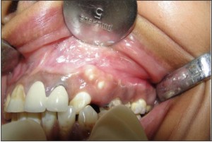 | Fig 1 - Introral Photograph Showing Miniature Tooth Like Structures
 |
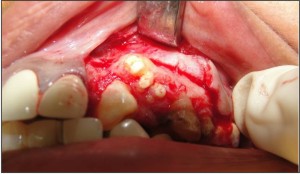 | Fig 2 - Surgical Exposure After Reflecting The Flap
|
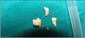 | Fig 3 - Surgical Specimen
|
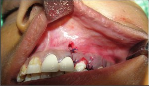 | Fig 4 - Postoperative Closure
|
healing took place uneventfully and the missing teeth were replaced by placing implants.
Discussion
Odontomas are classified as benign, mixed, calcified odontogenic tumors. Although classified as odontogenic tumors they lack proliferative potentiation. The etiology of odontoma is unknown, but has been attributed to various pathological conditions like local trauma, infections, hereditary anomalies (Gardners syndrome, Hermanns syndrome), odontoblastic activity and alteration in the genetic component responsible for controlling dental development. Hitchin suggested that odontomas are either inherited or due to a mutant gene or interference with genetic control of tooth development postnatally. On the other hand, Levy reported the experimental production of this lesion in rat by traumatic injury. Of all the odontoma, 67% occurred in the maxilla and 33% in the mandible. The compound odontoma has a predilection towards the anterior maxilla (61%) whereas only 34% of complex odontomas occurred in this site. In general, complex odontoma has a predilection for the posterior jaws (59%). In this case, compound odontoma was found in the anterior maxilla which is in accordance with the reported literature. Interestingly both types of odontoma occur more frequently on the right side of the jaw than on the left (Compound 62%, Complex 68%), whereas in our case, the odontoma occurred on the left side. Cystic odontomas slowly increase in size and cause expansion of bone which is not seen in this case. In 70% of the odontomas, pathologic alterations are observed in the neighboring teeth, such as devitalization, malformation, aplasia and malposition. In a clinical setting, an odontoma should be removed because it contains various tooth formulation that can predispose to cystic change, interfere with tooth eruption, cause considerable bone expansion. Usually, surgical treatment of odontoma is conservative and curative with no possibility of recurrence, if, the lining epithelium is removed intact. But one case of recurrent odontoma has been reported and the reason stated for recurrence includes-1) repeated episodes of infection in same region. 2) Multiple schizodontia due to dental lamina activity. 3) odontogenic rests left behind. Kaban states that odontoma can be easily enucleated and the adjacent teeth are seldom harmed by surgical excision because they are separated by septum of bone. Thereby early diagnosis and surgical removal of odontoma will aid in better prognosis of teeth in relation to odontomaand there is little probability of recurrence.[1],[8] When the odontomas are associated with unerupted teeth, orthodontic traction of the impacted tooth soon after removal of the lesion may be needed, especially if it is not diagnosed and treated early.[5],[12]
Conclusion
Odontomas rarely erupt into the mouth and tend to be associated with impacted teeth .In a clinical setting, an odontoma should be removed because it contains various tooth formulation that can predispose to cystic change, interfere with tooth eruption, cause considerable bone expansion. Usually, surgical treatment of odontoma is conservative and curative with no possibility of recurrence, if the epithelial lining is removed intact.
References
1. Neville BW, Damm DD, Allen CM, Bouquot JE: Oral and Maxillofacial Pathology. Philadelphia: Saunders, 1995, pp 531-33.
2. Cawson RA, Binnie WH, Eveson JW: Color Atlas of Oral Disease. Clinical and Pathological Correlations. Hong Kong: Mosby-Wolfe, 1993, pp 6-19.
3. Owens BM, Schuman NJ, Mincer HH, Turner JE, Oliver FM: Dental odontomas: a retrospective study of 104 cases. J Clin Pediatr Dent 1997; 21: 261-4. .
4. Katz RW: An analysis of compound and complex odontomas. ASDC J Dent Child1989; 56:445-9
5. Budnick SD: Compound and complex odontomas. Oral Surg Oral Med Oral Path 1976; 42:501-6
6. Shafer WG, Hine MK, Levy BM: A Textbook of Oral Pathology, 4th Ed. Philadelphia: Saunders, 1983, pp 308-11.
7. Areal-López L, Silvestre DF, Gil LJ: Compound odontoma erupting in the mouth: 4 year follow-up of a clinical case. J Oral Pathol 1992; 21:285-8.
8. Brunetto AR, Turley PK, Brunetto AP, Regattieri LR, Nicolau GV: Impaction of a primary maxillary canine by an odontoma: surgical and orthodontic management. Pediatr Dent1991; 13:301-2
9. Oliver RG, Hodges CGL: Delayed eruption of a maxillary central incisor associated with an odontome: report of case. ASDC J Dent Child 1988; 55:368-71.
10. Giunta JL, Kaplan MA: Peripheral, soft tissues odontomas. Oral Surg Oral Med Oral Pathol 1990; 69:406-11.
11. Kaban, LB: Pediatric Oral and Maxillofacial Surgery. Philadelphia: Saunders, 1990, pp 111-2. |