Introduction
According to Moyers[1], the tooth most frequently lost to caries or periodontal disease is the permanent first molar. Various treatment options are available for the closure of this space. If the second or third molar is present, a fixed partial denture (FPD) is usually the treatment of choice. But, loss of adjacent tooth structure, hypersensitivity, chances of caries on the abutments and food lodgement beneath improperly fabricated pontics are some of the drawbacks of an FPD. A prosthetic implant is a better option but the success of even an implant can be hindered by peri-implantitis.
Molar protraction can be an alternative to restoration with posterior dental implants or fixed partial dentures. When compared with the maxillary molars, the mandibular molars are more difficult to move mesially because of the structural differences between the two jaws. The posterior maxilla is composed of uniformly thin cortices interconnected by a network of spacious trabeculae[2], while the posterior mandible consists of thicker cortical bone with dense, radially oriented trabeculae[3]. Therefore avoiding anchorage loss is considerably more challenging in the mandible than in the maxilla. Furthermore, if the buccal and lingual cortical plates in the edentulous region have collapsed, safe and effective protraction may be impossible.
Recently, titanium screws have become popular for absolute anchorage during various types of tooth movement[4], [5], [6], [7].
In the case presented here, we demonstrate titanium screws placed in the buccal alveolar bone for the protraction of the mandibular second molars into atrophic first molar extraction sites.
Diagnosis
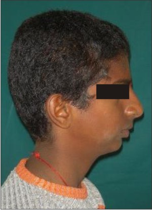
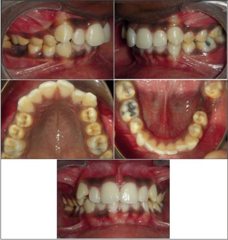
A 14 year old male reported with a chief complaint of forwardly placed upper front teeth. On extraoral examination he was found to have a convex profile due to retrognathic mandible, a deep mentolabial sulcus and competent lips (Figure 1). Intraoral examination showed increased overjet and overbite, Angle's class III molar relation on the right side, class II canines and mild crowding in upper and lower anterior teeth (Figure 2). 35 was congenitally absent and the patient also gave a history of extracted 46 because of caries 18 months ago (Figure 2). Cephalometric examination showed a skeletal class II with horizontal growth pattern. Hand wrist radiograph showed the patient to be in post pubertal growth phase with 10-25% of mandibular growth left according to Bjork, Grave and Brown method[8]. The patient was diagnosed as Angle's class III malocclusion on skeletal class II jaw bases with horizontal growth pattern.
Treatment Planning
To correct the mandibular retrognathism, it was decided to advance the mandible using Churro jumper fixed functional appliance[9]. However the main concern
 | Figure 1
 |
 | Figure 2
 |
 | Figure 3
 |
 | Figure 4
 |
was the missing lower molar. As the alveolar ridge from where the molar had been extracted showed sufficient thickness, it was decided to protract the second molar into the extraction site. On the left side it was planned to extract the upper second premolar and protract the molar to get a bilateral class II molar relationship. To preserve anterior anchorage it was decided to use mini implants for protraction of both the molars (26 and 46). In the lower arch proximal stripping would suffice to relieve the mild crowding.
Treatment Progress
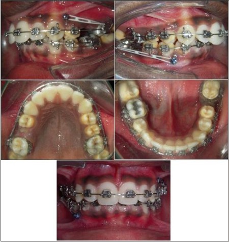
After extraction of 25 and proximal stripping in the lower arch, alignment of teeth was achieved in both the arches. A mini implant (diameter 0.8 mm, length 8 mm) was placed in the mandible between 43 and 44. Another mini implant (diameter 0.8 mm, length 11 mm) was placed in the maxilla between 23 and 24. Elastic chains were used to apply force for protraction of 26 and 46 using anchorage from these mini implants (Figure 3).
Protraction was done on 0.019" X 0.025" stainless steel wire to preserve the arch form during this protraction.
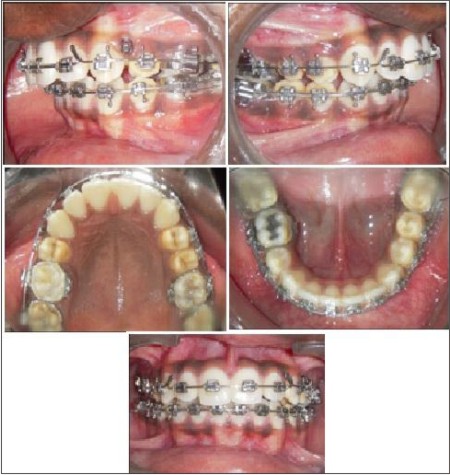
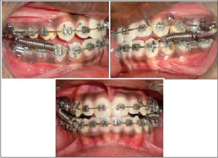
After molar protraction was completed in 5 months, bilaterally Angle's class II
 | Figure 5
 |
 | Figure 6
 |
molar relation was achieved with well aligned arches (Figure 4).
At this stage mandibular advancement was carried out using Churro jumper fixed functional appliance (Figure 5).
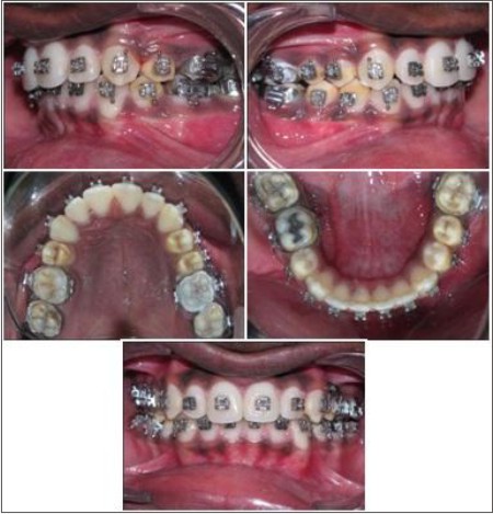
After 6 months of wearing the Churro jumper, a well settled occlusion with normal overjet and overbite was achieved with molars and canines in class I relation (Figure 6).
Even the profile showed a favourable change after mandibular advancement (Figure 7).
Discussion
Previously, Graber[10] stated that clinicians can seldom close molar spaces with limited orthodontic therapy. The large root surfaces of molars make their movement uncertain and simultaneously cause unwanted tooth movements such as lingual tipping of the incisors. However, now with skeletal anchorage, it is possible to solve anchorage problems that could not be addressed previously. Titanium screws have gained wider acceptability; they have several advantages over dental implants, such as simpler placement, lower costs, minimal surgical trauma, and immediate loading[4],[5],[6]. In addition, their small size allows them to be placed in most anatomic locations so that force can be applied in any direction. In our patient, we placed the titanium screws on the buccal alveolar bone between the roots of the premolars for easier accessibility and better oral hygiene maintenance.
Although the titanium screws remained stable throughout the protraction phase, discomfort from mild chronic inflammation is possible around the screw sites. These problems can be prevented if the screws are accurately positioned and careful oral hygiene is maintained with brushing and chlorhexidine treatment.
According to Kessler[11], mesial movement of mandibular molars should not be attempted because their roots are wider than the adjacent edentulous ridge and can cause loss of osseous support. However, a couple of reports in the orthodontic literature have refuted that statement[12],[13]. Hom and Turley[12] reported that mandibular space closure was not only possible, but it could even provide great benefits to some patients. They proposed space closure as potential therapy when the mandibular first molars are missing.
Root resorption was minimal for both molars; even though they were translated more than 8 mm. Stepovitch[13] studied the changes in edentulous ridge before and after space closure of mandibular first molar spaces. He concluded that clinicians can close spaces of 10 mm or more in adults, but maintaining the closed spaces is difficult. For the same reason, fixed buccal retainers are advocated from molar to premolar in the mandibular arch to prevent the spaces from reopening during retention.
Conclusion
Although we completely agree that bone loss must be avoided in edentulous patients, moderate bone loss should not in any way prevent the closure of edentulous spaces. A fixed prosthesis has always been the preferred option for these patients. However, prostheses have certain limitations: initial cost, partial destruction of abutment teeth, secondary caries, and mechanical failures. Hence, both space closure and a fixed prosthesis should be considered as solutions for missing teeth. From a clinical perspective, this case demonstrates that titanium screw anchorage is an effective means for protracting the mandibular second molars into the first molar extraction sites.
References
1. Moyers RE. Handbook of orthodontics. Chicago: Year Book Medical Publishers; 1988.
2. Adell, R.; Lekholm, U.; Rockler, B.; and Brånemark, P.I.: A 15-year study of osseointegrated implants in the treatment of the edentulous jaw, Int. J. Oral Surg. 10:387-416, 1981.
3. Deguchi, T.; Nasu, M.; Murakami, K.; Yabuuchi, T.; Kamioka, H.; and Takano-Yamamoto, T.: Quantitative evaluation of cortical bone thickness with computed tomographic scanning for orthodontic implants, Am. J. Orthod. 129:721.e7-12, 2006.
4. Creekmore TD, Eklund MK. The possibility of skeletal anchorage. J Clin Orthod 1983;17:266-9.
5. Costa A, Raffaini M, Melsen B. Miniscrews as orthodontic anchorage: a preliminary report. Int J Adult Orthod Orthognath Surg 1998;13:201-9.
6. Park HS, Bae SM, Kyung HM, Sung JH. Micro-implant anchorage for treatment of skeletal Class I bialveolar protrusion. J Clin Orthod 2001;35:417-22.
7. Kuroda S, Katayama A, Takano-Yamamoto T. Severe anterior open-bite case treated using titanium screw anchorage. Angle Orthod 2004;74:558-67.
8. Grave KC, Brown T. Skeletal ossification and the adolescent growth spurt. Am. J. Orthod. 1976; 69 (6): 611-9.
9. Castañon R, Mario S, Larry W. Clinical Use of the Churro Jumper. J Clin Orthod 1998; 32 (12): 731.
10. Graber TM. Orthodontics: principles and practice. Philadelphia: Saunders; 1972.
11. Kessler M. Interrelationships between orthodontics and periodontics. Am J Orthod 1976;70;154-72.
12. Hom BM, Turley PK. Effects of space closure of mandibular first molar area in adults. Am J Orthod 1984;85:457-69.
13. Stepovitch MI. A clinical study on closing edentulous spaces in the mandible. Angle Orthod 1979;49:227-33. |