Introduction
Normally, first deciduous tooth eruption in the oral cavity occurs when the child is approximately six-months old.[1] Teeth present at the time of birth are called Natal teeth. Teeth which erupt in the neonatal period, within thirty days of birth are Neonatal teeth.[2] Teeth erupting beyond the natal period of thirty days (i.e., erupting within 1-3.5 months) are usually referred to as early infancy teeth.[3],[4]
Incidence of neonatal teeth is very low. In previous studies, it has been estimated to be between 1: 1,000 and 1: 30,000. [3],[4] Terms such congenital teeth, fetal teeth, pre-deciduous teeth, premature teeth, precociously erupted teeth or dentitia praecox have been used to refer to this condition.[5] Natal and neonatal teeth erupt commonly in mandibular anterior region, but several reports show their unusual occurrence in the mouth. It has been reported that natal and neonatal teeth erupt 85% in mandibular incisor region, 11 % in maxillary incisor region, 3% in mandibular canine region and 1% in maxillary canine and molar region.[6]
Case Report
A thirteen days old male infant was referred with complaint of presence of mobile teeth in the lower jaw since birth and difficulty in breast feeding. Oral examination revealed grade-II mobile teeth in the mandibular anterior region (Figure 1). The crown size was normal; the gingiva was of normal appearance. A diagnosis of natal tooth in mandibular anterior region was made. The medical
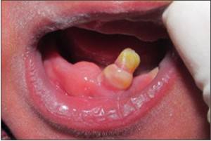 | Fig: 1 - Photograph of neonate mouth showing, natal tooth in mandibular anterior region.
 |
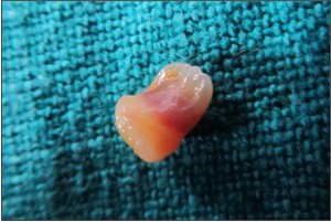 | Fig: 2 - Extracted natal tooth with pulp tissue
|
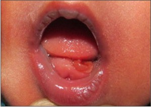 | Fig: 3 - Photograph of neonate mouth showing, post-operative area after extraction of natal teeth.
|
history was non-contributory. As the teeth were the suspected cause of the chief complaint, they were extracted under topical anaesthesia. The extracted teeth had crowns but were devoid of roots (Fig: 2). Post extraction socket appeared normal (Fig:3). Four weeks later, intraoral examination revealed complete and uneventful healing of the extraction socket and the mother reported that, feeding was normal.
Historical Review
During the earlier days (59 BC), appearance of natal and neonatal teeth was related to mixed superstitious beliefs. It was claimed to bring fortune or misfortune.[1],[4] The folklore and misconceptions surrounding natal and neonatal teeth vary; in some cultures like Malaysian communities, a natal tooth is believed to herald good fortune; in others, its occurrence is considered bad omen. In China, Poland, India, and Africa, the affected children are considered to be monsters and bearers of misfortune. In England, the belief was that this condition would guarantee the conquest of the world.[7]
Prevalence
The prevalence of natal teeth is 1:1000 to 1:30,000 depending on the type of the study; the highest prevalence is found in the only study that relies on personal examination of patients. Other reports reveal it to be around 1 in 2000-3500 live births[2], [8], [9], [10], [11]
Discussion
For past hundreds of years many cases of infants born with natal and neonatal teeth have been reported in dental literature. The majority of natal teeth represent early eruption of primary dentition. They are more common in mandibular arch than maxillary arch and more commonly occur in the incisor region than the canine and molar regions.
Classification
Spoug and Feasby (1966) have suggested that clinically, natal and neonatal teeth are classified according to their degree of maturity.[9]
1. A mature natal or neonatal tooth is one which is nearly or fully developed and has relatively good prognosis for maintenance.
2. The term immature natal or neonatal tooth, on the other hand, implies a tooth with incomplete or substandard structure; it implies a poor prognosis.
The appearance of each natal tooth into the oral cavity can be classified into four categories as the teeth emerge into the oral cavity.[9],[12]
1. Shell-shaped crown, poorly fixed to the alveolus by gingival tissue and absence of a root.
2. Solid crown, poorly fixed to the alveolus by gingival tissue and little or no root.
3. Eruption of the incisal margin of the crown through the gingival tissues.
4. Edema of gingival tissue with an unerupted but palpable tooth.
Etiology
Following hypothesis have been suggested for the occurrence of natal or neonatal teeth:
Hereditary transmission of a dominant autosomal gene appears to be an important factor.[13] According to Bodenhoff and Gorlin 15% of children with natal and neonatal teeth had parents, siblings, or close relatives with a history of same condition.[8]
Endocrine disturbances: Due to excessive secretion of the pituitary, thyroid or gonads.
Infection: For example, congenital syphilis [13]
Nutritional deficiency, e.g., hypo vitaminosis (which in turn is caused by poor maternal health, endocrine disturbances, febrile episodes, pyelitis during pregnancy, and congenital syphilis).[14]
Environmental factors: Poly chlorinated biphenyls (PCB) and dibenzofurans seem to increase the incidence of natal teeth. These children usually show other associated symptoms, such as dystrophic finger nails, hyper pigmentation, etc.[13]
The most acceptable theory is based upon the result of superficial localization of the dental follicles, probably related to hereditary factor.[5]
Associated Syndromes
Natal teeth and neonatal teeth are frequently found associated with developmental abnormalities and recognized syndromes. These syndromes include Adrenogenital syndrome, Cleft lip and palate, Craniofacial dysostosis, Craniosynostosis syndromes, Ectodermal dysplasia, Ellis-van Creveld syndrome, Epidermolysis bullosa simplex, Hallerman-Streiff syndrome, Jadassohn-Lewandowsky syndrome, Multiple steacystoma, Pallister-Hall syndrome, Pfeiffer syndrome, Pierre-Robin syndrome, Polydactyly type II, Rubinstein-Taybi syndrome, Sotos syndrome, Steatocystoma multiplex, Van der Woude syndrome, Walker-Warburg syndrome and Wiedeman-Rautenstrauch syndrome[14],[15],[16].
Clinical Aspects
Clinically, natal teeth are small or normal size, conical or of normal shape. They may reveal an immature appearance with enamel hypoplasia and small root formation. Natal teeth may exhibit a brownish yellow or yellowish brown or whitish opaque colour. They are attached to a pad of soft tissue above the alveolar ridge.[17], [18], [19] The dimensions of the crown of these teeth are smaller than those of the primary teeth under normal conditions.
Complications
Complications related to natal and neonatal teeth include discomfort during suckling causing irritation and trauma to infants' tongue, sublingual ulceration (Riga- Fédé disease) with laceration of the mother's breasts and aspiration of the teeth.[20] Presence of neonatal teeth may cause interruption of breastfeeding. Prolonged gingival irritation from natal or neonatal teeth may cause localized inflamed gingiva or fibrous hyperplasia.[12]
During neonatal period, clinical and radiographic examination helps to differentiate the premature eruption of a primary deciduous tooth from a supernumerary tooth. Additionally, presence or absence of a tooth germ in the primary dentition would determine whether or not the latter belongs to the normal dentition.
Cysts of the dental lamina and Bohn nodules could be confused with neonatal teeth, and therefore the diagnosis can be confirmed by radiographic examination. Another condition which should be considered is epulis, which is a tumor-like growth of the gingiva, and reactive rather than neoplastic lesion. Other differential diagnosis includes lymphangioma and hamartoma of the alveolar ridge.[1]
Treatment and Management
No intervention is necessary if the tooth does not interfere with breastfeeding, if it is not very mobile or if it is a part of deciduous dentition. The management of natal and neonatal teeth is largely aimed at their preservation for aesthetics and maintenance of space for eruption of successor tooth.
In light of this knowledge, extraction of natal and neonatal teeth is reserved until they cause difficulty to the infant and mother. Occasionally, they may exfoliate spontaneously or require extraction because of excessive mobility, concerns regarding aspiration or the loss of attachment with subsequent development of abscess. Extraction may be indicated if there is feeding difficulties or complications like Riga- Fédé disease. Extraction of natal teeth can be advised if child's age is 10 days or above and child has appropriate amounts of Vitamin K in the blood. Otherwise prophylactic administration of vitamin K (0.5-1.0mg, i.m.) is advised before and after extraction [21] because of the risk of haemorrhage, since vitamin K is essential for the production of prothrombin in the liver for clot formation at extraction site.
To prevent continued development of the cells of the dental papilla, extraction of the tooth should be followed by curettage of the socket. Failure to curette the socket may cause eruption of odontogenic remnants and necessitate future treatment. Alternatively, to prevent injury to maternal breast, grinding or smoothening of the incisal edges of the teeth is also recommended. Placement of composite resin over the incisal edges has also been suggested[7].
Conclusion
It must be considered that although rare, natal and neonatal teeth are conditions of fundamental importance to pediatric dentistry and paediatricians since that their presence may lead to numerous complications. The child must be monitored to restore the function and esthetics of the normal primary dentition.
References
1. Alexander K.C. Leung, William Lane M. Robson. Natal Teeth: A Review. Journal of the national medical association 2006; 98 (2):226-228.
2. Massler M, Savara BS. Natal and neonatal teeth; a review of 24 cases reported in the literature. J Pediatr. 1950;36(3):349-59.
3. Kates GA, Needleman HL, Holmes LB. Natal and neonatal teeth: a clinical study. JADA 1984, 109: 441-3.
4. Anegundi RT, Sudha P, Kaveri H, Sadanand K. Natal and neonatal teeth: a report of 4 cases. J Indian Soc Pedo Prev Dent 2002;20(3): 86-92.
5. Portela MB, Damasceno L, Primo LG. Unusual case of multiple natal teeth. Journal of Clinical Pediatric Dentistry 2004 ; 29(1): 37-40.
6. Kamboj M, Cougule R. Neonatal Tooth-How Dangerous Can it Be? The Journal of Clinical Pediatric Dentistry 2009;34(1): 59-60.
7. Rao RS, Mathad SV. Natal teeth: Case report and review of literature. J Oral Maxillofac Pathol 2009;13:41-6.
8. Bodenoff J, Gorlin RJ. Natal and neonatal teeth. Folklore and fact. Pediatrics 1963, 1087-93.
9. Spouge JD, Feasby WH. Erupted teeth in new born. Oral Surg Oral Med Oral Pathol 1966, 22: 198-208
10. Rao BB, Mamatha GR, Jameera KM, Hegde RB. Natal and neonatal teeth: A case report. J Indian Soc Pedo Prev Dent 2001;19:110-2.
11. Limos LV and Shintome LK. Natal and Neonatal teeth. Einstein. 2009; 7(1):112-3.
12. Singh S, Subbba Reddy VV, Dhananjaya G, Patil R. Reactive fibrous hyperplasia associated with a natal tooth: A case report. J Indian Soc Pedo Prev Dent 2004;22:183-6.
13. McDonald RD, Avery DR, Dean JA. Dentistry for the Child and Adolescent. In:Eruption of the teeth. 8th edition. Mosby publications.2005 pp183-184.
14. Alvarez MP, Crespi PV, Shanske AL. Natal molars in Pfeiffer syndrome type 3: A case report. J Clin Pediatr Dent 1993;18:21-4.
15. Darwish S, Sastry RH, Ruprecht A. Natal teeth, bifid tongue and deaf mutism. J Oral Med 1987;42:49-53.
16. Uzamis M, Olmez S, Ozturk H, Celik H. Clinical and ultrastructural study of natal and neonatal teeth. J Clin Pediatr Dent 1999;23:173-7.
17. Masatomi Y, Abe K, Ooshima T. Unusual multiple natal teeth: case report. Pediatr Dent 1991; 13: 170-72.
18. Tay WM. Natal canine and molar in an infant. Oral Surg Oral Med Oral Pathol 1970; 29: 598-602.
19. Goncalves FA, Birmani EG, Sugayai NN, Melo AM. Natal teeth: Review of literature and report of an unusual case. Braz Dent J 1998; 9: 53-6.
20. Goho C. Neonatal sublingual traumatic ulceration (Riga-Fede disease): reports of cases. J Dent Child 1996; 63: 362-364.
21. Groeneveld X, Damme VP. Natal teeth inperspective: Literature review and report of two cases. Ned Tijdschr Tandheelkd 1993; 100(2): 49-51. |