Introduction
Periradicular surgery encompasses surgical procedures performed to remove the causative agents of periapical pathosis and to restore the periodontium to a state of biological and functional health.
According to [Rud et al.,][1] inefficient retrograde sealing of the root canal following root- end resection is a major factor in surgical endodontic failure. A large number of invitro studies dealing with the marginal adaptation and sealing ability (leakage) of various root- end filling materials have been published. However, the results of these studies have often been inconsistent, contradictory, confusing and questioned as to their clinical relevance.
Amalgam, which till recently was the most favored root end filling material is falling out favor because of its disadvantages.[2],[3] In vitro studies have shown that a durable bond can be affected between amalgam and the tooth structure using unset glass ionomer luting cement [4],[5],[6]. The use of such a bonded amalgam restoration as a retrofill has not found any mention in scientific literature.
Super EBA (Bosworth) provided a viable option as a root end filling material. Conflicting views prevail as to the efficacy of this material[7],[8]. The nature of this material for root end fillings hence merits further evaluation. Torabinejad and associates developed Mineral trioxide aggregate (ProRoot Densply). It has been extensively analyzed for its sealing properties at the root end and investigators have demonstrated superior results with this in comparison to any other material[9],[10]. It therefore appears to be a promising material for use as a root end filling.
Hence the purpose of this study was first to reinvestigate amalgam with modified cavity design and bonding of amalgam with luting glass ionomer (Fuji 1) as a bond mediating agent and second to compare the apical sealing ability of bonded amalgam with amalgam over varnish, Super EBA and Mineral trioxide aggregate as root end filling materials using 1%methylene blue as a tracer in a dye penetration method.
Materials And Methods
72 recently extracted single rooted maxillary anterior teeth were used for this study. The anatomic crown of each tooth was removed at the level of the cemento-enamel junction to get a standardized length. The canals were instrumented and enlarged to an ISO size 40 K-File. All canals were obturated with guttapercha and AH-26 sealer with lateral compaction technique. Most of the specimen apical 2-3mm was resected so as to ensure adequate width of dentin around the root-end cavity to avoid micro fractures during condensation of the root-end filling materials. The access opening was sealed with glass ionomer cement GC Fuji II (GC Corporation Tokyo, Japan). Each root was then placed in a relative humidity & allowed to set at 370c for 1 week. Apical root resections were performed on all roots by removing 3 mm of each apex at 900 to the long axis of the tooth with #701 fissure bur. A 3 mm deep, tapered root end cavity was prepared with diamond points by marking on bur head corresponding to 3 mm. The teeth were randomly divided into four experimental groups of 15 roots each and two control groups having six roots each.
In fifteen teeth bonded amalgam used as a root end filling (Group I). In this technique luting glass ionomer cements was used as an adhesive liner between amalgam and tooth structure. Amalgam condensation was accomplished with a condensor before glass ionomer sets.
Another fifteen teeth, high copper amalgam and dental varnish used as a root end filling material. (Group II). Two coats of varnish were applied prior to the condensation of amalgam.
In another group of fifteen teeth Super EBA in putty consistency was used as a root end filling material (Group III). Mineral Tri oxide aggregate used as a root end filling (Group IV) in putty like consistency in another 15 teeth. Two coats of nail paint were then applied to the whole surface of the total length of each root in the experimental group except the tip of the root.
12 teeth were randomly assigned as controls which include positive controls (Group V), received no root end filling nor was it coated with nail paint and negative controls (Group VI) in which sticky wax used as root end filling material and completely covered with nail paint.
The apical one third of each specimen was suspended in a solution of 1 % methylene blue dye for 72 hours at 370c in individual containers. The teeth were then sectioned by buccolingually using a cylindrical diamond point and observed under a stereo microscope (17x) for the extent of linear dye penetration at the interface between dentin and root-end filling material. The longitudinal measurements of dye penetration was then made using profile projector 20:1 (Fig. 1). All the measurements for dye penetration were divided by a factor of 17 as the photographs were taken in the microscope at this magnification. The values so obtained (in mm) gave the actual apical leakage occurring at the tooth apices in the specimens under investigations.
Results and Analysis
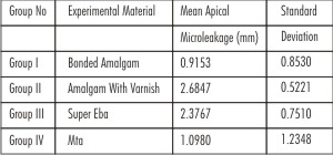
The mean apical microleakage and standard deviation for the test materials is given in (Table No.1 & Fig. 2).The
 | Table.1 Group wise Mean, And Standard Deviation of Specimens
 |
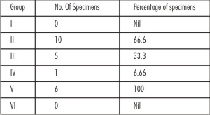 | Table 2.Percentage of Specimens Showing Complete dye penetration in each of the Groups(Group I-IV)
|
 | Table 3. Comparison between intergroups
|
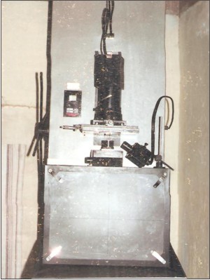 | Fig.1. Profile Projector
|
Bonded Amalgam (Group 1) Showed an apical dye penetration ranging from 0.29mm-2.64mm with the mean of 0.9153 (Fig. 3). Dye penetration in the Amalgam with varnish group (Group 2) was from 1.54mm-36mm with the mean of 2.6847mm (Fig. 4). Measurements for super EBA (Group 3) ranged from 1.05mm- 3mm with the mean of 2.3767mm (Fig. 5).The mineral trioxide group (Group 4) measurements varied from 0.00-3 with mean being 1.0980mm(Fig. 6). The statistical analysis of the results was done using the Non-parametric test "Kruskal Wallis test" (Table No. 2) to compare the groups. The results of this test H=27.434 (p=0.000) indicated that there was very high significant difference amongst the four groups. The significance of difference in the mean apical microleakage values between any two groups was evaluated using the non parametric Mann-Whitney' U' tests(Table No. 3).
Discussion
According to Wiene[11], the most common cause of endodontic failure is the lack of apical seal .A study conducted by Hirsh and colleagues[12] found that the quality of root-end filling was an important determinant of healing after periapical surgery.
Clinically, silver amalgam is the most extensively used root-end filling material. Recently, many authors have questioned the suitability of amalgam as a root-end filling material. Seltzer has stated that amalgam, regardless of time, leak more than other newer root - end filling materials. Whereas Frank et al[13] reported 60% success of such restorations.
The important aspects that was not considered by Frank et al includes Size and shape of root end cavity, Root end bevel, Type of amalgam used, whether it was bonded or not. This experiment is based on the fundamental improvement in Cavity design and Bonding.
Studies have shown that affective bond can be mediated between amalgam and tooth structure using unset luting glass ionomer cement to eradicate marginal percolation[4],[6]. Initially when the cement paste is fluid, there are many free pendant -COOH available for hydrogen bond formation that enhances the wetting
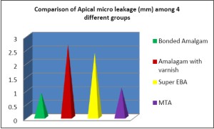 | Fig 2. Comparison of Apical micro leakage (mm) among 4 different groups
|
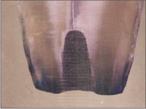 | Fig.3. specimen From Group 1 Showing Least Microleakage
|
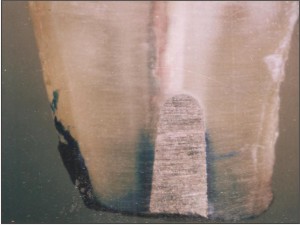 | Fig.4. Specimen From Group 2 Showing Least Microleakage
|
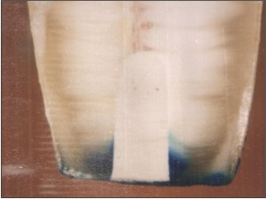 | Fig.5. Specimen From Group 3 Showing Least Microleakage
|
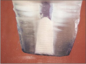 | Fig.6. Specimen From Group 4 Showing Least Microleakage
|
of the active substrates. As the reaction proceeds, these hydrogen bridges will be progressively replaced by the metal bridges, thus providing more rigid attachment[14]. As amalgam alloy contains tin, silver, copper and zinc, it can be expected to bond with glass ionomer.
Available literature shows Polyacrylic acid is a more effective conditioning agent and was used in this study. It also did not disturb the smear layer, which was rich in calcium and phosphate and played an important role in adhesion to dentin[15].
Dorn and Gartner[7] reported 94% success for Super-EBA as a root-end filling material. Therefore this material was chosen for comparison. Reports suggest MTA to be a promising root-end filling material. MTA[9],[10] was therefore selected for comparison in this study.
This investigation was thus conceived to reinvestigate amalgam with suitable modifications in cavity design and bonding and compare the apical microleakage of amalgam with varnish, super-EBA and Mineral trioxide aggregate using a dye-penetration method.
According to literature resection angle of 30 or 40 exposed many dentinal tubules as compared to a flat, 0 degree cut[16]. Mattison et al[17] and Gangliani[18] have recommended that an apical cavity depth of 3 mm or more along the vertical axis produce a safe and effective seal.
Microleakage tests have been criticized because tracers are not representative of 'disease-causing' agents in the canal and often attempt to standardize conditions of tests obviate the clinical limitations of access and artificially improve the chances of favorable results. Despite this, microleakage tests will remain the easiest and most useful means of comparing the efficacy of different materials and techniques in achieving this goal.[19]
Methylene blue is water soluble, easily penetrates the water compartment of the tooth, and does not adsorb to the dental matrix or apatite crystals. Apical 1/3rd of all specimens were immersed in 1% methylene blue for 72 hours, to allow maximum penetration. To observe dye penetration at the filling material- dentin interface stereomicroscope along with profile projector was used for making linear measurements. Literature review indicates hardly any studies that have utilized profile projector. This could become an additional or alternative supplement, which can be used while making the linear measurements of dye penetration.
From the analysis, it is evident that Group I, bonded amalgam, showed the least mean apical microleakage (0.9153). Scrutiny of published research indicates hardly any studies that have performed a comparative evaluation between the sealing abilities of bonded amalgam and other advocated root-end filling materials.
Mineral trioxide aggregate, also showed promise in this present dye leakage study. Torabinejad et al have stated that the sealing ability and marginal adaptation of MTA was superior to that of silver amalgam with or without varnish or to super-EBA cement in both dye and bacterial leakage methods. The mean apical microleakage of MTA (1.0980mm) was more than bonded amalgam (0.9153mm) but was not statistically significant.
From the analysis it is evident that there is no significant difference in the mean apical microleakage of high copper amalgam with varnish and super-EBA cement. This finding is in concurrence with various studies. However, this is in contrast with studies done by Szermeta Browar et al[20], Bondra[21], which might be due to difference in depth of apical preparation.
Mean apical microleakege of amalgam with varnish (Group II) was very highly statistically significant compared to bonded amalgam (Group I) and MTA (Group IV). This difference can be explained as Fuji 1 provides mechanical and chemical bond in the intermingled unset Glass ionomer and amalgam. In this study, the roots were kept moist to simulate clinical conditions, since MTA sets in the presence of moisture; this helps in enhancing the sealing property of MTA. Whereas when varnish was used it prevented microleakage only temporarily because of its eventual solubility and also by occupying space and not by bonding.
Similarly, differences between bonded amalgam (Group I) and super-EBA (Group III) were very highly statistically significant. This difference can be attributed to variations of temperature and humidity within the operatory, which effected the setting time of super-EBA. In contrast, amalgam- working time was more predictable.
The differences between super-EBA (Group III) and MTA (Group IV) were highly statistically significant. These differences could be related to physical and chemical properties of the material, as well as to handling during mixing and insertion.
The present study revealed excellent sealing ability of bonded amalgam as a root-end filling material when compared with materials in other experimental groups. However, further studies with better evaluation criteria with different clinical situations are required to substantiate the results of the present study.
Conclusion
Based on the data presented herein, it is not possible to make a recommendation about the best material to be used. Yet, bonded amalgam showed promise in this study. This proves the hypothesis that conservative cavity preparation and bonding does play a significant role in decreasing microleakage. So this can be considered as one of the potential alternatives along with MTA for root- end filling.
Acknowledgement
We thank Dr. Raj Shekar Patil, Mangalore University, Dr. Venkatesh Murthy, MIT & Manipal College of Dental Sciences, Manipal.
References
1. Rud J, Andreasen JO, Moller JJE .Multivariate Analysis of the Influence Of various Factors upon Healing after Endodontic Surgery. Oral Surg 1972; 1:258-71.
2. Moodnik RM, Levey MH, Besen MA, Borden BG. Retrograde Amalgam Filling: A scanning electron Microscopic study. J. Endod 1975; 1: 28-31.
3. Kos WL, Aulozzi DP, Gerstein H. A Comparative Bacterial Microleakage Study Of Retrofilling Materials. J. Endod 1982; 8:355-8.
4. Kurien S. Bhat KS. An Invitro Comparison Of The Sheer Bond Strength Of Amalgam To Tooth Structure Using Two Bonding Agents- Luting Glass Ionomer And 4-META. 1993 CODS Thesis.
5. Moayad MAI, Aboush YE, Elderton RJ. Bonded Amalgam Restorations: A comparative Study OF Glass-IonomerAnd Resin Adhesives. Br.Dent J 1993;175:363..
6. Chen RS, Liu CC, Cheng MR, Lin CP. Bonded amalgam restorations: Using a glass-ionomer as an adhesive liner. Oper Dentistry, 2000; 25:411-17.
7. Dorn SO, Gartner AH. Retrograde filling materials a retrospective success-failure study of amalgam,EBA and IRM. J.Endod 1990; 16:391-93
8. Tuggle ST, Anderson RW, Pantera EA, A dye penetration study of retro filling materials. J Endod 1989;15:122-124.
9. TorabinejadM , Watson TF , Pittford TR . Sealing ability of a mineral trioxide aggregate when used as a root-end filling material. J Endod 1993; 19 ; 12:591-5.
10. Aqrabawi J. Sealing ability of amalgam , Super EBA cement and MTA when used as retrograde filiing material . Br Dent J 2000; 189 , 9:469-471
11. Weine FS. Endodontic therapy.3rd edition. St. Louis:CV Mosby , 1982 ; 419-30.
12. Hirsch JM ,Ahlstorm Ulf ,Henrikson , Hayden G, Peterson LE. Periapical surgery.Int . J Oral surgery 1979 :8 :173-85.
13. Frank AL , Glick DH , Patterson SS, Weine FS . long term evaluation of surgically placed amalgam fillings J. Endod 1992; 18 : 391-98.
14. AboushVE , Jenkins CB. The bonding of glass- ionomer cements to dental amalgam . Br Dent J 1989 ; 166 :225 -257. [15] Martell B, Chandler NP . Electrical dye leakage comparison of three root-end restorative materials . Quintessence Int 2002 , 33 , 1:30-4
15. PowisDR ,Folleras T, Merson SA , Wilson AD. Improve adhesion of glass ionomer to dentin and enamel .JrDent . Res. 1982;61:1416-1422.
16. Gilheny PA, Figdor D, Tyas MJ. Apical dentin permeability and microleakage associated with root-end resection and retrograde filling, J. Endod 1994;20:22-6
17. Mattison D, Anthony I, Philip D, Anderson. Microleakage of retrograde amalgams. J Endod 1985; 11:340-5
18. Gagliani M, Taschieri S, Molinari R. Ultrasonic root-end preparations: influence of cutting angle on the apical seal. J Endod 1998; 24,11:726-30.
19. Matloff IR, Jensen JR, Singer L, Tabibi A. A comparison of methods used in root canal Sealability studies. Oral Surg 1982: 53: 203-08
20. Szeremeta - BrowarTL , Van Cura JE, Zaki AE. A comparison of the sealing properties of different retrograde techniques: an auto radiographic study. Oral surg 1985; 59:82-7.
21. Bondra DL, Hartwell GR, Macpherson MG, Portell FR. Leakage in vitro with IRM, high copper amalgam and EBA cement as retro filling materials. J.Endod 1989; 15: 157-60. |