Introduction
By definition, supernumerary teeth are extra teeth in comparison to normal dentition. The most common type of supernumerary tooth as indicated by Alberti et al, is mesiodens. [1], [2] They may be single or multiple, unilateral or bilateral, inverted, erupted or impacted and in one or both the jaws. [1],[3],[4] A combination of numerous genetic and environmental influences also may have an effect on tooth number and morphology. [5] Several reported cases show a familial incidence of mesiodens. In some cases, more than one sibling has been affected.[6]
This paper presents mesiodens in twins, one twin presenting with erupted double mesiodens in the midline and the other shows palatally erupted single mesiodens.
Case Report
An oral health survey in a primary school was conducted by the department of Oral Pathology. During examination of the students, we came across monozygotic twin brothers 14 years old with mesiodens. The twins were of similar facial appearance with many shared physical features. Their medical history was non- contributory and there was no history of similar anomalies in their family.
Intra oral examination of first twin revealed permanent dentition with mal-aligned teeth. Two conical shaped mesiodens were erupted labially displacing the maxillary central incisors (Figure 1). Complete root formation of both the mesiodens was evident in maxillary occlusal radiograph (Figure 2). Intraoral examination of the second twin revealed palatally placed single conical erupted mesiodens in the maxilla and slight overlapping of central insicors (Figure 3). Intraoral periapical radiograph showed complete root formation of the mesiodens (Figure 4). Radiographs revealed no associated pathologies.
Both the twins were referred to dental college for further treatment. Mesiodens were extracted in both the cases and the first twin was treated orthodontically for mal-aligned teeth.
Discussion
Mesiodens is one of the developmental anomalies commonly seen in dental clinics and can cause esthetic or pathologic problems. Therefore, early detection is the most important measure for prevention of complications. [2]
The etiology of mesiodens is not well understood but several theories have been postulated regarding the causes of supernumerary teeth, including atavism, dichotomy of the tooth bud, and hyperactivity of the dental lamina.[1],[2] Genetics is thought to contribute to the development of mesiodens as such teeth have been diagnosed in twins, siblings, and sequential generations of a family [7] and this could be the possible etiology in the present case report. Supernumerary teeth may occur in isolation or as a part of syndrome, such as, cleido- cranial, gardener's and cleft lip & palate syndrome.[1]
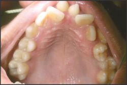 | Fig. 1: Intraoral photograph showing labially placed erupted double mesiodens in first twin .
 |
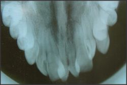 | Fig. 2: Intraoral occlusal radiograph showing complete root formation of both the mesiodens in first twin.
|
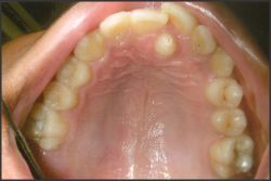 | Fig. 3: Intraoral photograph showing single palatally erupted mesiodens in the second twin.
|
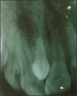 | Fig. 4: Intraoral periapical radiograph showing complete root formation of the mesiodens in the second twin.
|
The reported prevalence of mesiodens in general population ranges between 0.15- 1.9%. [1] They are more common in males than in females.[1],[3],[7]
Mesiodens has been reported commonly in maxilla specifically in pre maxillary region.[1] Earlier studies have shown predominantly, a palatal position for the mesiodens.[2] A study by Rajab LD and Hamdan MAM showed approximately 25% of mesiodens are located in the midline.[8] Mesiodens can significantly alter both occlusion and appearance by altering the eruption path and the position of the permanent incisors.[2] The site and location of the mesiodens in the present report is similar to earlier reported studies.
Mesiodens can occur individually or as multiples (mesiodentes), may appear unilaterally or bilaterally, and often do not erupt. According to the shape and size, two subclasses are considered in the classification of mesiodens; namely, eumorphic and dysmorphic. The eumorphic (supplemental) subclass is usually similar to a normal-sized central incisor, whereas the dysmorphic teeth have different shapes and sizes and are categorized into conical, tuberculate, supplemental and odontomes,[1],[4] of which the conical form is the most common type.[4] Conical mesiodentes usually occur singly. They are generally peg-shaped and are usually located palatally between the maxillary central incisors, tending to displace the erupting permanent central incisors. They often have a completely formed root and can erupt into the oral cavity.[4] Foster and Taylor examined this relationship and found tuberculate types more commonly produced delayed eruption, whereas, conical types more commonly produced displacement of the adjacent dentition.[9]
The present paper reported mesiodens in monozygotic twins. The occurrence of mesiodens in twins is, therefore, of interest which is an unusual and not a rare event. Such cases may shed some light on the etiology of this common anomaly which has been supposed to have a genetic basis, possibly inherited as autosomal dominant gene with incomplete penetrance.[5] Seddon RP, Johnstone SC and Smith PB reported a case of unerupted mesiodens in monozygotic twins. They also reviewed 8 cases and stated that monozygotic predominate dizygotic twins. Of the eight cases reported, two had described monozygous twins with bilateral mesiodens. Schon in 1974, described German twin boys with bilateral unerupted mesiodens and Choi et al in 1990 described monozygous Chinese twin boys both with an inverted mesiodens on the right side and a normally oriented mesiodens on the left side. In one twin the normally oriented mesiodens had erupted.[5] In our case report, first twin had double mesiodens and second twin had single. All three were erupted, being of conical variety.
The complications associated with mesiodens include delayed eruption in children with mixed dentition or permanent dentition, midline diastema, displacement or rotation of adjacent permanent incisors, cyst formation or cystic changes, resorption or dilacerations of the adjacent roots and nasal eruption.[2],[3]
Early diagnosis and proper line of treatment is necessary to prevent associated complications.
There are two methods for extraction of mesiodens; early extraction before root formation of the permanent incisors and late extraction after root formation of the permanent incisors. Some authors recommend extraction of mesiodens in the early mixed dentition in order to facilitate spontaneous eruption and alignment of the incisors.[1] In order to promote eruption and proper alignment of adjacent teeth, it is recommended to extract mesiodens in the early mixed dentition, which may reduce the need for orthodontic treatment.[1] The later the extraction of the mesiodens, the greater the chance that the permanent tooth either will not spontaneously erupt or will be mal-aligned when it does erupt.[4]
References
1. Meighani G, Pakdaman A . Diagnosis and Management of Supernumerary (Mesiodens): A Review of the Literature. Journal of Dentistry, Tehran University of Medical Sciences 2010; 7: 41-49.
2. Hyun HK, Lee SJ, Lee SH, Hahn SH, Kim JW. Clinical Characteristics and Complications Associated With Mesiodentes. J Oral Maxillofac Surg 2009; 67: 2639-2643.
3. Roychoudhury A, Gupta Y, Parkash H. Mesiodens: A retrospective study of fifty teeth. J Indian Soc Pedo Prev Dent 2000; 18:144-146.
4. Russell KA, Folwarczna MA. Mesiodens - Diagnosis and Managementof a Common Supernumerary Tooth. J Can Dent Assoc 2003; 69:362-366.
5. Seddon RP, Johnstone SC, Smith PB. Mesiodens in twins: a case report and review of the literature. Int J of Pediat Dent 1997; 7:177-184.
6. Sedano HO, Gorlin RJ. Familial occurrence of mesiodens. Oral Surg Oral Med Oral Pathol Oral Radiol Endod 1969;27: 360-362.
7. Pavithra S, Babu A. Mesiodens with an unusual morphology and multiple impacted supernumerary teeth in a non-syndromic patient. Indian J Dent Res 2007; 18: 137-140.
8. Rajab LD, Hamdan MAM. Supernumerary teeth: review of the literature and a survey of 152 cases. Int J of Pediat Dent 2002; 12:244-254.
9. Scheiner MA, Sampson WJ. Supernumerary teeth: A review of the literature and four case reports. Aust Dent J 2007;42:160-65. |